Báo cáo y học: " Mesothelioma of the testis and nephrotic syndrome: a case report" potx
Bạn đang xem bản rút gọn của tài liệu. Xem và tải ngay bản đầy đủ của tài liệu tại đây (1.79 MB, 4 trang )
Case report
Open Access
Mesothelioma of the testis and nephrotic syndrome: a case report
Justine Bacchetta
1
, Dominique Ranchère
1
, Frédérique Dijoud
2
and
Jean-Pierre Droz
1
*
Addresses:
1
Departments of Medical Oncology and Pathology, Centre Léon Bérard, Lyon and Université Claude Bernard Lyon 1, France and
2
Department of Pathology, Hôpital Femme Mère Enfant, Bron and Université Claude Bernard Lyon 1, France
Email: JB - ; DR - ; FD - ; JPD* -
* Corresponding author
Published: 5 June 2009 Received: 7 February 2008
Accepted: 23 January 2009
Journal of Medical Case Reports 2009, 3:7248 doi: 10.1186/1752-1947-3-7248
This article is available from: />© 2009 Bacchetta et al; licensee Cases Network Ltd.
This is an Open Access article distributed under the terms of the Creative Commons Attribution License (
/>which permits unrestricted use, distribution, and reproduction in any medium, provided the original work is properly cited.
Abstract
Introduction: Paraneoplastic glomerulopathies are rare manifestations of neoplastic disease to be
distinguished from iatrogenic renal damage. Solid tumors are preferentially associated with
membranous nephropathy, whereas Hodgkin’s lymphomas are associated with minimal change
disease.
Case presentation: We report a 63-year-old Caucasian male diagnosed with a mesothelioma of
the tunica vaginalis testis who, secondary to this, also presented with a nephrotic syndrome due to
minimal change disease. In the present case, the paraneoplastic etiology of the nephrotic syndrome
can be discussed on four unusual elements: minimal change lesions were found; the glomerulopathy
was very sensitive to corticosteroids; the nephrotic syndrome occurred 11 months after the
diagnosis of the primary malignancy, but concomitantly with the recurrence; and the nephrotic
syndrome did not decrease with tumor control and did not recur when the mesothelioma escaped
treatment. No other etiologies could nevertheless explain this phenomenon.
Conclusion: Paraneoplastic nephrotic syndrome is often associated with membranous nephropathy
in patients with solid tumors, especially in patients with lung and gastrointestinal tract neoplasia. The
management of these patients is associated with a symptomatic treatment such as sodium and water
restriction, diuretics and ACE inhibitors and a prophylaxis of specific complications of nephrotic
syndrome including thromboembolism, infections and lipid abnormalities. Treatment of neoplasia
must be undertaken rapidly, treatments must be regularly analyzed and drugs binding to albumin may
be used with precaution.
Introduction
The term ‘paraneoplastic syndrome’ refers to clinical
manifestations not directly related to tumor burden,
invasion or metastasis, but caused by the secretion of
tumor cell products such as hormones, cytokines, growth
factors and tumor antigens. Paraneoplastic glomerulopa-
thies are rare manifestations of neoplastic disease to be
distinguished from iatrogenic renal damage. Solid tumors
Page 1 of 4
(page number not for citation purposes)
are preferentially associated with membranous nephro-
pathy, whereas Hodgkin’s lymphomas are associated
with minimal change disease. Paraneoplastic glomerulo-
pathies are well known entities rarely associated with
mesothelioma.
Case presentation
We report a 63-year-old Caucasian man diagnosed with a
tumor of the right side of the scrotum in September 2002.
He was operated through an inguinal incision. The aspect
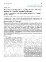
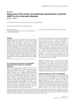
and histologic pattern were indicative of a tubulopapillar,
malignant and well differentiated mesothelioma of the
tunica vaginalis testis (Figures 1 and 2). Resection margins
were in vaded, thus a second surgical procedure was
performed, with both a scrotectomy and an orchiectomy.
A staging work-up, with thoraco-abdominal CT scan and
standard blood chemistry showed no abnormality. The
patient was then referred to our occupational medicine
clinic. No exposure to asbestos was found, but he had a
prolonged history of tobacco exposure for 30 years, until
1994. In August 2003, he experienced weight gain and
generalized edema which regressed with furosemide and
spironolactone diuretics. He was then referred to our
institution for evolution of retroperitoneal lymph nodes
on whole-body CT scan. The clinical examination was
uninformative and there was no hypertension. The only
abnormalities on the CT scan were 18 mm transversal inter
aortico-cava and retro-cava lymph-nodes. Major biological
abnormalities were seen in routine laboratory tests: low
total serum protein (48 g/l), low serum albumin (9·7 g/l)
and elevated chol esterol ( 5·59 g/l). However, serum
creatinine was normal at 70 umol/l; liver enzymes,
serum ionogram and triglycerides were within normal
limits. Proteinuria was 9 g/24 hours without microscopic
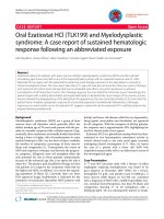
hematuria. The diagnosis of pure nephrotic syndrome led
to performing a percutaneous renal biopsy in October
2003. It showed minimal change disease with neither
immunoglobulins nor complement deposits (Figure 3).
Other etiologies of nephrotic syndrome were eliminated:
antibodies against hepatitis B and C viruses and against
HIV, antinuclear antibodies and antineutrophil cytoplas-
mic antibodies were negative. Serum levels of C3, C4 and
CH50 were normal. Oral prednisolone administration
was initiated at a dose of 1 mg/kg/day. Furosemide was
continued at a dose of 40 mg/day. His proteinuria
disappeared in December 2003, so the prednisolone
dosage was progressively decreased. While the nephrotic
Figure 1. Papillary pattern of mesothelioma, Hemalun Eosin
Safran (HES).
Figure 2. Immunohistochemistry of mesothelioma, calretinin
antibody, Zymec DC8, dilution 1/50.
Figure 3. Minimal change disease on the renal biopsy, Masson
Trichrome (x200).
Page 2 of 4
(page number not for citation purposes)
Journal of Medical Case Reports 2009, 3:7248 />syndrome became controlled, retr operitoneal disease
progressed both in size and localisation. A control CT
scan performed in February 2004 showed increased
lombo-aortic lymph nodes, with a suspicion of extension
to the retrocrural area. A percutaneous retroperitoneal
lymph node biopsy showed tissue invasion by mesothe-
lioma. As the nephrotic syndrome was well controlled by
treatment, the strategy was to perform radical bilateral
retroperitoneal lymph node dissection. There was no
peritoneal involvement. The histologic aspect was epithe-
lioid mesothelioma with necrosis and invasion of both the
capsule and small vessels. A recurrence of the nephrotic
syndrome was observed 10 days after the surgery.
Prednisolone was then increased to 1 mg/kg/day for
1 month, and then decreased to 0.75 mg/kg/day, and
then 0.5 mg/kg/day to allow for postoperative healing. In
May 2004, a CT scan showed a disease progression with
suprarenal lymph nodes of 20 mm maximal diameter.
A combination of cisplatin 75 mg/m
2
and pemetrexed
500 mg/m
2
was initiated. Six cycles were given from June
2004 to September 2004. In June 2004, the renal function
was normal, with a normal proteinuria, tota l serum
protein of 59 g/l and serum creatinine of 75 umol/l. A
slow decrease in prednisolone dosage over 6 months was
decided. In September 2004, an elevated blood pressure
was observed for the first time (systolic at 150 mmHg and
diastolic at 95 mmHg), and serum creatin ine levels
increased to 125 umol/l. This was attributed to cisplatin
renal toxicity, and then prednisolone decrease was
continued. In October 2004, the CT scan showed disease
progression in the retroperitoneum; FDG-PET examina-
tion revealed a unique site of radio-isotope fixation on the
eleventh dorsal vertebra. A percutaneous biopsy showed
involvement of mesothelioma, but the histologic pattern
of the lesion was undifferentiated. Radiotherapy on the
lumbar area was then decided. In November 2004, the
renal function was normal and the nephrotic syndrome
did not recur while he was receiving a daily dose of 0.1 mg/
kg prednisolone. In February 2005, he developed disease
recurrence with ascites. Cytological examination of the
ascites showed mesothelioma involvement. Performance
status declined, palliative treatment was given and the
patient eventually died of disease progression in March
2005.
Discussion
Paraneoplastic glomerulopathy is a well known entity [1]
rarely associated with mesothelioma. A PubMed search
using the keywords “mesothelioma”, “paraneoplastic
glomerulopathy” and every histopathologic subtype of
glomerul opathy was perfor med. Only five cases of
concurrent mesothelioma and nephrotic syndrome have
been reported in the literature. All were associated with
pleural mesothelioma [2-6] and all five patients were
men. A d iagnosis of nephrotic syndrome preceded
mesothelioma in two cases and was concomittant in the
three other cases. Membranous nephropathy, minimal
change disease and membrano-proliferative glomerulone-
phritis were observed. T hree patients had asbestosis
exposure and only one patient with nephrotic syndrome
was treated successfully with corticosteroids. All the five
patients died of disease progression. As far as we know,
this is the first report of a mesothelioma of the tunica
vaginalis testis associated with nephrotic syndrome [7].
Nephrotic syndrome is often associated with membranous
glomerulopathy in patients with solid tumors, especially
in patients with lung and gastrointestinal tract neoplasias
[8]. IgA nephropathy, minimal change disease and
glomerulosclerosis are less frequent. The nephrotic syn-
drome usually precedes the tumor by several months
according to Lee et al. [9] and renal disease antedates the
diagnosis of cancer in two-thirds of the surveyed popula-
tion. The diagnosis of paraneoplastic nephrotic syndrome
may be evoked when the following criteria are present
[10]: no evidence of other etiology of the nephrotic
syndrome; time relationship between the diagnosis of
nephrotic syndrome and cancer; tumor treatment asso-
ciated with a decrease o f renal symptoms; tumor
recurrence associated with an increase in renal symptoms
and proteinuria. A causal relationship is suggested if
nephrotic proteinuria develops either 6 months before or
after the diagnosis of malignancy. In the present case, we
can discuss the paraneoplastic etiology of the nephrotic
syndrome on four unusual elements: we found minimal
change lesions, not a membranous nephropathy; the
glomerulopathy was very sensitive to corticosteroids; the
nephrotic syndrome occurred eleven months after the
diagnosis of the primary malignancy, but concomitantly
with the recurrence and; the nephrotic syndrome did not
decrease with tumor control and did not recur when the
mesothelioma escaped treatment. No other etiologies
could explain this phenomenon.
Conclusion
We conclude that this minimal change nephropathy is a
casual event in the history of a very rare tumor. A person
known to suffer from malignancy and who develops a
nephrotic syndrome, should undergo renal biopsy if their
general condition allows. The management of people with
cancer and paraneoplastic nephropathy should focus on
the following elements [8]. First, symptomatic treatment
of the nephrotic syndrome with sodium and water
restriction and diuretic therapy is justified. In the majority
of patients, the use of a distal diuretic is sufficient. To our
knowledge, there are no studies of corticotherapy in
paraneoplastic glomerulopathies in the literature. Prophy-
laxis and the early treatment of complications of the
nephrotic syndrome such as thromboembolism, infec-
tions and lipid abnormalities are useful. ACE inhibitors
can be used to decrease blood pressure and proteinuria,
Page 3 of 4
(page number not for citation purposes)
Journal of Medical Case Reports 2009, 3:7248 />controlling hyperkalemia and renal function. Second, a
systematic search for associated electrolyte abnormalities
is legitimate. Third, all treatments should be regularly
analyzed to avoid further toxicity; drugs binding albumin
may be used with caution. And last, the treatment of
neoplasia should be undertaken rapidly. Patients with
cancer may be screened daily for proteinuria at diagnosis
and during the course of the disease.
Competing interests
The authors declare that they have no competing interests.
Abbreviations
HES, Hemalun Eosin Safran.
Consent
As the patient died three years ago, we could not obtain his
consent for publication of results. We tried but were
unable to trace his next of kin. However, the patient
cannot be identified and we see no reason why his next of
kin would object to publication of this case report.
Authors’ contributions
JB wrote the manuscript and reviewed the literature about
paraneoplastic glomerulopathies. DR and FD perfomed
pathology examinations and provided figures (mesothe-
lioma and kidney biospy, respectively). JPD perfomed the
revisions and the final approval of the manuscript.
Acknowledgements
Mrs. Marie Dominique Reynaud for her help in editing the
manuscript.
References
1. Ronco PM: Paraneoplastic glomerulopathies: new insights into
an old entity. Kidney Int 1999, 56:355-377.
2. Schroeter NJ, Rushing DA, Parker JP et al.: Minimal-change
nephrotic syndrome associated with malignant mesothe-
lioma. Arch Intern Med 1986, 146:1834-1836.
3. Venzano C, Di Marco E, Garbero M et al.: Nephrotic syndrome
associated with pleural mesothelioma. An unusual paraneo-
plastic event. Recenti Prog Med 1988, 81:325-326.
4. Tanaka S, Oda H, Satta H et al.: Nephrotic syndrome associated
with malignant mesothelioma. Nephron 1994, 67:510-511.
5. Galesic K, Bozic B, Heinzl et al.: Pleural mesothelioma and
membranous nephropathy. Nephron 2000, 84:71-74.
6. Sakamoto K, Suzuki H, Jojima T: Membranous glomerulone-
phritis associated with diffuse malignant pleural mesothe-
lioma: report of a case. Surg Today 2000, 30:1124-1126.
7. WinstanleyAM,LandonG,BerneyD,MinhasS,FischerC,
Parkinson MC: The immunohistochemical profile of malignant
mesotheliomas of the tunica vaginalis. A report of 20 cases.
Am J Surg Pathol 2006, 30:1-6.
8. Davison AM: Renal diseases associated with malignancies.
Nephrol Dial Transplant 2001, 16:13-24.
9. Lee JC, Yamauchi H, Hopper J: The association of cancer and the
nephrotic syndrome. Ann Intern Med 1966, 64:41-51.
10. Burstein DM, Korbet SM, Schwartz MM: Membranous glomer-
ulonephritis and malignancy. Am J Kidney Dis 1993, 9:23-26.
11. Edgar JD, Rooney DP, McNamee P, McNeill TA: An association
between ANCA positive renal disease and malignancy. Clin
Nephrol 1993, 40:22-25.
Page 4 of 4
(page number not for citation purposes)
Journal of Medical Case Reports 2009, 3:7248 />Do you have a case to share?
Submit your case report today
• Rapid peer review
• Fast publication
• PubMed indexing
• Inclusion in Cases Database
Any patient, any case, can teach us
something
www.casesnetwork.com