Kỹ thuật xâm lấn tối thiểu trong điều trị thoát vị đĩa đệm doc
Bạn đang xem bản rút gọn của tài liệu. Xem và tải ngay bản đầy đủ của tài liệu tại đây (238.95 KB, 6 trang )
Journal of the American Academy of Orthopaedic Surgeons
80
Open hemilaminectomy to treat
symptomatic intervertebral disk her-
niation, described by Mixter and
Barr in 1934,
1
set the standard for
subsequent surgical techniques. The
trend since has been to develop less
invasive surgical procedures for the
treatment of radiculopathy second-
ary to herniated disk. The concept
of minimally invasive spine surgery
is to provide surgical options that
optimally address the disk pathol-
ogy without producing the types of
morbidity commonly associated
with open surgical procedures. Min-
imally invasive techniques are not,
however, a panacea for all lumbar
disk pathology. These techniques
are designed to treat nerve root com-
pression alone as the source of ra-
diculopathy in patients with acute
primary disk herniations.
Radiculopathy has been attrib-
uted to the production of chemical
mediators that result from the com-
pression and/or leakage of degen-
erative nuclear material through
annular tears. These chemical me-
diators may result in inflammation
and may affect the large and small
sensory afferent nerve fibers.
2,3
Phospholipase A
2
and nitric oxide
synthase from extruded or migrated
disk fragments have been specifi-
cally cited as possible agents related
to the pathophysiology of radicu-
lopathy.
4
Minimally Invasive
Techniques: An Overview
First-generation minimally invasive
methods were blind percutaneous
techniques, including chemonucleo-
lysis, percutaneous nucleotomy,
automated percutaneous nucleot-
omy, and laser disk decompression.
The development of fiberoptic visu-
alization with rigid discoscopes and
flexible endoscopes allowed more
advanced methods to be undertak-
en, including biportal arthroscopic
intradiscal diskectomy and percuta-
neous intradiscal and epidural uni-
portal techniques through postero-
lateral, posterior interlaminar, or
foraminal approaches. The stan-
dard for evaluation of percutaneous
techniques became open microdisk-
ectomy, considered the benchmark
for comparison. A recently devel-
oped percutaneous variation of the
standard laminotomy technique is
endoscopic diskectomy. Laparo-
scopic transperitoneal and retroperi-
toneal approaches to herniated
nuclear pathology also have been
introduced.
Dr. Mathews is Associate Clinical Professor,
Department of Orthopaedic Surgery, Virginia
Commonwealth University, Medical College of
Virginia, Richmond, VA. Ms. Long is Clinical
Researcher, MidAtlantic Spine Specialists,
Richmond.
One or more of the authors or the departments
with which they are affiliated has received
something of value from a commercial or other
party related directly or indirectly to the sub-
ject of this article.
Reprint requests: Dr. Mathews, Suite 200,
7650 Parham Road, Richmond, VA 23294.
Copyright 2002 by the American Academy of
Orthopaedic Surgeons.
Abstract
Hemilaminectomy with diskectomy, the original surgical option to address
intervertebral disk herniation, was superseded by open microdiskectomy, a less
invasive technique recognized as the surgical benchmark with which minimally
invasive spine surgery techniques have been compared as they have been devel-
oped. These minimally invasive surgical techniques for patients with herniated
nucleus pulposus and radiculopathy include laser disk decompression, arthro-
scopic microdiskectomy, laparoscopic techniques, foraminal endoscopy, and
microendoscopic diskectomy. Each has its own complications and requires a
long learning curve to develop familiarity with the technique. Patient selection,
and especially disk morphology, are the most important factors in choice of tech-
nique. The optimal candidate has a previously untreated single-level herniation
with limited migration or sequestration of free fragments.
J Am Acad Orthop Surg 2002;10:80-85
Minimally Invasive Techniques for the Treatment of
Intervertebral Disk Herniation
Hallett H. Mathews, MD, and Brenda H. Long, MS, RN
Perspectives on Modern Orthopaedics
Hallett H. Mathews, MD, and Brenda H. Long, MS, RN
Vol 10, No 2, March/April 2002
81
Advances in minimally invasive
surgery relate to a number of factors:
understanding of a technique’s abili-
ty to effect nerve root decompres-
sion, development of approaches
that are based on the location of the
disk pathology, and the refinement
of diagnostic modalities to aid in lo-
cating specific disk pathology. Ad-
vances in fiberoptic visualization
have been a factor, as has refined
surgical instrumentation. Better
patient selection has resulted from
experience with individual tech-
niques as well as an appreciation of
technique-related complications and
outcomes. In addition, improved
fluoroscopic imaging and navigation
systems have enhanced the safety
and predictability of minimally inva-
sive techniques when performed by
experienced endoscopic surgeons.
Perhaps the most notable advan-
tage of minimally invasive tech-
niques is the ability they provide to
surgically address and resolve her-
niated nuclear pathology without
the morbidity associated with inci-
sion of the paraspinal muscle in tra-
ditional open techniques. Enhanced
visualization of the surgical field
allows the pathology to be seen and
permits both identification and
avoidance of injury to the neurovas-
cular structures. The surgical field
can be surveyed before conclusion of
the procedure, and the diskectomy
itself can be inspected and docu-
mented on videotape. In addition,
these procedures generally are done
on an outpatient basis. Patients usu-
ally require minimal analgesic med-
ication and have a timely return to
activities of daily living, including
work. Little, if any, postoperative
rehabilitative therapy is necessary.
Consequently, the overall economic
impact of minimally invasive tech-
niques in most instances is less than
that of open techniques.
Disadvantages to minimally
invasive surgery for the herniated
disk are few. Primarily, the learn-
ing curve for the surgeon and his
or her staff is steep. Mastery of the
neurovascular anatomy is required,
and familiarity with the spatial ori-
entation of the endoscopic field is
critically important.
The goal of minimally invasive
techniques is either disk debulking
or selective fragment removal to
alter disk morphology and subse-
quently abate nerve root compres-
sion. Selective fragmentectomy
may remove an obstructive disk
herniation mechanically. However,
intradiscal depressurization and
lavage also may improve symptoms
without significant change in neural
anatomy. Good results have been
achieved without significant change
in neural anatomy following the
procedure. The governing factor in
considering a minimally invasive
procedure is patient selection.
5
Indications for Minimally
Invasive Spine Surgery
Except in emergent circumstances,
such as rapidly progressive neuro-
logic deficits or the threat of cauda
equina syndrome, 6 to 8 weeks of
nonsurgical treatment with appro-
priate medication and conservative
care is routine before proceeding
with surgical intervention. The ideal
candidate should have unilateral
radicular pain radiating into the
foot, with leg pain greater than back
pain. Positive straight leg raising is
often present. The radicular pain
may be described as lancinating
and/or aching. Other complaints
can include numbness, tingling, and
weakness, along with decreased sen-
sation to light touch and pin prick.
Because herniation can result in
canal stenosis relative to the size of
the herniation, some patients com-
plain of pseudoclaudication.
Location of the herniation dic-
tates the appropriate minimally
invasive approach. Magnetic reso-
nance imaging is the most effective
radiologic test for visualizing disk
pathology and achieving a defini-
tive correlation with patient presen-
tation. Diskography, although con-
troversial, may be especially helpful
in the diagnosis when symptoms
are equivocal and the pain genera-
tor can be isolated through symp-
tom provocation.
6
Optimal candidates for minimal-
ly invasive access are those with a
single-level herniation that has not
previously undergone surgery and
occupies <50% of the spinal canal,
with limited migration or sequestra-
tion of free fragments. Scarring or
other deviations in normal anatomy
that may have resulted from previ-
ous surgical intervention at a de-
fined level are a relative contrain-
dication to minimally invasive revi-
sion surgery for disk herniation
recurrence. Developmental spinal
stenosis and minimal disk hernia-
tion presenting as only a small bulge
also are relative contraindications.
Techniques, Complications,
and Results
Early Techniques
Open microdiskectomy, the
benchmark procedure with which
percutaneous and minimally inva-
sive techniques are compared, uti-
lizes a small incision and a micro-
scope or loupe magnification rather
than an endoscope. The technique is
similar to minimally invasive tech-
niques in regard to patient selection
and indications. Compared with
percutaneous techniques, especially
foraminal epidural endoscopy,
diskectomy is limited in its ability to
address sequestered free fragments
with significant migration. Open
diskectomy allows the surgeon to
visualize the pathology and neu-
rovascular anatomy, but in this tech-
nique, the anatomic structures often
must be gently manipulated (rather
than avoided) for optimal access to
the disk–nerve root compression
interface. The overall rate of suc-
Treatment of Intervertebral Disk Herniation
Journal of the American Academy of Orthopaedic Surgeons
82
cessful outcomes for diskectomy has
been reported to range from 76% to
100%.
7
Other studies demonstrate
that the rate and type of complica-
tions are similar to those of mini-
mally invasive spine surgery.
8
The
most common complications are
neurovascular trauma, diskitis, and
cerebrospinal fluid leak.
8,9
Diskec-
tomy, like minimally invasive tech-
niques used to treat lumbar disk
herniations, has a learning curve
that, once achieved, must be main-
tained through regular application.
Percutaneous techniques currently
have few applications, given the
more advanced procedures now ac-
cepted. These early blind techniques
addressed central and posterocentral
pathology and were nonselective:
their basic mechanism of action was
a debulking of the nucleus pulposus
without directly addressing the her-
niation. The result was depressur-
ization and relief of tension on annu-
lar fibers as well as involution of the
nucleus, essentially withdrawing the
compressive nerve pathology back
into the annular confines.
10
Chemonucleolysis caused dena-
turization of the intervertebral
nucleus and a relative disk debulk-
ing. Allergic reactions and even
anaphylaxis occurred in approxi-
mately 1% of patients treated with
chymopapain.
11
Postoperatively,
radiographs of treated patients
often demonstrated disk space col-
lapse.
12
In some centers, chemo-
nucleolysis currently is used in con-
junction with epiduroscopy in the
treatment of migrated or seques-
tered free fragments. The technique
is more popular in Europe than in
the United States.
11
Gogan and
Fraser
13
reported an 80% success
rate at 10-year follow-up with
chemonucleolysis, and similar suc-
cess has recently been reported in a
European study.
14
However, nota-
ble neurovascular complications
and transverse myelitis, probably
resulting from the inadvertent intro-
duction of chymopapain intrathe-
cally, has substantially decreased
the use of chemonucleolysis.
Percutaneous nucleotomy and
automated percutaneous nucleotomy
are both blind, intradiscal, nonselec-
tive techniques for disk debulking
or deflation with disk involution
from the site of neural compression.
Complications have included trau-
ma or injury to neural or vascular
structures, diskitis, and cerebro-
spinal fluid leaks, as well as the po-
tential for bowel perforation. In one
multicenter study,
15
success rates
for automated percutaneous diskec-
tomy in carefully selected patients
ranged from 55% to 85%. Because
these results are less satisfactory
than those achieved with other tech-
niques, this procedure has largely
fallen out of favor.
Laser disk decompression is a
blind, nonspecific disk depressur-
ization procedure resulting in grad-
ual withdrawal of disk compression
on the nerve root. Because it is a
blind procedure, complications have
paralleled those of the blind nu-
cleotomy techniques. An additional
concern related to complications
early in the use of laser disk decom-
pression was the heat associated
with direct-firing wavelengths
delivered by probes in proximity to
neurovascular structures.
16
In cen-
ters where this technique is still in
use, successful outcomes have
ranged from 50% to 89%,
10,17
imply-
ing various outcomes in nonprospec-
tive, nonrandomized studies with
resulting inconsistent data. Recent
application of the holmium:YAG
cool, side-firing laser in conjunction
with endoscopic visualization
appears to be a promising option in
selected patients.
16
Recent Techniques
The combination of fiberoptic
technology with arthroscopic can-
nulae led to the development of
rigid discoscopes and rod-lens
endoscopes, and thus to visualized
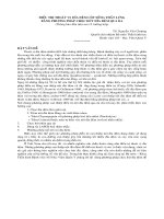
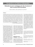
selective microdiskectomy. Arthro-
scopic microdiskectomy can be
either uniportal or biportal (Fig. 1),
depending on the targeted herniated
pathology. Small central herniations
can be approached uniportally; bi-
portal access is dictated for large
central herniations and subligamen-
tous and sequestered herniations.
Dedicated instrumentation sized to
arthroscopic application allows for
manual or automated selective disk
decompression at the pathologic
Figure 1 Arthroscopic microdiskectomy technique: the biportal approach to a paracentral
disk herniation. The instrument enters through the right foramen to allow access to the disk
herniation. Triangulation occurs within the disk nucleus. (Adapted with permission.
12
)
R
Nucleus
L
Hallett H. Mathews, MD, and Brenda H. Long, MS, RN
Vol 10, No 2, March/April 2002
83
site. Complications with this tech-
nique are minimal, but infection
(two cases), transient peroneal neu-
ropraxia (two cases), and transient
skin hypersensitivity (five cases)
have been reported, for a complica-
tion rate of 3% in one large series of
patients spanning 10 years.
18
Theo-
retic complications related to trau-
ma to neurovascular structures and
perineural/intraneural fibrosis have
not been reported. The success rate
for this technique ranges from 75%
to 98%.
18-21
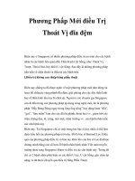
The laparoscopic anterior ap-
proach to the lumbar spine for pri-
mary disk herniations began to be
used in the mid-1980s and early
1990s. This technique allows access
to contained disk herniations, as
well as to some extruded and mi-
grated fragments, through either a
transperitoneal or retroperitoneal
approach (Fig. 2). Complications
including diskitis and segmental
instability have been reported.
22
Trauma to major vascular structures
is a potential complication that can
result in marked morbidity. This
laparoscopic procedure requires an
approach surgeon as well as unique,
expensive instrumentation. Sur-
geons also must anticipate a steep
learning curve. These factors con-
tributed to long surgical times with-
out any decrease in hospital lengths
of stay. This has led to surgical
costs that far exceed those of other
minimally invasive techniques.
23
With an early success rate of only
69% for treatment of disk hernia-
tion, the transperitoneal approach
has now been adapted for use in
anterior lumbar interbody fusion at
L5/S1.
22
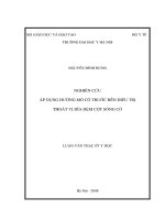
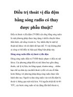
Foraminal epidural endoscopy is
a diskectomy technique that ad-
dresses paramedian, foraminal, and
extraforaminal disk herniations. It
can also access migrated and se-
questered free fragments in the
epidural space when they are limit-
ed to confines of the axilla and the
pedicle. Such access is facilitated by
appropriately sized endoscopes
with varied lens angles; foraminal
and extraforaminal disk herniations
are technically demanding for stan-
dard microscopic techniques. The
foraminal endoscopic technique
allows visualization of the patholo-
gy and avoidance of neurovascular
structures at risk, as well as visual-
ization of the selective diskectomy
and documentation of the surgical
effect at the time of the procedure
(Fig. 3). As with arthroscopic micro-
diskectomy, manual and automated
instrumentation sized to the work-
ing channel of the endoscope allows
Figure 2 Laparoscopic diskectomy technique. R = retroperitoneal space. The instruments
are inserted on the left side, with the smooth pituitary instrument traversing the retroperi-
toneum through the psoas muscle. The trochar needle is placed through the posterolateral
approach. (Adapted with permission.
22
)
Figure 3 Foraminal endoscopic diskectomy technique compared with the extraforaminal
approach. The foraminal approach allows direct dissection and removal of herniated
material. (Adapted with permission.
12
)
R
R
Exiting root
Herniated disc
Dura mater
Traversing
root
Foraminal approach
Extraforaminal
approach
Transpsoas
retroperitoneal
approach
Posterolateral
needle
Treatment of Intervertebral Disk Herniation
Journal of the American Academy of Orthopaedic Surgeons
84
for diskectomy or fragment removal
tailored to the morphology respon-
sible for the radiculopathy. The
complication rate for endoscopic
foraminal diskectomy in one large
study was 5%.
24
There is potential
for trauma to neurovascular struc-
tures, diskitis, and cerebrospinal
fluid leak, although the risk is mini-
mized by the excellent visualization.
The success rate has recently been
reported at 78%.
25
Key to the suc-
cess of foraminal epidural endo-
scopic surgery are patient selection
and, at surgery, familiarity with the
spatial orientation and with the
anatomy at risk. The learning curve
is steep, and success with this tech-
nique requires regular use.
26
For-
aminal epidural endoscopic surgery
has been equated to open micro-
diskectomy as “microdiscectomy
through a cannula.”
26
Microendoscopic diskectomy
through the interlaminar approach
allows endoscopic intervention for a
broad range of disk pathology. This
technique is indicated for all forms
of disk herniation (Fig. 4) as well as
for associated pathology, such as lat-
eral recess or central canal stenosis.
Microendoscopic diskectomy is per-
formed through a slightly larger
tubular distractor and thus closely
approximates open microdiskec-
tomy. It requires an approach to the
pathology through the paraspinous
musculature. Dilators are placed in
succession until the optimum win-
dow for surgical exposure is
achieved. A tubular retractor is then
placed that allows use of a working
channel endoscope, through which
both disk and bony pathology can
be addressed. The surgical system
and technique allow for attention
either intradiscally or extradiscally
in an area that can span from the
pedicle to the midline. Complica-
tions are similar to those of arthro-
scopic microdiskectomy and forami-
nal epidural endoscopy; in addition,
there is the potential for cauda
equina syndrome, epidural scarring,
and segmental instability. This tech-
nique is appropriate not only for
disk pathology previously not treated
surgically but also for recurrence. In
their preliminary series, the devel-
opers of this technique reported a
complication rate of one patient in
41 (3%), with all patients reporting
good to excellent results in follow-up
based on modified MacNab crite-
ria.
27
This series included patients
who underwent surgery for lateral
herniations, herniations within the
spinal canal, and free-fragment
pathology.
27
Summary
Early blind, nonspecific intradiscal
techniques have been superseded
by a variety of low-morbidity, min-
imally invasive surgical options
that offer treatment for patients
with radiculopathy secondary to
disk pathology tailored to their re-
spective pathologies. These proce-
dures provide results comparable
to those of microdiskectomy done
with magnification and may poten-
tially have advantages for some
specific indications.
Dural sac
Exiting
nerve root
Intervertebral
disk
L5
Herniation
Central
Paramedian
Foraminal
Extraforaminal
Figure 4 Disk herniations approachable by interlaminar techniques and selectively by
other minimally invasive techniques. Central: open microdiskectomy, microendoscopic
diskectomy, biportal approach. Paramedian: open microdiskectomy; microendoscopic
diskectomy; uniportal, biportal, or foraminal approach. Foraminal: open foraminal
approach, microendoscopic diskectomy, endoscopic foraminal approach. Extraforaminal:
open far lateral approach, microendoscopic diskectomy, extraforaminal endoscopy.
L4
Hallett H. Mathews, MD, and Brenda H. Long, MS, RN
Vol 10, No 2, March/April 2002
85
References
1. Mixter WJ, Barr JS: Rupture of inter-
vertebral disc with involvement of
spinal canal. N Engl J Med 1934;211:
210-215.
2. Nygaard OP, Mellgren SI: The func-
tion of sensory nerve fibers in lumbar
radiculopathy: Use of quantitative sen-
sory testing in the exploration of dif-
ferent populations of nerve fibers and
dermatomes. Spine 1998;23:348-353.
3. Saifuddin A, Mitchell R, Taylor BA:
Extradural inflammation associated
with annular tears: Demonstration
with gadolinium-enhanced lumbar
spine MRI. Eur Spine J 1999;8:34-39.
4. Kawakami M, Tamaki T, Hayashi N,
Hashizume H, Nishi H: Possible
mechanism of painful radiculopathy
in lumbar disc herniation. Clin Orthop
1998;351:241-251.
5. Andreshak TG, An HS, Hall J, Stein B:
Lumbar spine surgery in the obese
patient. J Spinal Disord 1997;10:376-379.
6. Tehranzadeh J: Discography 2000.
Radiol Clin North Am 1998;36:463-495.
7. Mayer HM: Principles of microsurgi-
cal discectomy in lumbar disc hernia-
tions, in Mayer HM (ed): Minimally
Invasive Spine Surgery: A Surgical
Manual. Berlin, Germany: Springer-
Verlag, 2000, pp 73-77.
8. Mayer HM, Brock M: Percutaneous
endoscopic discectomy: Surgical tech-
nique and preliminary results com-
pared to microdiscectomy. J Neurosurg
1993;78:216-225.
9. McCulloch JA: Microsurgery for lum-
bar disc disease, in An HS (ed): Prin-
ciples and Techniques of Spine Surgery.
Baltimore, MD: Williams & Wilkins,
1998, pp 747-764.
10. Choy DS: Percutaneous laser disc de-
compression (PLDD): Twelve years’
experience with 752 procedures in 518
patients. J Clin Laser Med Surg 1998;16:
325-331.
11. Javid MJ, Nordby EJ: Lumbar chymo-
papain nucleolysis. Neurosurg Clin N
Am 1996;7:17-27.
12. Mathews HH, Mathern BE: Percuta-
neous procedures in the lumbar spine,
in An HS (ed): Principles and Tech-
niques of Spine Surgery. Baltimore, MD:
Williams & Wilkins, 1998, pp 731-745.
13. Gogan WJ, Fraser RD: Chymopapain:
A 10-year, double-blind study. Spine
1992;17:388-394.
14. Riquelme C, Tournade A, Cerfon JF:
Efficacy of lumbar chemonucleolysis
in the treatment of foraminal and
extra-foraminal hernias [French]. J
Neuroradiol 1999;26:35-48.
15. Quigley MR, Maroon JC: Automated
percutaneous discectomy. Neurosurg
Clin N Am 1996;7:29-35.
16. Casper GD, Hartman VL, Mullins LL:
Percutaneous laser disc decompres-
sion with the holmium: YAG laser. J
Clin Laser Med Surg 1995;13:195-203.
17. Siebert W, Kaiser J, Pfeil U: Percutane-
ous laser disc decompression: Personal
experience and outlook, in Mayer HM
(ed): Minimally Invasive Spine Surgery:
A Surgical Manual. Berlin, Germany:
Springer-Verlag, 2000, pp 233-242.
18. Kambin P: Arthroscopic microdiscec-
tomy, in Mayer HM (ed): Minimally
Invasive Spine Surgery: A Surgical
Manual. Berlin, Germany: Springer-
Verlag, 2000, pp 187-199.
19. Kambin P: Diagnostic and therapeutic
spinal arthroscopy. Neurosurg Clin N
Am 1996;7:65-76.
20. Kambin P, Zhou L: Arthroscopic disc-
ectomy of the lumbar spine. Clin
Orthop 1997;337:49-57.
21. Kambin P, O’Brien E, Zhou L, Schaffer
JL: Arthroscopic microdiscectomy and
selective fragmentectomy. Clin Orthop
1998;347:150-167.
22. Obenchain TG, Cloyd D: Laparo-
scopic lumbar discectomy: Description
of transperitoneal and retroperitoneal
techniques. Neurosurg Clin N Am
1996;7:77-85.
23. Mathews HH, Long BH: The laparo-
scopic approach to the lumbosacral
junction, in Mayer HM (ed): Minimally
Invasive Spine Surgery: A Surgical
Manual. Berlin, Germany: Springer-
Verlag, 2000, pp 207-216.
24. Porchet F, Chollet-Bornand A,
deTribolet N: Long-term follow up of
patients surgically treated by the far-
lateral approach for foraminal and
extraforaminal lumbar disc herniations.
J Neurosurg 1999;90(1 Suppl):59-66.
25. Haag M: Transforaminal endoscopic
microdiscectomy: Indications and
short-term to intermediate-term results
[German]. Orthopade 1999;28:615-621.
26. Mathews HH: Transforaminal endo-
scopic microdiscectomy. Neurosurg
Clin N Am 1996;7:59-63.
27. Foley KT, Smith MM: Microendo-
scopic discectomy. Tech Neurosurg
1997;3:301-307.