Báo cáo y học: " Intracystic papillary carcinoma in a male as a rare presentation of breast cancer: a case report and literature review" potx
Bạn đang xem bản rút gọn của tài liệu. Xem và tải ngay bản đầy đủ của tài liệu tại đây (1.33 MB, 4 trang )
BioMed Central
Page 1 of 4
(page number not for citation purposes)
Journal of Medical Case Reports
Open Access
Case report
Intracystic papillary carcinoma in a male as a rare presentation of
breast cancer: a case report and literature review
Laszlo Romics Jr*
1
, M Emmet O'Brien
2
, Norma Relihan
1
,
Fionnuala O'Connell
3
and H Paul Redmond
1
Address:
1
Department of Surgery, Cork University Hospital, University College Cork, Wilton Road, Cork, Ireland,
2
Faculty of Medicine and Health,
University College Cork, Cork, Ireland and
3
Department of Pathology, Cork University Hospital, University College Cork, Wilton Road, Cork,
Ireland
Email: Laszlo Romics* - ; M Emmet O'Brien - ;
Norma Relihan - ; Fionnuala O'Connell - ; H
Paul Redmond -
* Corresponding author
Abstract
Introduction: The term "intracystic papillary ductal carcinoma in situ" has recently changed and is
now more appropriately referred to "intracystic papillary carcinoma". Intracystic papillary
carcinoma in men is an extremely rare disease with only a few case presentations published in the
literature so far.
Case presentation: We discuss a case of a 44-year-old Caucasian man with an intracystic
papillary carcinoma treated with simple mastectomy, sentinel lymph-node biopsy and contralateral
risk-reducing mastectomy. These were followed by adjuvant radiotherapy of the breast.
Conclusion: Triple assessment (i.e. clinical examination and radiological and histological
assessment) with a high level of clinical suspicion is necessary to diagnose intracystic papillary
carcinoma in men due to its rarity. Furthermore, genetic testing and risk-reducing mastectomy
should also be considered in cases of a strong family history for male breast cancer.
Introduction
Breast carcinoma in men is rare; it represents 0.6% of all
breast carcinomas and less than 1% of all malignancies in
men. Male breast cancer has an incidence of one per
100,000 per annum. Overall survival rates for men with
breast carcinoma, stratified by stage of disease, are lower
than for women with breast carcinoma. However, these
differences are most likely due to the higher age distribu-
tion of male patients and the lower life expectancy of men
in the general population [1].
Intracystic papillary carcinoma (IPC) is a rare form of
breast cancer, accounting for 0.5–1% of all breast cancers.
It typically occurs in older women and has an excellent
prognosis. The reported 10-year survival rate for IPC is
100%, the recurrence-free survival rate is 96% and 77% at
2 and 10 years, respectively [2].
Here, we report the case of intracystic papillary ductal car-
cinoma in situ (DCIS)/carcinoma of the breast in a 44-
year-old male patient.
Published: 13 January 2009
Journal of Medical Case Reports 2009, 3:13 doi:10.1186/1752-1947-3-13
Received: 7 May 2008
Accepted: 13 January 2009
This article is available from: />© 2009 Romics et al; licensee BioMed Central Ltd.
This is an Open Access article distributed under the terms of the Creative Commons Attribution License ( />),
which permits unrestricted use, distribution, and reproduction in any medium, provided the original work is properly cited.
Journal of Medical Case Reports 2009, 3:13 />Page 2 of 4
(page number not for citation purposes)
Case presentation
A 44-year-old Caucasian man presented to the breast
clinic with a 3-week history of a swelling in his left breast.
He also had a significant family history for breast cancer
including a maternal grandmother, two of his maternal
aunts and a maternal first cousin diagnosed with breast
cancer. On examination, a well-circumscribed, 2.5 cm
swelling was palpable within an area of gynecomastia on
the left chest wall. Sonographically, a cystic mass with
internal echoes was present without posterior acoustic
shadowing. Aspiration of the lesion revealed uniformly
blood-stained fluid and a residual swelling persisted.
Cytology analysis of the aspirate confirmed the presence
of atypical cells. Mammography showed a circumscribed
mass in the sub-areolar region of the left breast with par-
tially obscured margins. An irregular outline was noted on
cranio-caudal view, but no spiculation or suspicious inter-
nal micro-calcifications were found. A core biopsy of the
lesion revealed atypical ductal hyperplasia, but no evi-
dence of malignancy was seen. In view of the atypical cells
and residual swelling the lesion was excised.
Histological analysis revealed a lesion 2.5 cm × 1.8 cm ×
1.2 cm in size. The lesion comprised a papillary and solid
proliferation of atypical cells within a large cystic space
with a thick fibrous capsule (Figures 1, 2, 3) Haemorrhage
was also noted within the cyst with changes consistent
with the prior biopsy. The margins were clear; there was
no evidence of stromal or fibrovascular invasion. The
lesion displayed features of papillary DCIS and a diagno-
sis of intracystic papillary DCIS was made. Immunohisto-
chemistry showed oestrogen receptor (ER) and
progesterone receptor (PR) positivity.
Completion left mastectomy with sentinel lymph-node
mapping was carried out. Contralateral risk-reducing mas-
tectomy was also performed in view of the strong family
history for breast cancer. No evidence of further disease
was detected in the mammary tissue and the sentinel node
was clear. Adjuvant radiotherapy (40 Gy in 25 fractions)
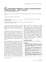
Hematoxylin-eosin stain in excised tumoursFigure 1
Hematoxylin-eosin stain in excised tumours. Low-
power view illustrating a 2.5 × 1.8 × 1.2 cm lesion within a
large cystic space surrounded by a thick fibrous capsule.
Haemorrhage was also noted within the cyst with changes
consistent with the prior biopsy.
Hematoxylin-eosin stain in excised tumours, At higher power, a papillary and solid proliferation of atypical cells of a uniform population were observedFigure 2
Hematoxylin-eosin stain in excised tumours, At
higher power, a papillary and solid proliferation of
atypical cells of a uniform population were observed.
Fibrovascular cores were well visualized.
Hematoxylin-eosin stain in excised tumours, At higher power, a papillary and solid proliferation of atypical cells of a uniform population were observedFigure 3
Hematoxylin-eosin stain in excised tumours, At
higher power, a papillary and solid proliferation of
atypical cells of a uniform population were observed.
Fibrovascular cores were well visualized.
Journal of Medical Case Reports 2009, 3:13 />Page 3 of 4
(page number not for citation purposes)
was advised due to tumour extension close to deep mar-
gin.
Due to his significant family history for breast cancer,
genetic testing was offered. Breast-cancer gene 2 (BRCA2)
mutation had been identified in a maternal aunt recently
on the exon 24 insertion called c9481_9482insA. It was
very likely that this patient was a carrier of this BRCA2
mutation, which poses a small risk for him to develop
prostate cancer. He was therefore advised to undergo pros-
tate-specific antigen (PSA) blood testing on a yearly basis.
Discussion
IPC is a rare malignancy of the breast; however, a rela-
tively higher incidence range of 5–7.5% has been reported
in men [3-5]. IPC in men is usually reported among those
of an older age group (68 to 84 years) [4,6-9]; however, in
our patient, IPC developed at a significantly younger age,
which might be due to this patient likely carrying the
BRCA2 mutation. The prognosis for this type of tumour is
excellent [3,7]. In a study of 77 patients with IPC all
patients were alive 10 years after their diagnosis and
metastases occurred only in 4% of patients, but none of
the patients with low-grade tumours were in this group
[10].
The terminology applied to describe papillary breast
lesions in the literature is relatively confusing. The tradi-
tional term "intracystic papillary carcinoma" generally
refers to a localized lesion, in situ in a cystically dilated
duct. Given the often marked stromal response surround-
ing these lesions, the distinction between in situ and inva-
sive papillary carcinoma can be very difficult to make.
Therefore, IPC had been divided it into three subgroups,
which seems to correlate with the prognosis: IPC alone,
IPC plus DCIS, and IPC with invasion [5]. In this manner,
the term "papillary DCIS" would refer to a more diffuse
process that involves multiple ducts as opposed to a local-
ized lesion [5].
Recently, Hill et al., using myoepithelial cell staining, sug-
gest a spectrum of progression from in situ disease to inva-
sive disease, signifying that what appears to be DCIS on
histology may potentially cause distant metastases [11].
The lack of an intact basal myoepithel cell layer can be
identified by calponin, smooth-muscle myosin heavy
chain (SMM-HC) cytoplasmic stains and by p63 nuclear
stains. This "gold standard" method has a relatively high
sensitivity and denotes the invasiveness of the tumour
cells in malignant papillary breast lesions [11].
The diagnosis of IPC of the male breast should be made
carefully. Triple assessment is essential and the goal is to
achieve a preoperative diagnosis prior to surgery. The radi-
ological diagnosis of IPC is relatively challenging. The typ-
ical sonographical appearance of IPC is a hypoechoic area
with soft tissue echoes projecting from the wall of the cyst
[6,7]. However, a relatively large amount of variation
exists on ultrasounds from an intraductal (which might be
associated with ductal dilatation) and a predominantly
solid pattern with the intraductal or intracystic mass
totally filling the duct [12]. Importantly, IPCs are highly
vascular tumours demonstrating a characteristic flow pat-
tern on colour-flow studies, which are sensitive to identi-
fying even very small IPCs. A distinct vascular pedicle can
be identified within the central core with branching ves-
sels arborising within the mass [12].
The mammographic appearance of IPCs is less specific.
Small IPCs are often mammographically negative, while
larger lesions may resemble any other focal well-circum-
scribed dense mass on mammography [12]. Both can
cause a minimal to moderate duct dilatation in a tapering
band-like density pattern from the nipple towards the
parenchyma. In addition, one report suggested the use of
pneumocystography [8], and another MRI [9], in combi-
nation with mammography and ultrasound to diagnose
IPC.
Fine-needle aspiration cytology and core biopsy are usu-
ally performed; however, the false negative results with
cytology are relatively frequent [13]. Therefore, excisional
biopsy should be carried out in all cystic lesions of the
male breast which are suspicious on any of the above
diagnostic modalities.
There are no clear guidelines about the management of
IPC, which is due to various factors. On one hand, IPC is
a rarity; on the other hand, the histopathological classifi-
cation and detection of invasiveness in IPC is rather con-
fusing [3,5]. In a recent review, Grabowski et al. [3]
confirmed that surgery is the mainstay of treatment,
which can be either conservation or mastectomy. Since
the prognosis of IPC is excellent with low locoregional
and distant recurrence rates, mastectomy is usually not
necessary, unless it is technically unavoidable [3]. Axillary
node metastasis can occur in up to 14% of the cases [3];
therefore, an axillary staging procedure or clearance is rec-
ommended by most authors [3,4,9]. Others argue that
IPC should be generally regarded as an in situ disease;
therefore axillary surgery is not recommended by these
authors [6-8]. There has been no clear indication for adju-
vant endocrine therapy, even among patients with oestro-
gen-receptor-positive tumours. The addition of hormonal
treatment does not appear to have impacted the outcome
[3]. On the contrary, Fayanju et al. recently reviewed the
usual adjuvant treatment applied for IPC and found that
patients with DCIS or microinvasive disease in association
with IPC were more likely to receive radiotherapy and
tamoxifen [14].
Publish with BioMed Central and every
scientist can read your work free of charge
"BioMed Central will be the most significant development for
disseminating the results of biomedical research in our lifetime."
Sir Paul Nurse, Cancer Research UK
Your research papers will be:
available free of charge to the entire biomedical community
peer reviewed and published immediately upon acceptance
cited in PubMed and archived on PubMed Central
yours — you keep the copyright
Submit your manuscript here:
/>BioMedcentral
Journal of Medical Case Reports 2009, 3:13 />Page 4 of 4
(page number not for citation purposes)
Between 4% and 40% of male breast cancers might result
from autosomal dominant mutations, primarily BRCA1
or BRCA2 mutations [15]. Due to the high risk to our
patient, contralateral risk-reducing mastectomy was also
carried out. This reduces the incidence of contralateral
breast cancer by approximately 95% but will not have an
impact on overall survival of the patient [15].
Conclusion
Triple assessment with a high level of clinical scepticism-
scepticism is necessary to diagnose IPC in a man, due to
the rarity of the condition. The treatment of choice for this
tumour is ample local excision. However, genetic testing
and risk-reducing mastectomy should also be considered
in cases of male breast cancer with a strong family history.
Abbreviations
BRCA1 or BRCA2: breast-cancer gene 1 or 2; DCIS: ductal
carcinoma in situ; ER: oestrogen receptor; IPC: intracystic
papillary carcinoma; PR: progesterone receptor; PSA:
prostate-specific antigen; SMM-HC: smooth-muscle
myosin heavy chain
Competing interests
The authors declare that they have no competing interests.
Authors' contributions
LR reviewed the case notes and wrote up the manuscript.
ME, OB and NR did the literature review and contributed
to the completion of the manuscript. FOC carried out the
histopathological analysis and HPR created the final ver-
sion of the manuscript.
Consent
Written informed consent was obtained from the patient
for publication of this case report and accompanying
images. A copy of the written consent is available for
review by the Editor-in-Chief of this journal.
References
1. Anderson WF, Devesa SS: In situ male breast carcinoma in the
Surveillance, Epidemiology, and End Results database of the
National Cancer Institute. Cancer 2005, 104:1733-1741.
2. Solorzano CC, Middleton LP, Hunt KK, Mirza N, Meric F, Kuerer HM,
Ross MI, Ames FC, Feig BW, Pollock RE, Singletary SE, Babiera G:
Treatment and outcome of patients with intracystic papil-
lary carcinoma of the breast. Am J Surg 2002, 184:364-368.
3. Grabowski J, Salzstein SL, Sadler GR, Blair S: Intracystic papillary
carcinoma: a review of 917 cases. Cancer 2008, 113:916-920.
4. Dragoumis DM, Tsiftsoglou AP: Intracystic papillary carcinoma
associated with ductal carcinoma in situ in a male breast. J
Postgrad Med 2008, 54:39-40.
5. Collins LC, Carlo VP, Hwang H, Barry TS, Gown AM, Schnitt SJ:
Intracystic papillary carcinomas of the breast: a reevaluation
using a panel of myoepithelial cell markers. Am J Surg Pathol
2006, 30:1002-1007.
6. Sinha S, Hughes RG, Ryley NG: Papillary carcinoma in a male
breast cyst: a diagnostic challenge. Ann R Coll Surg Engl 2006,
88:W3-5.
7. Kinoshita T, Fukutomi T, Iwamoto E, Takasugi M, Akashi-Tanaka S,
Hasegawa T: Intracystic papillary carcinoma of the breast in a
male patient diagnosed by core needle biopsy: a case report.
Breast 2005, 14:322-324.
8. Andrés B, Aguilar J, Torroba A, Martínez-Gálvez M, Aguayo JL: Intra-
cystic papillary carcinoma in the male breast. Breast J 2003,
9:249-250.
9. Blaumeiser B, Tjalma WA, Verslegers I, De Schepper AM, Buytaert P:
Invasive papillary carcinoma of the male breast. Eur Radiol
2002, 12:2207-2210.
10. Lefkowitz M, Lefkowitz W, Wargotz ES: Intraductal (intracystic)
papillary carcinoma of the breast and its variants: a clinico-
pathological study of 77 cases. Hum Pathol 1994, 25:802-809.
11. Hill CB, Yeh IT: Myoepithelial cell staining patterns of papillary
breast lesions: from intraductal papillomas to invasive papil-
lary carcinomas. Am J Clin Pathol 2005, 123:36-44.
12. Ganesan S, Karthik G, Joshi M, Damodaran V: Ultrasound spec-
trum in intraductal papillary neoplasms of breast. Br J Radiol
2006, 79:843-849.
13. Levine PH, Waisman J, Yang GC: Aspiration cytology of cystic
carcinoma of the breast. Diagn Cytopathol 2003, 28:39-44.
14. Fayanju OM, Ritter J, Gillanders WE, Eberlein TJ, Dietz JR, Aft R, Mar-
genthaler JA: Therapeutic management of intracystic papillary
carcinoma of the breast: the roles of radiation and endocrine
therapy. Am J Surg 2007, 194:497-500.
15. Fentiman IS, Fourquet A, Hortobagyi GN: Male breast cancer.
Review. Lancet 2006, 367:595-604.