Báo cáo y học: " Relapsing massive metal bezoar: a case report" pptx
Bạn đang xem bản rút gọn của tài liệu. Xem và tải ngay bản đầy đủ của tài liệu tại đây (491.41 KB, 4 trang )
BioMed Central
Page 1 of 4
(page number not for citation purposes)
Journal of Medical Case Reports
Open Access
Case report
Relapsing massive metal bezoar: a case report
Manuel Rodrigo Prieto-Aldape
1
, Francisco Issac Almaguer-García
2
,
Sandra Edith Figueroa-Jiménez
2
, Oscar Fernández-Díaz
1
, José Antonio Mora-
Huerta
2
and Alejandro González-Ojeda*
1
Address:
1
Surgical Division, Medical Research Unit, Clinical Epidemiology, Western National Medical Center, Mexican Institute of Social Security,
Belisario Domínguez 1000, Guadalajara, Jalisco, Mexico and
2
Department of Surgery, Civil Hospital of Guadalajara "Fray Antonio Alcalde",
Hospital 278, Guadalajara, Jalisco, Mexico
Email: Manuel Rodrigo Prieto-Aldape - ; Francisco Issac Almaguer-García - ;
Sandra Edith Figueroa-Jiménez - ; Oscar Fernández-Díaz - ; José Antonio Mora-
Huerta - ; Alejandro González-Ojeda* -
* Corresponding author
Abstract
Introduction: Bezoars are uncommon findings in the gastrointestinal tract and are composed of
a wide variety of materials. We report a case of a relapsing metal bezoar in a man with
schizophrenia.
Case presentation: A 34-year-old man presented with a history of sub-acute onset of mild diffuse
abdominal pain and abdominal distention. Physical examination revealed dullness to percussion in
the upper and lower left quadrants. Past medical history was remarkable for epilepsy, schizophrenia
and previous abdominal surgery for intestinal occlusion. Plain radiographs revealed objects of metal
density contained within a dilated stomach. Celiotomy was performed revealing more than 350
metal objects inside the stomach. The patient was discharged and referred to a psychiatric facility.
Conclusion: Intestinal occlusion in patients with psychiatric disorders can result from rare causes
such as bezoars. This report alerts surgeons to rule out bezoars in the differential diagnosis of
intestinal occlusion in people with mental health problems.
Introduction
A bezoar is a conglomeration of partially digested or non-
digested foreign material in the gastrointestinal (GI) tract,
most commonly found in the stomach [1]. Less com-
monly, bezoars are found in the small intestine and the
colon and only a few in the rectum are reported in the lit-
erature [2]. Bezoars may cause a wide variety of signs and
symptoms depending on their location and can range
from asymptomatic to occlusion and perforation. Bezoars
are classified into several main types and are named
according to the materials from which they are composed:
phytobezoars, trichobezoars, pharmacobezoars and lacto-
bezoars. Other rare, less frequent bezoars are unclassified
and include materials such as plastic and metal.
The occurrence of relapse is rare and will reappear in 14%
of cases, more often associated in psychiatric patients [3].
Only two cases of metal bezoar have been reported in the
international literature [4,5]. To the best of our knowl-
edge, we report the first case of a relapsing massive metal
Published: 10 February 2009
Journal of Medical Case Reports 2009, 3:56 doi:10.1186/1752-1947-3-56
Received: 22 August 2008
Accepted: 10 February 2009
This article is available from: />© 2009 Prieto-Aldape et al; licensee BioMed Central Ltd.
This is an Open Access article distributed under the terms of the Creative Commons Attribution License ( />),
which permits unrestricted use, distribution, and reproduction in any medium, provided the original work is properly cited.
Journal of Medical Case Reports 2009, 3:56 />Page 2 of 4
(page number not for citation purposes)
bezoar, managed at the Department of General Surgery of
the "Hospital Civil de Guadalajara Fray Antonio Alcalde",
University of Guadalajara.
Case presentation
A 34-year-old man presented to the emergency room with
sub-acute onset of mild diffuse abdominal pain and
abdominal distention, accompanied with vomiting of
gastric and metal fragment contents. His social history
was significant for tobacco and cocaine consumption. The
patient was under treatment for long-term epilepsy and
schizophrenia. He had an abdominal celiotomy two years
earlier for removal of foreign metal contents in the stom-
ach. Physical examination revealed an afebrile patient
with tachycardia and tachypnea. The abdomen was dis-
tended and tender to palpation without peritoneal signs
of irritation. A palpable mass was felt in the upper and
lower left quadrants both being dull to percussion. Liver
dullness was not obliterated and rectal examination
revealed an empty rectum. Laboratory tests revealed leu-
kocytosis with left shift, other tests (liver function tests,
serum electrolytes and blood gases) were unremarkable.
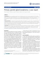
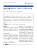
Plain radiographs (anteroposterior (AP) and lateral) of
the abdomen showed multiple objects of metal density
contained within the stomach (Figure 1).
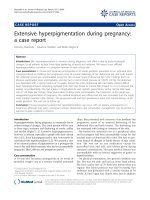
The patient was taken to the operating room (OR) for an
exploratory celiotomy through a midline incision. He was
found to have a grossly dilated stomach. A longitudinal
gastrostomy was made revealing multiple metal objects
including: nails, copper wires, stones, plastic rosary beads
and the remains of partially digested food (389 objects)
(Figure 2). After emptying the stomach, it was closed with
a double layer suture in a custom fashion leaving in place
a nasogastric tube for gastric drainage. Before closing the
abdomen, a complete exploration of the small and large
bowel was made to avoid remnants of metal particles to
prevent any postoperative complication. Postoperative
recovery was uneventful. The patient was discharged and
transferred to a psychiatric facility 8 days after surgery. A
6-month follow-up showed no recurrence or any postop-
erative complication.
Discussion
Bezoars are classified according to the materials which
they are composed of (named in order of frequency): Phy-
tobezoar made of vegetable fibers or plant material, trichob-
ezoar are a result of ingestion of human hair, drug-bezoar
contain accumulated masses of medication, lactobezoars
made of undigested milk described in premature infants
and in full term infants and other less frequent materials
are named miscellaneous bezoars or polybezoars [6]. Gas-
Plain radiographs (anteroposterior and lateral) of the abdomen showing multiple objects of metal density contained within the stomachFigure 1
Plain radiographs (anteroposterior and lateral) of the abdomen showing multiple objects of metal density con-
tained within the stomach.
Journal of Medical Case Reports 2009, 3:56 />Page 3 of 4
(page number not for citation purposes)
trointestinal bezoar is uncommon and is reported to
occur in 4% of all admissions for small-bowel obstruction
[7]. Reports of bezoars causing obstruction of the gas-
trointestinal tract have existed since the late 18th century.
Around 400 cases of trichobezoar and a larger number of
phytobezoars have been reported in the literature [8].
After reviewing the literature, we found only two previous
reports of "metal bezoar", the first in 1956 by Salb [4] and
the second reported by Kaplan et al. [5] in 2005, none
being relapsing or massive. Approximately 10% of
patients show psychiatric abnormalities or mental retar-
dation [8] therefore psychiatric evaluation and therapy are
needed to prevent a recurrence [9]. As in our patient, it is
obvious that inadequate psychiatric follow-up can lead to
a life-threatening recurrence.
This entity occurs in normal stomachs caused by the
ingestion of materials that cannot pass the pylorus such as
plastic, metal and wooden foreign bodies. Other bezoars
occur as a complication of gastric motility, usually prior
gastric surgery such as vagotomy, leading to reduced gas-
tric acidity; gastric stasis and loss of pyloric function; pep-
tic ulcer disease/stenosis; chronic gastritis; Crohn's
disease; carcinoma of the stomach, duodenum, or pan-
creas; dehydration and hypothyroidism [10]; diabetic
patients with neuropathy or myotonic dystrophy [11].
Also to be considered is the fact that certain medications
that decrease GI motility, such as anticholinergic agents,
ganglionic blocking agents, and opiates, may also give rise
to a bezoar [10]. With time, undigested foreign bodies are
retained by mucus and become enmeshed, creating a
mass in the shape of the stomach where they are usually
found. They may attain large sizes owing to the chronicity
of the problem and delayed reporting by the patients [12].
Bezoars have been known to cause a wide variety of symp-
toms. In the stomach, they are associated with anorexia,
bloating, early satiety, dyspepsia, malaise, weakness,
weight loss, headaches, and a feeling of fullness or heavi-
ness in the epigastrium [13]. They may also present with
gastrointestinal bleeding (6%) and intestinal obstruction
or perforation (10%) [14]. Gastrointestinal bezoars can
be easily diagnosed in most patients. Plain X-rays, like the
radiograph in our patient, are unique and lead to the diag-
nosis, however diagnostic difficulties arise in patients with
radiolucent bezoars, and contrast studies of the GI tract by
radiography and computed tomography (CT) scan are
necessary in such circumstances. Upper GI endoscopy is
the method of choice in detecting esophageal, gastric and
duodenal foreign bodies. Occasionally, bezoars are found
incidentally when an emergency laparotomy is done sec-
ondarily to bowel obstruction. Several treatments have
been proposed for bezoars and they depend upon the
clinical presentation as well as on the composition of the
bezoar. Chemical and enzymatic compounds have been
used for dissolution of esophageal and gastric phytobez-
oars and lactobezoars [15]. For small bezoars, endoscopy
has been the treatment of choice. Once the obstruction
occurs, surgery is the only way to solve the problem. Fre-
quently, synchronous bezoars are found in the stomach or
other areas of the gastrointestinal tract; therefore it is man-
datory to carry out a thorough exploration of the small
intestine and colon [11] to avoid recurrence of intestinal
obstruction due to a retained bezoar. After discharge,
recurrence has been reported in up to 14% of cases, espe-
cially in patients with psychiatric disturbances and with
previous gastric surgery [3].
Objects obtained from the stomachFigure 2
Objects obtained from the stomach.
Publish with BioMed Central and every
scientist can read your work free of charge
"BioMed Central will be the most significant development for
disseminating the results of biomedical research in our lifetime."
Sir Paul Nurse, Cancer Research UK
Your research papers will be:
available free of charge to the entire biomedical community
peer reviewed and published immediately upon acceptance
cited in PubMed and archived on PubMed Central
yours — you keep the copyright
Submit your manuscript here:
/>BioMedcentral
Journal of Medical Case Reports 2009, 3:56 />Page 4 of 4
(page number not for citation purposes)
Conclusion
In our patient, the diagnosis was simple because there
were not many options; a patient with intestinal obstruc-
tion, psychiatric disturbances, previous gastric surgery and
the radiographic findings took him immediately to the
OR. The gastrostomy with foreign material removal and
thorough exploration of the rest of the GI tract was cura-
tive. We conclude that, in the face of an intestinal occlu-
sion in a patient with a psychiatric disorder and a history
of GI surgery, the physician must suspect, in addition to
adhesions or hernias, the possibility of a bezoar.
Plain and contrast enhanced radiographic studies are
important for both diagnosis and treatment. It is also
important to consider GI endoscopy as part of the diagno-
sis and treatment plan. In cases where bezoars cannot be
treated by dissolving agents such as enzymes or chemicals
and endoscopy is not able to remove the obstruction, sur-
gery is the last option.
Consent
Written informed consent was obtained from the patient's
mother for publication of this case report and any accom-
panying images. A copy of the written consent is available
for review by the Editor-in-Chief of this journal.
Competing interests
The authors declare that they have no competing interests.
Authors' contributions
MRPA participated in the surgical team and the care of the
patient during his hospitalization, and was also involved
in drafting and review of the manuscript. FIAG made the
evaluation in the emergency department, the diagnostic
and treatment protocol, and the postoperative care, and
also the review of the literature. SEFJ participated in the
surgical team and the care of the patient, and also took the
pictures, carried out the review of the literature and partic-
ipated in the drafting of the manuscript. OFD and JAMH
performed the case review, completed the documentation
and were major contributors in writing the manuscript.
AGO reviewed the case, evaluated the available literature,
and made a substantive intellectual contribution to the
final version. All authors read and approved the final
manuscript.
References
1. Davis RN, Rettmann JA, Christensen B: Relapsing altered mental
status secondary to a meprobamate bezoar. J Trauma 2006,
61:990-991.
2. Steinberg JM, Eitan A: Prickly pear fruit bezoar presenting as
rectal perforation in an elderly patient. Int J Colorectal Dis 2003,
18:365-367.
3. Robles R, Parrilla P, Escamilla C, Lujan JA, Torralba JA, Liron R,
Moreno A: Gastrointestinal bezoars. Br J Surg 1994,
81:1000-1001.
4. Salb RL: Metallic bezoar. Med Radiogr Photogr 1956, 32:32-33.
5. Kaplan R, Celebi F, Guzey D, Celik AS, Erozgen F, Firat N: Medical
image. Metal bezoar. N Z Med J 2005, 118(1219):U1588.
6. Hall JD, Shami VM: Rapunzel's syndrome: gastric bezoars and
endoscopic management. Gastrointest Endosc Clin N Am 2006,
16:111-119.
7. Ho TW, Koh DC: Small-bowel obstruction secondary to bez-
oar impaction: a diagnostic dilemma. World J Surg 2007,
31:1073-1079.
8. Sharma RD, Kotwal S, Chintamani , Bhatnagar D: Trichobezoar
obstructing the terminal ileum. Trop Doct 2002, 32:99-100.
9. Kuroki Y, Otagiri S, Sakamoto T, Tsukada K, Tanaka M: Case report
of trichobezoar causing gastric perforation. Dig Endosc 2000,
12:181-185.
10. LaFountain J: Could your patient's bowel obstruction be a bez-
oar. Todays Surg Nurse 1999, 21:34-37.
11. Campos RR, Paricio PP, Albasini JLA, et al.: Gastrointestinal bez-
oars. Presentation of 60 cases. Dig Surg 1990, 7:39-44.
12. Chintamani , Durkhure R, Singh JP, Singhal V: Cotton bezoar – a
rare cause of intestinal obstruction: case report. BMC Surg
2003, 4:3-5.
13. Goldstein SS, Lewis JH, Rothstein R: Intestinal obstruction due to
bezoars. Am J Gastroenterol 1984, 79:313-318.
14. Andrus CH, Ponsky JL: Bezoars: classification, pathophysiology,
and treatment. Am J Gastroenterol 1988, 83:476-478.
15. Gupta R, Share M, Pineau BC: Dissolution of an esophageal bez-
oar with pancreatic enzyme extract. Gastrointest Endosc 2001,
54:96-99.