Báo cáo y học: "Perivascular epitheloid cell tumour (PEComa) of the retroperitoneum – a rare tumor with uncertain malignant behaviour: a case report" ppsx
Bạn đang xem bản rút gọn của tài liệu. Xem và tải ngay bản đầy đủ của tài liệu tại đây (1.34 MB, 5 trang )
BioMed Central
Page 1 of 5
(page number not for citation purposes)
Journal of Medical Case Reports
Open Access
Case report
Perivascular epitheloid cell tumour (PEComa) of the
retroperitoneum – a rare tumor with uncertain malignant
behaviour: a case report
Alexandra M Koenig*
1
, Alexander Quaas
2
, Thorsten Ries
3
, Emre F Yekebas
1
,
Karim A Gawad
1
, Yogesh K Vashist
1
, Christoph Burdelski
1
, Oliver Mann
1
,
Jakob R Izbicki
1
and Andreas Erbersdobler
2
Address:
1
Department of General, Visceral and Thoracic Surgery, University Medical Centre of Hamburg-Eppendorf, Martinistraße 52, Hamburg,
Germany,
2
Institute of Pathology, University Medical Centre of Hamburg-Eppendorf, Martinistraße 52, Hamburg, Germany and
3
Department of
Diagnostic and Interventional Radiology, University Medical Centre of Hamburg-Eppendorf, Martinistraße 52, Hamburg, Germany
Email: Alexandra M Koenig* - ; Alexander Quaas - ; Thorsten Ries -
hamburg.de; Emre F Yekebas - ; Karim A Gawad - ;
Yogesh K Vashist - ; Christoph Burdelski - ; Oliver Mann -
hamburg.de; Jakob R Izbicki - ; Andreas Erbersdobler -
* Corresponding author
Abstract
Introduction: Perivascular epitheloid cell tumours are rare mesenchymal neoplasms
characterized by a proliferation of perivascular cells with an epitheloid phenotype and expression
of myomelanocytic markers.
Case presentation: Here we present the case of a cystic perivascular epitheloid cell tumour of
the retroperitoneum associated with multifocal lung lesions. A 27-year-old woman underwent
laparotomy to remove a 10 × 6 × 4 cm sized retroperitoneal mass. The resected specimen was
subjected to frozen and permanent histological sections with conventional and
immunohistochemical stains, including antibodies against HMB45. The tumour displayed the typical
morphological and immunohistochemical features of a perivascular epitheloid cell tumour. Focal
necrosis and a proliferative index of 10% suggested a malignant potential. Moreover, postoperative
computed tomography scans demonstrated multiple lung lesions, which were radiologically
interpreted as being most likely compatible with lymphangioleiomyomatosis.
Conclusion: Since lymphangioleiomyomatosis, an otherwise benign condition, belongs to the
family of perivascular epitheloid cell tumours, it cannot be excluded that the lung lesions in this case
in fact represent metastases from the retroperitoneal perivascular epitheloid cell tumour rather
than independent neoplasms. More experience with this new and unusual tumour entity is clearly
needed in order to define reliable criteria for benign or malignant behaviour.
Published: 16 February 2009
Journal of Medical Case Reports 2009, 3:62 doi:10.1186/1752-1947-3-62
Received: 12 February 2008
Accepted: 16 February 2009
This article is available from: />© 2009 Koenig et al; licensee BioMed Central Ltd.
This is an Open Access article distributed under the terms of the Creative Commons Attribution License ( />),
which permits unrestricted use, distribution, and reproduction in any medium, provided the original work is properly cited.
Journal of Medical Case Reports 2009, 3:62 />Page 2 of 5
(page number not for citation purposes)
Introduction
Perivascular epitheloid cell tumours (PEComas) are mes-
enchymal tumours composed of distinctive, so-called
perivascular epitheloid cells, which were first described by
Bonetti in 1992 and were observed in "sugar tumours" of
the lung as well as in angiomyolipomas of the kidney [1].
These cells are characterized by an epitheloid shape, eosi-
nophilic cytoplasm, perivascular location and a coexpres-
sion of immunohistochemical markers indicating both
smooth muscle and melanocytic differentiation. PECo-
mas are related to the tuberous sclerosis complex (TSC),
characterized by mental retardation, seizures and cellular
proliferations. The PEComa family includes angiomyol-
ipomas, clear cell "sugar" tumours of the lung, pancreas
and uterus and lymphangioleiomyomatosis (LAM) [2,3].
The latter is a rare disease, which typically manifests as
multiple lung lesions in young women consisting of
tumour-like proliferations of lymphatic channels and
smooth muscle cells. Although considered a benign
tumour-like lesion, LAM may lead to a rapid deterioration
of lung function and the need for lung transplantation.
There are some important open questions about PECo-
mas: the histogenesis, the normal counterpart of PEC and
the identification of the histological criteria of malig-
nancy.
We report the unusual case of a patient with a malignant
retroperitoneal PEComa and subsequent detection of
multiple lung lesions compatible with LAM.
Case presentation
A 27-year-old woman, who first complained of upper
abdominal pain, was referred from a local clinic with the
impression of a retroperitoneal haematoma after blunt
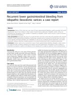
abdominal trauma 4 months ago. Magnetic resonance
tomography (MRT) of the abdomen revealed the presence
of a large, well-circumscribed, right-sided retroperitoneal
mass measuring 10 × 8 cm in size with an irregular echo-
genicity (Figure 1A). The mass compressed the right kid-
ney and the caval vein without renal involvement. No
chylous ascites was present. No clinical evidence of tuber-
ous sclerosis was present and there was no family history
of cancer or known genetic disorders. Based on the MRT,
the retroperitoneal mass was removed completely by
laparotomy (Figure 1B). During the postoperative course
the patient complained of exertional dyspnoea. The sub-
sequently performed computed tomography (CT) scan of
the lung showed the typical image of LAM with numerous
thin-walled cysts throughout both lungs, but without
spontaneous pneumothorax or chylous pleural effusions
(Figure 2).
Macroscopically, the 10 × 6 × 4 cm sized mass had a soft
consistency and was circumscribed, but not truly encapsu-
lated. On cut sections, large, central areas of haemorrhage
could be observed, giving it an impression of an old hae-
matoma. On frozen sections, it became obvious that the
wall of the cystic mass consisted of nests of tumour cells
with a uniform, spindle shape. There were no overt signs
of malignancy. On permanent sections, the tumour dis-
played fascicles and nests of elongated epitheloid tumour
cells with a clear to pale eosinophilic cytoplasm, arranged
around numerous ectatic blood vessels (Figure 3A).
Sometimes, the tumour cell proliferations seemed to
evolve directly from the walls of medium-sized blood ves-
sels. Occasional mitoses and foci of haemorrhage and
necrosis were present (Figure 3C). Immunohistochemi-
cally, most of the tumour cells showed a positive reaction
for alpha-smooth muscle actin (SMA), Desmin and
HMB45 (Figure 3B). About 50% of tumour cells showed
a weak positivity for the oestrogen receptor. Proliferative
activity, as measured by an antibody against the Ki-67
antigen, was 10% of tumour cells. Cytokeratins, epithelial
membrane antigen (EMA), synaptophysin, S100, and
Macroscopic TumourFigure 1
Macroscopic Tumour. A: MRT revealed the presence of a
large, well circumscribed right sided retroperitoneal mass
measuring 10 × 8 cm in size. B: Macroscopically the 10 × 6 ×
4 cm sized tumour was soft with focal areas of haemorrhage
circumscribed by a 2,5 × 1,5 × 0,3 cm sized capsule and with
central necrosis.
Journal of Medical Case Reports 2009, 3:62 />Page 3 of 5
(page number not for citation purposes)
CD117 (c-kit) were negative. CD31, CD34 and D2-40
decorated the endothelial linings of the numerous vessels.
The diagnosis of a tumour with perivascular epitheloid
cell differentiation, a so-called PEComa, was made. Based
on the histological findings on permanent sections, a
malignant potential was suggested. The margins of resec-
tion were free of tumour cells.
Discussion
Neoplasms with perivascular epitheloid cell differentia-
tion are a group of ubiquitous mesenchymal tumours
sharing morphological, immunohistochemical,
ultrastructural and genetically distinctive features [4].
These PEComas are characterized by cells with an epith-
eloid appearance, a clear to eosinophil cytoplasm and an
intimate relationship to blood vessels [5]. The cells are
consistently immunoreactive to the melanocytic marker
HMB45, variably immunoreactive to smooth muscle actin
and negative for epithelial markers. The histogenesis and
physiological counterparts of PEC are unknown. The
PEComa family comprises angiomyolipomas (AML),
clear cell "sugar" tumour of the lung (CCST), lymphangi-
oleiomyomatosis (LAM), clear cell myomelanocytic
tumour of the falciform ligament/ligamentum teres
(CCMMT) and unusual clear cell tumours of the pancreas,
rectum, abdominal serosa, uterus, vulva, thigh and heart.
The uterus is one of the most prevalent sites of involve-
ment [6].
Clinically, a subset of PEComas behaves in a malignant
fashion. Clear criteria for malignancy have not been elab-
orated in this very rare tumour entity until now, owing to
their rarity. Folpe et al. reported 26 cases of PEComas of
soft tissue and gynaecological origin proposing criteria for
the classification of these tumours as "benign", "of uncer-
tain malignant potential" and "malignant". In our patient
we observed a significant association between tumour size
>5 cm, infiltrative growth pattern, high nuclear grade,
necrosis and mitotic activity >1/50 HPF and subsequent
aggressive clinical behaviour of PEComas [7]. Surgery
seems to be the only approach for aggressive cases, as
chemo- and radiotherapy have not shown significant
results.
The above reported tumour is a rare case of a PEComa
arising in the retroperitoneum.
Based on the occasional foci of necrosis, the infiltrative
growth pattern on microscopic level, as well as the rela-
tively high proliferative activity suggested a malignant
potential in the present case. Even more unusual is the
subsequent occurrence of multiple pulmonary lesions,
which were radiologically described as being quite typical
for lymphangioleiomyomatosis (LAM). This rare disease
usually occurs in young women of childbearing age and is
characterized by a distinctive proliferation of lymphatic
and smooth muscle cells. The primary site of origin is the
lung and occurrence is usually associated with decreased
pulmonary function and chylous effusions [8]. The spec-
ulation that LAM is a female sex hormone dependent
tumour is supported by the high prevalence rate in
women of reproductive age and exacerbation of the dis-
ease in pregnancy. Several studies regarding clinical trials
of hormonal therapy have been reported [9,10]. Surgical
intervention is necessary in complications (thoracic drain-
age, pleurectomy for recurrent pneumothorax). If hormo-
nal therapy is not successful, a combined heart and lung
transplantation should be attempted as ultima ratio [11].
LAM can occur without evidence of other disease (spo-
radic LAM) or in conjunction with tuberous sclerosis com-
plex, an autosomal dominant tumour suppressor gene
syndrome characterized by seizures, mental retardation,
and tumours in the brain, heart, skin and kidney.
Therefore, a full work up for tuberous sclerosis is neces-
sary in these patients.
The association between LAM and PEComas as a family
and the co-occurrence in an individual patient is well
known. It could be speculated that these patients may
have a special predisposition to develop such tumours.
On the other hand, it cannot be excluded that the pulmo-
nary lesions actually represent metastases from the retro-
peritoneal PEComa. However the possibility that the lung
lesions represent metastases is doubtful. It is most likely
that they represent separate lesions as true LAM.
Postoperative Chest CT ScanFigure 2
Postoperative Chest CT Scan. A chest CT scan showing
diffuse small thin walled cystic lesions in the parenchyma of
both lungs.
Journal of Medical Case Reports 2009, 3:62 />Page 4 of 5
(page number not for citation purposes)
Histological FindingsFigure 3
Histological Findings. A: The perivascular epitheloid cells proliferate haphazardly around slit-like vascular channels, with
aggregation of lymphoid cells. B: Tumour cells with expression of the HMB45-antigen. (immunohistochemistry with the avidin-
biotin-peroxidase-complex method; counterstain haematoxylin; original magnification × 100). C: Focal coagulative necrosis of
tumour cells (H&E; original magnification × 200).
Publish with BioMed Central and every
scientist can read your work free of charge
"BioMed Central will be the most significant development for
disseminating the results of biomedical research in our lifetime."
Sir Paul Nurse, Cancer Research UK
Your research papers will be:
available free of charge to the entire biomedical community
peer reviewed and published immediately upon acceptance
cited in PubMed and archived on PubMed Central
yours — you keep the copyright
Submit your manuscript here:
/>BioMedcentral
Journal of Medical Case Reports 2009, 3:62 />Page 5 of 5
(page number not for citation purposes)
Since the pulmonary lesions were not biopsied, a histo-
logical comparison to the retroperitoneal tumour was not
possible. However, it is likely that even histological exam-
ination of the pulmonary tumours would not have been
able to solve the question of metastatic versus independ-
ent origin, since both metastasis of a PEComa and pri-
mary LAM could have the same histological appearance.
Clinical follow-up must show if the pulmonary lesions
will behave in the typical fashion of LAM.
Conclusion
This case report demonstrates the diagnostic, prognostic
and therapeutic dilemmas of a new and rare tumour
entity. The outcome of this disease can be devastating, yet
the aetiology and effective treatments are unknown. Firm
criteria for malignancy and proper subclassifications of
PEComas have yet to be established and should be vali-
dated by case reports and studies of clinical behaviour.
Abbreviations
AML: angiomyolipoma; CCMMT: clear cell myomelano-
cytic tumour of the falciform ligament/ligamentum teres;
CCST: clear cell "sugar" tumour of the lung; CT: computed
tomography; EMA: epithelial membrane antigen; LAM:
lymphangioleiomyomatosis; MRT: magnetic resonance
tomography; PEC: perivascular epitheloid cell tumor;
SMA: alpha-smooth muscle actin; TSC: tuberous sclerosis
complex
Consent
Written informed consent was obtained from the patient
for publication of this case report and accompanying
images. A copy of the written consent is available for
review by the Editor-in-Chief of this journal.
Competing interests
The authors declare that they have no competing interests.
Authors' contributions
AMK initiated the concept, literature search and write up
of the manuscript. AQ performed the pathological inves-
tigations and helped in the literature search. TR performed
and diagnosed the CT scans. EFY helped in revision of the
article. KAG contributed to the clinical management of
the patient and gave approval for the final write up. YKV
assisted in performing the surgery and helped in drafting
the article. CB helped in the revision of the article. OM
performed the surgery. JRI is the consultant surgeon
responsible for the patient's care and made final correc-
tions to the manuscript. AE diagnosed the specimens and
supervised the overall structure of the article. All authors
read and approved the final manuscript.
References
1. Bonetti F, Pea M, Martignoni G, Zamboni G: PEC and sugar. Am J
Surg Pathol 1992, 16:307-308.
2. Bonetti F, Martignoni G, Colato C, Manfrin E, Gambacorta M, Faleri
M, Bacchi C, Sin VC, Wong NL, Coady M, Chan JK: Abdominopel-
vic sarcoma of perivascular epithelioid cells. Report of four
cases in young women, one with tuberous sclerosis. Mod
Pathol 2001, 14:563-568.
3. Pea M, Martignoni G, Zamboni G, Bonetti F: Perivascular epithe-
lioid cell. Am J Surg Pathol 1996, 20:1149-1153.
4. Lai HY, Chen CK, Lee YH, Tsai PP, Chen JH, Shen WC: Multicentric
aggressive angiomyolipomas: a rare form of PEComas. AJR
Am J Roentgenol 2006, 186:837-840.
5. Cibas ES, Goss GA, Kulke MH, Demetri GD, Fletcher CD: Malig-
nant epithelioid angiomyolipoma ('sarcoma ex angiomyol-
ipoma') of the kidney: a case report and review of the
literature. Am J Surg Pathol 2001, 25:121-126.
6. Ruco LP, Pilozzi E, Wedard BM, Marzullo A, D'Andrea V, De Antoni
E, Silvestrini G, Bonetti F: Epithelioid lymphangioleiomyomato-
sis-like tumour of the uterus in a patient without tuberous
sclerosis: a lesion mimicking epithelioid leiomyosarcoma.
Histopathology 1998, 33:91-93.
7. Folpe AL, Mentzel T, Lehr HA, Fisher C, Balzer BL, Weiss SW:
Perivascular epithelioid cell neoplasms of soft tissue and
gynecologic origin: a clinicopathologic study of 26 cases and
review of the literature. Am J Surg Pathol 2005, 29:1558-1575.
8. Chu SC, Horiba K, Usuki J, Avila NA, Chen CC, Travis WD, Ferrans
VJ, Moss J: Comprehensive evaluation of 35 patients with lym-
phangioleiomyomatosis. Chest 1999, 115:1041-1052.
9. Matsui K, Takeda K, Yu ZX, Valencia J, Travis WD, Moss J, Ferrans
VJ: Downregulation of estrogen and progesterone receptors
in the abnormal smooth muscle cells in pulmonary lym-
phangioleiomyomatosis following therapy. An immunohis-
tochemical study. Am J Respir Crit Care Med 2000, 161:1002.
10. Logginidou H, Ao X, Russo I, Henske EP: Frequent estrogen and
progesterone receptor immunoreactivity in renal angiomy-
olipomas from women with pulmonary lymphangioleiomyo-
matosis. Chest 2000, 117:25-30.
11. Schonemann B, Merkle P: Differential diagnosis of spontaneous
pneumothorax-pulmonary lymphangioleiomyomatosis.
Clinical aspects, diagnosis and therapy. Chirurg 1990,
61:301-303.