báo cáo khoa học: " The impact of bisphosphonates on the osteoblast proliferation and Collagen gene expression in vitro" ppsx
Bạn đang xem bản rút gọn của tài liệu. Xem và tải ngay bản đầy đủ của tài liệu tại đây (529.16 KB, 6 trang )
HEAD & FACE MEDICINE
Koch et al. Head & Face Medicine 2010, 6:12
/>Open Access
RESEARCH
© 2010 Koch et al; licensee BioMed Central Ltd. This is an Open Access article distributed under the terms of the Creative Commons
Attribution License ( which permits unrestricted use, distribution, and reproduction in
any medium, provided the original work is properly cited.
Research
The impact of bisphosphonates on the osteoblast
proliferation and Collagen gene expression
in vitro
Felix Peter Koch
†1
, Sareh Said Yekta
†3
, Christina Merkel
1
, Thomas Ziebart
1
and Ralf Smeets*
2
Abstract
Background: Bisphosphonates are widely used in the clinical treatment of bone diseases with increased bone
resorption. In terms of side effects, they are known to be associated with osteonecrosis of the jaw (BONJ).
The objective of this study was to evaluate the effect of bisphosphonates on osteoblast proliferation by cell count and
gene expression analysis of cyclin D1 in vitro. Furthermore, the gene expression of the extracellular matrix protein
collagen type I was evaluated. Nitrogen-containing and non-nitrogen-containing bisphosphonates have been
compared on gene expression levels.
Methods: Human osteoblast obtained from hip bone were stimulated with zoledronate, ibandronate and clodronate
at concentrations of 5 × 10
-5
M over the experimental periods of 1, 2, 5, 10 and 14 days. At each point in time, the cells
were dissolved, the mRNA extracted, and the gene expression level of cyclin D1 and collagen type I were quantified by
Real-Time RT-PCR. The gene expression was compared to an unstimulated osteoblast cell culture for control.
Results: The proliferation appeared to have been influenced only to a small degree by bisphosphonates. Zolendronate
led to a lower cyclin D1 gene expression after 10 days. The collagen gene expression was enhanced by nitrogen
containing bisphosphonates, decreased however after day 10. The non-nitrogen-containing bisphosphonate
clodronate, however, did not significantly influence cyclin D1 and collagen gene expression.
Conclusions: The above data suggest a limited influence of bisphosphonates on osteoblast proliferation, except for
zoledronate. The extracellular matrix production seems to be initially advanced and inhibited after 10 days.
Interestingly, clodronate has little influence on osteoblast proliferation and extracellular matrix production in terms of
cyclin D1 and collagen gene expression.
Background
Bisphosphonates are widely used in the clinical treatment
of bone diseases with increased bone resorption [1] such
as Paget's disease, osteoporosis, and malignant diseases
like multiple myeloma or metastasis to the bone. The
increased bone mineral density has been attributed to a
decreased bone turnover [2-5] by the inhibition of osteo-
clastic bone resorption.
There is, however, increasing evidence, that bisphos-
phonates interact with osteoblasts. The bisphosphonates
are a family of pyrophosphate analogs that can further be
separated into nitrogen-containing and non-nitrogen-
containing bisphosphonates. Non-nitrogen-containing
bisphosphonates are build into ATP resulting in a non-
hydrolysable adenine containing metabolite, whereas
nitrogen-containing bisphosphonates interfere with the
mevalonate pathway by inhibition of farnesyl pyrophos-
phate (FPP) synthase enzyme [6,7]. This interference
causes a reduction in geranyl geranyl diphosphate
(GGPP), which is required for the prenylation of
guanosin triphosphate (GTP)-binding proteins such as
Rab, Rac, Ras, Rho and Cdc42 [8-12]. In contrast to older
in vivo studies that attribute higher bone density to
reduced bone turnover, newer studies have shown the
potential of bisphosphonates to enhance osteoblast pro-
liferation and differentiation in bone marrow-derived
mesenchymal stem cells (MSC) and osteoblasts [13-15].
These actions could cause an altered cell metabolism,
which is supposed to promote osteonecrosis that almost
always occurs in the jaw as a serious side effect with
* Correspondence:
2
Department of Oral and Maxillofacial Surgery, University Hospital Aachen,
Aachen, Germany
†
Contributed equally
Full list of author information is available at the end of the article
Koch et al. Head & Face Medicine 2010, 6:12
/>Page 2 of 6
exposed bone, fistulae and even pathological fractures
[16,17]. Especially after treatment by nitrogen containing
bisphosphonates intravenously an incidence of 5%-19%
has been reported [18-20]. In addition to a direct effect
on osteoclasts and osteoblasts, some authors suggest that
a bisphosphonate induced obliteration of the regional
blood vessels could lead to an avascular osteonecrosis of
the jaw [17,21,22].
The objective of this in vitro study was to illuminate the
impact of bisphosphonates on osteoblast proliferation
and extracellular matrix production over a period of 14
days. Therefore, the genes of cyclin D1 and collagen were
quantified by Real Time RT-PCR. The nitrogen-contain-
ing bisphosphonates zoledronate and ibandronate were
compared to the non-nitrogen-containing bisphospho-
nate clodronate.
Methods
Cell culture
Human hip bone osteoblasts (HOB-c, Promo Cell,
Heidelberg, Germany) between passages 5-9 were cul-
tured at a density of 200 000 cells per well using 6-well
plates. They were allowed to attach for two days using an
osteoblast specific medium (10% FCS/DMEM Dulbecco
modified medium (Invitrogen, Carlsbad, Ca/US) contain-
ing 1% L-glutamin, 1% penicillin/streptomycin/neomy-
cin, 1% ascorbic acid, and 20 μg/ml dexamethasone. The
cells were stimulated by osteoblast specific medium con-
taining zoledronate, ibandronate, or clodronate at a con-
centration of 5 × 10
-5
M. The osteoblast specific cell
culture medium without bisphosphonate supplement was
used for control. The media and bisphosphonates were
renewed every 4 days for a period of 14 days to guarantee
a constant stimulation und nutrition supply over the
experimental period.
mRNA extraction and reverse transcriptase polymerase
chain reaction (RT-PCR)
On day 1, 2, 5, 10, and 14 of cultivation, the osteoblasts
were detached with 0.05% trypsin-EDTA solution (Invit-
rogen, Carlsbad, Ca, US) and individually harvested.
MRNA was extracted using a silicate gel technique that
was provided by the Qiagen RNeasy extraction kit (Qia-
gen, Hilden, Germany). This included a DNAse digestion
step. The amount of extracted mRNA was measured by
extinction at 260nm; the contamination with proteins
was determinated with the 260/280 ratio.
To detect the mRNA of cyclin D1 and collagen type I in
osteoblasts, primers were designed using NCBI-nucle-
otide library and Primer3-design (Tab. 1). All primers had
been matched to the mRNA sequences of the target genes
(NCBI Blast software).
As housekeeping genes, human ribosomal protein
(HuPO), actin, glyceraldehyde-3-phosphate dehydroge-
nase (GAPDH) and ribosomal protein S18 (RPS18) were
evaluated. We were able to show the most stable expres-
sion for the actin, GAPDH and RPS18 genes by compar-
ing the bisphosphonate stimulated versus a non
stimulated cell-culture using a specialized freeware,
called GeNorm.
As a quantitative RT-PCR we used the SYBR Green
Real Time PCR (oneStep RT-PCR, Bio-Rad, Hercules,
CA/USA). This method enables reverse transcription
using the individual primers immediately before PCR
amplification and SYBR Green fluorescence measure-
ment for quantification of gene expression. Samples were
amplified in 96-well microplates in an IQ5-Cycler (Bio-
Rad, Hercules, CA/USA) with an annealing temperature
of 56°C and an elongation temperature of 71°C over 40
cycles. Background was to determine over 3-10 cycles
and the threshold was set above this fluorescence, cross-
ing the SYBR green fluorescence curve at the exponential
part. This method was applied to calculate the cycle
number and C
T
-value for quantitation. Furthermore, the
C
T
-values of actin, GAPDH and RPS18 housekeeping
genes and the individual primer efficacy were considered.
Single product formation was confirmed by melting point
analysis. For negative control, water instead of mRNA-
samples was used.
CDNA from individual cell experiments was analyzed
in triplicate PCR. The ΔΔC
T
method was applied [23,24]
for a statistical analysis of the C
T
-values. For each specific
primer and Real-Time PCR, the efficiency was calculated
on the basis of the SYBR Green fluorescence curves and
the standard dilution series. The relative gene expression
levels were standardized with those measured in the
unstimulated control, which was set to 100%. Each point
in time for relative mRNA is the mean +/- standard devi-
ation. (See Table 1)
Statistical analysis
The mean values and standard deviations were calculated
by the IQ5-software (BioRad, Hercules, CA/USA) to pro-
vide a descriptive data analysis.
Results
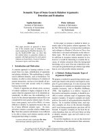
Effect of bisphosphonates on cyclin D1 gene expression
Time-course experiments were performed to determine
the effects of zoledronate, ibandronate and clodronate on
cyclin D1 gene expression. As shown in figure 1, treat-
ment of human hip bone osteoblast [hOB] cells with
ibandronate, zoledronate and clodronate did not signifi-
cantly influence the gene expression of cyclin D1 during
the first 6 days. Zoledronate, however, caused a decreas-
ing cyclin D1 gene expression after the 6th day whereas
ibandronate and clodronate did not significantly show
enhanced or decreased gene expression levels.
Koch et al. Head & Face Medicine 2010, 6:12
/>Page 3 of 6
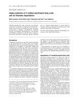
Effect of bisphosphonates on collagen gene expression
The collagen gene expression was stimulated to the most
extent by ibandronate, reaching a maximum of 400% at
day 10 compared to the non-stimulated control. Zole-
dronate also caused osteoblasts to increase their gene
expression to a maximum level of 330% on day 2. After 14
days of stimulation the gene expression of collagen type I
has decreased to a level of 12% for zoledronate, respec-
tively 30% for ibandronate compared to an unstimulated
control.
The non-nitrogen-containing clodronate, however, did
not cause a significant alteration of collagen gene expres-
sion (figure 2).
Discussion
Bisphosphonates are therapeutically applied to treat met-
abolic bone diseases, such as osteoporosis or metastasis
to the bone. Clinical studies have shown their potency to
increase bone density over an extended period of time
[25-28]. This effect is not only caused by a positive bone
turnover, but also by a direct stimulation of osteoblast
and osteoblast precursor cells by applying nitrogen-con-
taining bisphosphonates [15,29]. An anabolic effect to the
bone could be caused by proliferation and by extracellular
matrix production, mainly of collagen type I. With
respect to osteoblast proliferation, we examined cyclin
D1, an important regulator of the cell cycle and a surro-
gate of cell proliferation. Our results did not show a sig-
Table 1: Oligonucleotide primer sequences used for Real Time PCR
Sense Antisense
Cyclin D1 ATCTCTGTACTTTGCTTGCT AGTACATGGATATTCCCAAA
Collagen I AGAACTGGTACATCAGCAAG GAGTTTACAGGAAGCAGACA
GAPDH AAAAACCTGCCAAATATGAT CAGTGAGGGTCTCTCTCTTC
RPS 18 TCGGAACTGAGGCCATGA GAACCTCCGACTTTCGTTC
Figure 1 Quantitative RT-PCR-results of cyclin D1 gene expression as fold of unstimulated control gene expression (means +/- SD), that was
set at 1 (100%).
Koch et al. Head & Face Medicine 2010, 6:12
/>Page 4 of 6
nificant impact on osteoblast proliferation during the
first 6 days. However, after day 6 zoledronate led to a
reduced Cyclin D1 gene expression. As shown in other in
vitro studies, pamidronate, a nitrogen-containing bispho-
sphonate, decreased osteoblast proliferation in a dose
dependent manner [29]. In contrast, bisphosphonates
have been reported to induce proliferation of marrow
osteoprogenitors [30]. These anabolic effects are evi-
denced by a positive bone turnover, evaluated in clinical
studies of up to 10 years of bisphosphonate treatment
[25,31].
With respect to extracellular matrix production and
early bone differentiation, type I collagen is the most
important matrix protein. It is produced by osteoblasts
and permits bone mineralization. The nitrogen-contain-
ing bisphosphonates appeared to induce collagen type I
gene expression during the first 10 days. At day 14 the
collagen gene expression level was lowered by the nitro-
gen containing bisphosphonates below 30%. These
results are confirmed by Reinholz et al., who also found
an enhanced collagen production [29]. The bisphospho-
nate as well effect bone marrow stromal cells by an
enhanced collagen gene expression [15].
Our data suggest that nitrogen-containing bisphospho-
nate treatment enhances the differentiation of the osteo-
blasts from the proliferation stage into a nonproliferating
matrix maturation stage. The lower proliferation but
higher bone density through differentiation could explain
the missing regeneration potential of BONJs.
In contrast, the non-nitrogen-containing bisphospho-
nate clodronate did not have any significant impact on
osteoblasts cyclin D1 gene expression or type I collagen
gene expression. These results support the assumption,
that for the inhibition of farnesyl pyrophosphate (FPP)
synthase enzyme [6,7] non-nitrogen-containing bisphos-
phonates mainly effect the osteoclasts, but not the osteo-
blasts. This was also confirmed by the clinically higher
potency of nitrogen-containing bisphosphonates for bone
density evaluation and a lower incidence of BONJ.
Conclusions
Our data suggest that there is an antiproliferative effect of
bisphosphonates on osteoblasts. Bisphosphonates, how-
ever, appear to enhance extracellular matrix production
of collagen type I. The enhanced bone density mediated
by bisphosphonates appears to be caused by the stimula-
tion of osteoblast differentiation. Non-nitrogen-contain-
ing bisphosphonates do not appear to influence
osteoblast proliferation and extracellular matrix produc-
tion.
Figure 2 Quantitative RT-PCR-results of type I collagen gene expression as fold of unstimulated control gene expression (means +/- SD),
that was set at 1 (100%).
Koch et al. Head & Face Medicine 2010, 6:12
/>Page 5 of 6
Competing interests
The authors declare that they have no competing interests.
Authors' contributions
FK conceived of the study, organized and carried out the PCR studies, designed
the primers and drafted the manuscript. CK carried out the PCR studies as well.
TZ participated in the design of the study. RS and SSY participated in the study
design, supported by scientific consulting and coordination and helped to
draft the manuscript. All authors read and approved the final manuscript.
Acknowledgements
Thanks to the laboratory staff.
Author Details
1
Department of Oral and Maxillofacial Surgery, University medical centre of the
Johannes Gutenberg University Mainz, Augustusplatz 2, Mainz, Germany,
2
Department of Oral and Maxillofacial Surgery, University Hospital Aachen,
Aachen, Germany and
3
Department of Operative Dentistry, Periodontology
and Preventive Dentistry, University Hospital Aachen, Aachen, Germany
References
1. Russell RG, Rogers MJ: Bisphosphonates: from the laboratory to the
clinic and back again. Bone 1999, 25:97-106.
2. Glatt M, Pataki A, Evans GP, Hornby SB, Green JR: Loss of vertebral bone
and mechanical strength in estrogen-deficient rats is prevented by
long-term administration of zoledronic acid. Osteoporos Int 2004,
15:707-715.
3. Hornby SB, Evans GP, Hornby SL, Pataki A, Glatt M, Green JR: Long-term
zoledronic acid treatment increases bone structure and mechanical
strength of long bones of ovariectomized adult rats. Calcif Tissue Int
2003, 72:519-527.
4. Pataki A, Müller K, Green JR, Ma YF, Li QN, Jee WS: Effects of short-term
treatment with the bisphosphonates zoledronate and pamidronate on
rat bone: a comparative histomorphometric study on the cancellous
bone formed before, during, and after treatment. Anat Rec 1997,
249:458-468.
5. Balena R, Toolan BC, Shea M, Markatos A, Myers ER, Lee SC, Opas EE,
Seedor JG, Klein H, Frankenfield D: The effects of 2-year treatment with
the aminobisphosphonate alendronate on bone metabolism, bone
histomorphometry, and bone strength in ovariectomized nonhuman
primates. J Clin Invest 1993, 92:2577-2586.
6. Fisher JE, Rodan GA, Reszka AA: In vivo effects of bisphosphonates on
the osteoclast mevalonate pathway. Endocrinology 2000,
141:4793-4796.
7. Fisher JE, Rogers MJ, Halasy JM, Luckman SP, Hughes DE, Masarachia PJ,
Wesolowski G, Russell RG, Rodan GA, Reszka AA: Alendronate
mechanism of action: geranylgeraniol, an intermediate in the
mevalonate pathway, prevents inhibition of osteoclast formation,
bone resorption, and kinase activation in vitro. Proc Natl Acad Sci USA
1999, 96:133-138.
8. Dunford JE, Thompson K, Coxon FP, Luckman SP, Hahn FM, Poulter CD,
Ebetino FH, Rogers MJ: Structure-activity relationships for inhibition of
farnesyl diphosphate synthase in vitro and inhibition of bone
resorption in vivo by nitrogen-containing bisphosphonates. J
Pharmacol Exp Ther 2001, 296:235-242.
9. Luckman SP, Hughes DE, Coxon FP, Graham R, Russell G, Rogers MJ:
Nitrogen-containing bisphosphonates inhibit the mevalonate
pathway and prevent post-translational prenylation of GTP-binding
proteins, including Ras. J Bone Miner Res 1998, 13:581-589.
10. Luckman SP, Hughes DE, Coxon FP, Russell GG, Rogers MJ: JBMR
anniversary classic. Nitrogen-containing biophosphonates inhibit the
mevalonate pathway and prevent post-translational prenylation of
GTP-binding proteins, including Ras. Originally published in Volume 7,
number 4, pp 581-9 (1998). J Bone Miner Res 2005, 20:1265-1274.
11. Rogers MJ, Gordon S, Benford HL, Coxon FP, Luckman SP, Monkkonen J,
Frith JC: Cellular and molecular mechanisms of action of
bisphosphonates. Cancer 2000, 88:2961-2978.
12. van Beek ER, Cohen LH, Leroy IM, Ebetino FH, Löwik CW, Papapoulos SE:
Differentiating the mechanisms of antiresorptive action of nitrogen
containing bisphosphonates. Bone 2003, 33:805-811.
13. Fromigue O, Body JJ: Bisphosphonates influence the proliferation and
the maturation of normal human osteoblasts. J Endocrinol Invest 2002,
25:539-546.
14. Im GI, Qureshi SA, Kenney J, Rubash HE, Shanbhag AS: Osteoblast
proliferation and maturation by bisphosphonates. Biomaterials 2004,
25:4105-4115.
15. von Knoch F, Jaquiery C, Kowalsky M, Schaeren S, Alabre C, Martin I,
Rubash HE, Shanbhag AS: Effects of bisphosphonates on proliferation
and osteoblast differentiation of human bone marrow stromal cells.
Biomaterials 2005, 26:6941-6949.
16. Bamias A, Kastritis E, Bamia C, Moulopoulos LA, Melakopoulos I, Bozas G,
Koutsoukou V, Gika D, Anagnostopoulos A, Papadimitriou C, Terpos E,
Dimopoulos MA: Osteonecrosis of the jaw in cancer after treatment
with bisphosphonates: incidence and risk factors. J Clin Oncol 2005,
23:8580-8587.
17. Marx RE, Sawatari Y, Fortin M, Broumand V: Bisphosphonate-induced
exposed bone (osteonecrosis/osteopetrosis) of the jaws: risk factors,
recognition, prevention, and treatment. J Oral Maxillofac Surg 2005,
63:1567-1575.
18. Walter C, Al-Nawas B, du Bois A, Buch L, Harter P, Grötz KA: Incidence of
bisphosphonate-associated osteonecrosis of the jaws in breast cancer
patients. Cancer 2009, 115:1631-1637.
19. Walter C, Al-Nawas B, Grötz KA, Thomas C, Thüroff JW, Zinser V, Gamm H,
Beck J, Wagner W: Prevalence and risk factors of bisphosphonate-
associated osteonecrosis of the jaw in prostate cancer patients with
advanced disease treated with zoledronate. Eur Urol 2008,
54:1066-1072.
20. Walter C, Grötz KA, Kunkel M, Al-Nawas B: Prevalence of bisphosphonate
associated osteonecrosis of the jaw within the field of osteonecrosis.
Support Care Cancer 2007, 15:197-202.
21. Abu-Id MH, Açil Y, Gottschalk J, Kreusch T: [Bisphosphonate-associated
osteonecrosis of the jaw]. Mund Kiefer Gesichtschir 2006, 10:73-81.
22. Ruggiero SL, Mehrotra B, Rosenberg TJ, Engroff SL: Osteonecrosis of the
jaws associated with the use of bisphosphonates: a review of 63 cases.
J Oral Maxillofac Surg 2004, 62:527-534.
23. Vandesompele J, De Preter K, Pattyn F, Poppe B, Van Roy N, De Paepe A,
Speleman F: Accurate normalization of real-time quantitative RT-PCR
data by geometric averaging of multiple internal control genes.
Genome Biol 2002, 3:RESEARCH0034.
24. Pfaffl MW: A new mathematical model for relative quantification in real-
time RT-PCR. Nucleic Acids Res 2001, 29:45.
25. Bone HG, Hosking D, Devogelaer JP, Tucci JR, Emkey RD, Tonino RP,
Rodriguez-Portales JA, Downs RW, Gupta J, Santora AC, Liberman UA: Ten
years' experience with alendronate for osteoporosis in
postmenopausal women. N Engl J Med 2004, 350:1189-1199.
26. Devogelaer JP, Broll H, Correa-Rotter R, Cumming DC, De Deuxchaisnes
CN, Geusens P, Hosking D, Jaeger P, Kaufman JM, Leite M, Leon J,
Liberman U, Menkes CJ, Meunier PJ, Reid I, Rodriguez J, Romanowicz A,
Seeman E, Vermeulen A, Hirsch LJ, Lombardi A, Plezia K, Santora AC, Yates
AJ, Yuan W: Oral alendronate induces progressive increases in bone
mass of the spine, hip, and total body over 3 years in postmenopausal
women with osteoporosis. Bone 1996, 18:141-150.
27. Liberman UA, Weiss SR, Bröll J, Minne HW, Quan H, Bell NH, Rodriguez-
Portales J, Downs RW Jr, Dequeker J, Favus M: Effect of oral alendronate
on bone mineral density and the incidence of fractures in
postmenopausal osteoporosis. The Alendronate Phase III Osteoporosis
Treatment Study Group. N Engl J Med 1995, 333:1437-1443.
28. Mortensen L, Charles P, Bekker PJ, Digennaro J, Johnston CC Jr:
Risedronate increases bone mass in an early postmenopausal
population: two years of treatment plus one year of follow-up. J Clin
Endocrinol Metab 1998, 83:396-402.
29. Reinholz GG, Getz B, Pederson L, Sanders ES, Subramaniam M, Ingle JN,
Spelsberg TC: Bisphosphonates directly regulate cell proliferation,
differentiation, and gene expression in human osteoblasts. Cancer Res
2000, 60:6001-6007.
30. Klein BY, Ben-Bassat H, Breuer E, Solomon V, Golomb G: Structurally
different bisphosphonates exert opposing effects on alkaline
Received: 19 April 2010 Accepted: 9 July 2010
Published: 9 July 2010
This article is available from: 2010 Koch et al; licensee BioMed Central Ltd. This is an Open Access article distributed under the terms of the Creative Commons Attribution License ( ), which permits unrestricted use, distribution, and reproduction in any medium, provided the original work is properly cited.Head & Face Medicine 2010, 6:12
Koch et al. Head & Face Medicine 2010, 6:12
/>Page 6 of 6
phosphatase and mineralization in marrow osteoprogenitors. J Cell
Biochem 1998, 68:186-194.
31. Chavassieux PM, Arlot ME, Reda C, Wei L, Yates AJ, Meunier PJ:
Histomorphometric assessment of the long-term effects of
alendronate on bone quality and remodeling in patients with
osteoporosis. J Clin Invest 1997, 100:1475-1480.
doi: 10.1186/1746-160X-6-12
Cite this article as: Koch et al., The impact of bisphosphonates on the osteo-
blast proliferation and Collagen gene expression in vitro Head & Face Medicine
2010, 6:12