báo cáo khoa học: " Release kinetics of VEGF165 from a collagen matrix and structural matrix changes in a circulation model" pot
Bạn đang xem bản rút gọn của tài liệu. Xem và tải ngay bản đầy đủ của tài liệu tại đây (608.48 KB, 4 trang )
MET H O D O LO G Y Open Access
Reconstruction of a maxillary defect with a fibula
graft and titanium mesh using CAD/CAM
techniques
Bernd Lethaus
1*
, Peter Kessler
1
, Roland Boeckman
1
, Lucas J Poort
1
, Rene Tolba
2
Abstract
We present a case of maxillary and orbital floor reconstruction with a microvascular fibula graft and an individua-
lized titanium mesh. Both were planned virtually; templates were made by rapid prototyping. The postoperative
computertomography scans showed that the planned positions were achieved correctly. This case report illustrates
maxillary reconstruction performed with a special template technique and demonstrates the possibilities of compu-
ter aided design/compu ter aided manufacturing (CAD/CAM) applications in reconstructive surgery.
Background
Theuseofvirtualplanningtorestoretissuethatwas
lostduetotraumaortumorsurgeryisbecomingmore
popular in reconstructive surgery. Particularly in com-
plex anatomical situations in volving different sorts of
tissue, the use of CAD/CAM applications facilitates
planning and execution. This method is widespread in
craniomaxillofacial surgery, but also other specialties are
using this techniques in their clinical routine [1,2]. The
rapid prototyping approach allows the creation of any
desired three-dimensional design, which is created vir-
tually using computer software. Models and t emplates
built through rapid prototyping allow the surgeon to
bring the planning to the operating theatre and close
the gap between set-up and execution. Here, we report
a case of reconstruction with a special technique for vir-
tual planning and rapid prototyping. We also want to
demonstrate the ability to plan and execute the restora-
tion of an anatomically complex area with functional
demands.
Case presentation
A 25-year-old female was introduced to our department
seeking reconstruction of her left maxilla. At the age of
17, an ossifying cementoblastoma was diagnosed, and
the patient underwent hemimaxillectomy. The orbital
floor next to the maxilla had also been removed, which
resulted in an enophthalmus and a collapsed cheek. The
open connection between the nasal and oral cavities was
treated with a removable prosthesis. The patient com-
plained about the prosthesis size and its heaviness,
which made chewing difficult and gave the speech a
nasal tone. According to the patient, this was a massive
reduction of her quality of life. To reduce the defect and
to reconstruct the processus alveolaris, a microvascular
fibula flap was selected for transfer. An individually pre-
molded titanium mesh was used to reconstruct the floor
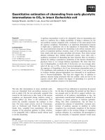
of the eye. “Backward” planni ng was used to find the
best position of the bony part. The position of the man-
dibula was predefi ned a s the ideal position for the
implants, which then predef ined the ideal position for
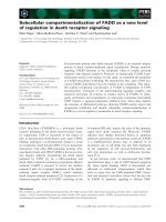
the transferred bone (Figure 1). A compu tertomography
(CT) scan of both legs was performed, and the necessary
bony shape was virtually matched w ith the patient’sleft
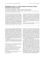
fibula (Figure 2). To achieve the desired lengths and
angles at the fibula’s resection and split sites, a rapid
prototyped template (figure 3) was manufactured by
Materialize
©
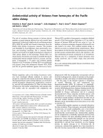
(Leuven, Belgium). To reconstruct the orbi-
tal floor, the intact site of the skull was mirrored, and
the missing bony part was identified (figure 4). Both
parts were produced with rapid prototyping b y IDEE
©
(Instrument Development Engineering & Evaluation,
Maastricht University, Maastricht, The Netherlands).
Thecompleteskullwasthenusedtopremoldatita-
nium mesh, which was sterilized before surgery.
* Correspondence:
1
Department of Cranio-Maxillofacial Surgery, Maastricht University Medical
Center, The Netherlands
Lethaus et al. Head & Face Medicine 2010, 6:16
/>HEAD & FACE MEDICINE
© 2010 Lethaus et al; licensee BioMed Centra l Ltd. T his i s an O pen Access article di stributed u nder the terms of th e Creat ive Commons
Attribution License ( which permits unrestricted use, dis tribution, and re production in
any medium, provided the original work is properly cited.
During the operation, the incision made previously
was used to open the site. As planned, the fibula and
the individualized titanium mesh were placed in the
sites selected preoperatively. Both were fixed with osteo-
synthesis screws of 2.0 diameter (KLS Martin Tuttlin-
gen, Germany). The fibula was reanastomized to the
vena jugularis interna and the arteria carotis externa.
Wound healing was uneventful for the following three
weeks. A CT scan obtained two days postoperatively
demonstrated the accuracy of the fibula insertion (figure
1, 5). The removal of the osteosynthesis material and
the placement of dental implants will be performed six
months after the operation.
Discussion and Conclusion
This case demonstrates that CAD/CAM techniques can
be of great value in planning and executing the recon-
struction of r esected or damagedtissue.Theboneand
the titanium mesh can be placed in the desired posi-
tions. Dental rehabilitation will take place after healing
of the bony junctions is complete.
Two groups have recently demonstrated the efficacy of
virtual planning and use of a rapid prototyped template
to reconstruct the mandible with a fibula graft. These
researchers presented favorable results concerning preci-
sion and outcome [3,4]. Compared to the mandible, the
maxilla presents an even more c omplex area for recon-
struction. Soft tissue covers most of the bony structures,
especially the remaining bone at the skull base region,
which is necessary for bone fixation. The anatomical
proximity to vital structures further complicates the pro-
cess of reconstruction.
We regard 3D models as a reasonable amendment in
craniofacial reconstruction that offers multiple advan-
tages. They facilitate surgical planning by demonstrating
theanatomicalcharacteristicsofthetissuetobeoper-
ated upon. By adding a haptic sensation, this approach
optimizes preoperative planning. The surgeon achieves a
bett er impression of the anatomical situation, the actual
amount of bone and the demands on the reconstruction,
which will result in a safer operation, shorter operation
time and a more predictable result. We also use the
models to explain and discuss the operation with our
Figure 1 Preoperative situation w ith prosthetic ideal bone
position.
Figure 2 Necessary bone needed to match with left fibula.
Lethaus et al. Head & Face Medicine 2010, 6:16
/>Page 2 of 4
patients, providing them with a better understanding o f
the process and its possible outcomes.
Virtual planning and the use of rapid prototyping have
been used mainly in craniomaxillofacial surgery. Because
of the use of specialized software systems, application of
this technique is limited to larger medical centers. The
disadvantages are additional costs for software and com-
puters and the additional time needed to plan the opera-
tion. Nevertheless, rapid prototyping is used in different
areas of medicine. In the context of spine surgery, tem-
plates can be used to position cervical screws to ensure
correct positioning that will avoid nerve damage [5,6].
In orthopedic surgery, templates can be used to navigate
endoprostheses. Both hip and knee implants were posi-
tioned correctly after virtual planning by means of rapid
prototyped templates [7,8]. Cardiosurgeons have
described the benefits of using rapid prototyped models
to visualize complex cardiac morphology or to build
aortic stents for training [9-11]. Those examples shoul d
encourage more surgical specialties to use these
techniques and to benefit from the advantages of
preoperative planning.
Consent
Written informed consent was obtained from the patient
for publication of this case report and any accompany-
ing images. A copy of the written consent is available
for review by the Editor-in Chief of this journal.
Author details
1
Department of Cranio-Maxillofacial Surgery, Maastricht University Medical
Center, The Netherlands.
2
Institute for Laboratory Animal Science and
Experimental Surgery, RWTH Aachen University, Aachen, Germany.
Authors’ contributions
BL was responsible for a part of the operation and drafted the manuscript.
LP was responsible for the planning and manufacturing of the templates
and the titanium mesh. PK and RB were responsible for a part of the
operation. RT conceived the report, participated in its coordination and
helped to draft the manuscript. All authors read and approved the final
manuscript.
Competing interests
The authors declare that they have no competing interests.
Received: 1 June 2010 Accepted: 19 July 2010 Published: 19 July 2010
References
1. Bill JS, Reuther JF: Rapid prototyping in planning reconstructive surgery
of the head and neck. Review and evaluation of indications in clinical
use. Mund Kiefer Gesichtschir 2004, 8:135-153.
2. Metzger MC, Hohlweg-Majert B, Schon R, Teschner M, Gellrich NC,
Schmelzeisen R, Gutwald R: Verification of clinical precision after
computer-aided reconstruction in craniomaxillofacial surgery. Oral Surg
Oral Med Oral Pathol Oral Radiol Endod 2007, 104:e1-10.
3. Hirsch DL, Garfein ES, Christensen AM, Weimer KA, Saddeh PB, Levine JP:
Use of computer-aided design and computer-aided manufacturing to
Figure 3 Rapid prototyped template, which defined the graft
length and angle of junction.
Figure 4 Rapid prototyped skull with the mirrored intact
zygomatic region inserted (marked with dots).
Figure 5 Postoperativ CT 3D reconstruction.
Lethaus et al. Head & Face Medicine 2010, 6:16
/>Page 3 of 4
produce orthognathically ideal surgical outcomes: A paradigm shift in
head and neck reconstruction. J Oral Maxillofac Surg 2009, 67:2115-2122.
4. Juergens P, Krol Z, Zeilhofer HF, Beinemann J, Schicho K, Ewers R, Klug C:
Computer simulation and rapid prototyping for the reconstruction of
the mandible. J Oral Maxillofac Surg 2009, 67:2167-2170.
5. Lu S, Xu YQ, Lu WW, Ni GX, Li YB, Shi JH, Li DP, Chen GP, Chen YB,
Zhang YZ: A novel patient-specific navigational template for cervical
pedicle screw placement. Spine (Phila Pa 1976). 2009, 34:E959-966.
6. Owen BD, Christensen GE, Reinhardt JM, Ryken TC: Rapid prototype
patient-specific drill template for cervical pedicle screw placement.
Comput Aided Surg 2007, 12:303-308.
7. Del Gaizo D, Soileau ES, Lachiewicz PF: Value of preoperative templating
for primary total knee arthroplasty. J Knee Surg 2009, 22:284-293.
8. Hananouchi T, Saito M, Koyama T, Hagio K, Murase T, Sugano N,
Yoshikawa H: Tailor-made surgical guide based on rapid prototyping
technique for cup insertion in total hip arthroplasty. Int J Med Robot
2009, 5:164-169.
9. Kalejs M, von Segesser LK: Rapid prototyping of compliant human aortic
roots for assessment of valved stents. Interact Cardiovasc Thorac Surg
2009, 8:182-186.
10. Greil GF, Wolf I, Kuettner A, Fenchel M, Miller S, Martirosian P, Schick F,
Oppitz M, Meinzer HP, Sieverding L: Stereolithographic reproduction of
complex cardiac morphology based on high spatial resolution imaging.
Clin Res Cardiol 2007, 96:176-185.
11. Sodian R, Schmauss D, Schmitz C, Bigdeli A, Haeberle S, Schmoeckel M,
Markert M, Lueth T, Freudenthal F, Reichart B, Kozlik-Feldmann R: 3-
dimensional printing of models to create custom-made devices for coil
embolization of an anastomotic leak after aortic arch replacement. Ann
Thorac Surg 2009, 88:974-978.
doi:10.1186/1746-160X-6-16
Cite this article as: Lethaus et al.: Reconstruction of a maxillary defect
with a fibula graft and titanium mesh using CAD/CAM techniques. Head
& Face Medicine 2010 6:16.
Submit your next manuscript to BioMed Central
and take full advantage of:
• Convenient online submission
• Thorough peer review
• No space constraints or color figure charges
• Immediate publication on acceptance
• Inclusion in PubMed, CAS, Scopus and Google Scholar
• Research which is freely available for redistribution
Submit your manuscript at
www.biomedcentral.com/submit
Lethaus et al. Head & Face Medicine 2010, 6:16
/>Page 4 of 4