báo cáo khoa học: " Release kinetics of VEGF165 from a collagen matrix and structural matrix changes in a circulation model" pptx
Bạn đang xem bản rút gọn của tài liệu. Xem và tải ngay bản đầy đủ của tài liệu tại đây (2.32 MB, 7 trang )
HEAD & FACE MEDICINE
Kleinheinz et al. Head & Face Medicine 2010, 6:17
/>Open Access
RESEARCH
© 2010 Kleinheinz et al; licensee BioMed Central Ltd. This is an Open Access article distributed under the terms of the Creative Com-
mons Attribution License ( which permits unrestricted use, distribution, and reproduc-
tion in any medium, provided the original work is properly cited.
Research
Release kinetics of VEGF
165
from a collagen matrix
and structural matrix changes in a circulation
model
Johannes Kleinheinz*
1
, Susanne Jung
1
, Kai Wermker
1
, Carsten Fischer
2
and Ulrich Joos
1
Abstract
Background: Current approaches in bone regeneration combine osteoconductive scaffolds with bioactive cytokines
like BMP or VEGF. The idea of our in-vitro trial was to apply VEGF
165
in gradient concentrations to an equine collagen
carrier and to study pharmacological and morphological characteristics of the complex in a circulation model.
Methods: Release kinetics of VEGF
165
complexed in different quantities in a collagen matrix were determined in a
circulation model by quantifying protein concentration with ELISA over a period of 5 days. The structural changes of
the collagen matrix were assessed with light microscopy, native scanning electron microscopy (SEM) as well as with
immuno-gold-labelling technique in scanning and transmission electron microscopy (TEM).
Results: We established a biological half-life for VEGF
165
of 90 minutes. In a half-logarithmic presentation the VEGF
165
release showed a linear declining gradient; the release kinetics were not depending on VEGF
165
concentrations. After
12 hours VEGF release reached a plateau, after 48 hours VEGF
165
was no longer detectable in the complexes charged
with lower doses, but still measurable in the 80 μg sample. At the beginning of the study a smear layer was visible on
the surface of the complex. After the wash out of the protein in the first days the natural structure of the collagen
appeared and did not change over the test period.
Conclusions: By defining the pharmacological and morphological profile of a cytokine collagen complex in a
circulation model our data paves the way for further in-vivo studies where additional biological side effects will have to
be considered. VEGF
165
linked to collagen fibrils shows its improved stability in direct electron microscopic imaging as
well as in prolonged release from the matrix. Our in-vitro trial substantiates the position of cytokine collagen complexes
as innovative and effective treatment tools in regenerative medicine and and may initiate further clinical research.
Background
Osteogenesis
The human skeleton is subject to permanent remodelling
processes: 5% of the human skeleton is rebuilt per year.
This remodelling is an integral part also of the mecha-
nism of bone healing and regeneration of bony defaults.
In the process of bone healing and regeneration, bio-
chemical procedures follow a well-defined temporal and
territorial pattern. Resting chondrocytes start to prolifer-
ate, differentiate into hypertrophic chondrocytes, and
synthesise collagen and extracellular matrix.
Then blood vessels invade; osteogenesis takes place in
the vicinity of neo-vessels that mediate the delivery of
osteoprogenitors, secrete mitogen for osteoblasts, and
transport nutrients and oxygen. The cartilage matrix is
degraded and replaced with the typical trabecular bone
matrix produced by osteoblasts. Blood vessels provide a
conduit for the recruitment of cells involved in cartilage
resorption and bone deposition and are therefore a cru-
cial condition for any regeneration [1,2]. The process is
operated by a variety of cytokines as bone morphogenetic
proteins (BMPs) or vascular endothelial growth factor
(VEGF) [3,4].
* Correspondence:
1
Department of Cranio-Maxillofacial Surgery, Research Unit "Vascular Biology
of Oral, Structures (VABOS)", University Hospital Muenster, Waldeyerstrasse 30,
D-48149, Muenster, Germany
Full list of author information is available at the end of the article
Kleinheinz et al. Head & Face Medicine 2010, 6:17
/>Page 2 of 7
There are two basic options to support bone formation:
to enhance the remodelling processes by optimizing the
vascularization via application of potent angiogenetic
cytokines as VEGF or to implant a scaffold to provide a
matrix that induces bone regeneration [5,6].
VEGF
165
VEGF is an important cytokine in the process of endo-
chondral bone development and mediating bone vascu-
larisation for normal differentiation of chondrocytes and
osteoblasts. An increase in VEGF is an indication of
increased vascular permeability and microvascular activ-
ity, including angiogenic growth of new blood vessels [7-
9].
VEGF is a homodimer glycoprotein, its family includes
6 related proteins; VEGF
165
is most common and biologi-
cally active [10]. It is released by many cell populations as
fibroblasts, monocytes, macrophages or lymphocytes
[11]. The corresponding receptors belong to the tyrosine
kinase family. VEGF
165
induces angiogenesis on different
levels: it acts as mitogen especially on endothelial cells,
raises the vessel permeability and dilatation by releasing
NO and has chemotactic impact on other growth pro-
moting cell populations [12]. The most potent stimulus
for VEGF
165
synthesis is lack of oxygen. Under hypoxia an
increase in VEGF
165
mRNA was shown and, in addition,
the RNA's half-life was extended. This effect is translated
by the hypoxia sensitive transcription factor HIF1. The
instantaneous angiogenetic effect of VEGF
165
is the
increase in vessel permeability and mitogenic stimulation
of endothelial cells. According to its potential VEGF
165
is
also involved in pathophysiological processes like tumour
growth; mainly in hypoxic tumour regions raised
VEGF
165
levels were scored [13,14]. Disadvantageous for
a routine use are a difficult handling of the liquid applica-
tion form, its short half-life and susceptibility to light and
temperature.
Bone graft substitutes and collagen
Some of the common methods used to repair bony skele-
tal defects are autografts, allografts, or synthetic implant
materials. Yet, imperfections persist in these methods,
such as limited harvesting, the possibility of disease
transmission, poor biocompatibility, and the risk of pros-
thetic implantation failure. Therefore, alternative strate-
gies, such as tissue engineering approaches, are needed to
improve the treatment and quality of life of all patients.
The minimum requirements for bone graft substitutes
are:
• No cancerogenic effect
• No water-solubility
• Non-immunogenic effect
• Lacking of an inflammatory response
• Defined bio-degradation and
• Biocompatibility, namely of the surface.
Widely-used materials are hydroxylapatite and trical-
cium phosphate as synthetic inorganic bone graft substi-
tutes. They come with good biocompatibility and
osteoconductivity. Yet, they are brittle and not resilient in
functionally stressed areas [15-17]. The advantage of col-
lagen as a natural substitute is the fact that collagen is the
main constituent of organic bone matrix. Fitted in bony
defaults it is not degraded by but incorporated into the
regenerating tissue. It accelerates the healing process and
reduces the side effects of decomposition products
[18,19].
In innovative approaches the osteoconductive collage-
nous scaffold is combined with the osteoinductive impact
of cytokines like BMP or VEGF
165
. The objectives of our
study were to apply VEGF
165
in gradient concentrations
to an equine collagen carrier and to study the complex in
a circulation model. The VEGF
165
release kinetics should
be quantified and the morphological degradation of the
collagen-cytokine complex should be visualized.
Methods
VEGF
165
-collagen complex
Collagen I was purchased (Resorba, Nuernberg, Ger-
many) and liquefied. Human recombinant VEGF
165
(R&D
Systems, Wiesbaden, Germany) was added in different
concentrations. The complexes were formed in hemi-
spheres and drugged with aldehyde to avoid the cross-
linking of collagen fibrils.
The total quantity of collagen was 5.6 mg/cm
3
per
application, VEGF
165
was added in 0.8 μg, 10 μg or 80 μg
quantities.
Circulation model
We used a digitally controlled peristaltic pump that deliv-
ered the medium with a mean flow rate of 27 ml per min-
ute (Cole Parmer Masterlex Console Drive Pump). As
aqueous solution a 0.2 mol PBS buffer was utilized in a
total quantity of 80 ml. Circulation was simulated under
constant conditions of 20°C and pH 7.2.
Lab report
The complexes were charged with VEGF
165
in three dif-
ferent concentrations: 0.8 μg, 10 μg and 80 μg. Three
complexes of each concentration were incubated for 5
days. As a sample, the total volume of buffer medium was
extracted and analysed to avoid saturation of the buffer
medium with free VEGF
165
. To differentiate between the
initial degradation of our collagen complexes with a quick
VEGF
165
release and the slow long-term saturation pro-
cess, we adopted an asymmetrical test pattern:
Kleinheinz et al. Head & Face Medicine 2010, 6:17
/>Page 3 of 7
On day one we took samples after 30 min, 1, 2, 4, 8, 12
and 24 hours. The next specimens were taken after day 2,
3, 4 and 5. VEGF
165
-free collagen complexes served as
negative controls and were analysed identically.
ELISA
VEGF
165
concentrations were assessed by performing a
solid-phase VEGF
165
Immunoassay (VEGF
165
Quantikine,
DVE00, R&D Systems GmbH, Wiesbaden-Nordenstadt,
Germany). The ELISA was performed according to the
manufacturer's protocol; its sensitivity was described as <
9 pg/ml. The concentration of VEGF
165
was expressed as
pg/ml.
VEGF
165
was quantified by using a standard curve made
by human VEGF
165
ranging from 31.2 pg/ml to 2000 pg/
ml. The chromogenic reaction was read at 415 nm
(Molecular Devices).
Light microscopy
Collagen samples were processed according to a standard
protocol. In short, they were fixed, dehydrated in increas-
ing gradients of ethanol and embedded in paraffin. Thin
sections were sliced, stained according to an azan stan-
dard procedure and fixed in methacrylate.
The sections were evaluated with a light microscope
(Zeiss Axioscop, Jena, Germany).
Scanning electron microscopy (SEM)
Samples were fixed in 3% glutaraldehyde in 0.1 mol phos-
phate buffered saline and then washed in the buffer (0.1
mol PBS). After rinsing, the samples were dehydrated in a
graded ethanol series and dried with a critical point dry-
ing. All dried samples were mounted on aluminium stubs
and sputter coated with coal to a coating thickness of 8
nm.
For immunohistochemical SEM analysis the sections
were fixed in 4% paraformaldehyde solution, rinsed with
0.1 mol PBS buffer and incubated with primary VEGF
165
-
specific antibodies at room temperature for 1 hour. After-
wards, the secondary immunogold-labelled antibody was
incubated at room temperature for 1 hour. Between incu-
bation steps phosphate buffered saline rinses were per-
formed. All antibodies were diluted according to the
manufacturers' instructions.
The gold particles as spheres of a 10 nm diameter were
easily detectable in scanning electron microscopy.
Transmission electron microscopy (TEM)
For TEM analysis the collagen samples were fixed in 3%
glutaraldehyde for 24 hours, rinsed in 0.1 mol phosphate
buffered saline and incubated in osmium acid for 1 hour.
Afterwards, the samples were dehydrated in a graded
ethanol series, embedded in araldite and sliced thin sec-
tions (1 μm). The slices were stained with tolouidin blue
following a standard procedure. Representative areas
were cut in ultra-thin slices of 70 nm, placed on copper
nets and analysed in transmission electron microscopy.
Immunohistochemical staining was performed as
described before; the gold spheres in TEM presented as
dark areas.
Results
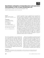
VEGF
165
half-life
To determine biological half-life of VEGF
165
its dissolu-
tion in aqueous solution at room temperature was analy-
sed. VEGF
165
collagen complexes charged with 10 μg of
VEGF
165
were probed over 12 hours. Our results provide
a half-life of free VEGF
165
of 90 minutes (Fig. 1).
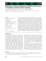
VEGF
165
release kinetics
In a half-logarithmic presentation the observed VEGF
165
concentration showed a characteristic linear decline over
time. The gradients of the three VEGF
165
doses were par-
allel and independent of VEGF
165
concentration. VEGF
165
release reached a plateau after 12 hours and was no lon-
ger detectable in the applications of 0.8 μg and 10 μg after
48 hours, whereas the complex charged with 80 μg of
VEGF
165
still showed measurable cytokine release after
over 50 hours. Saturation effects of the buffer medium
were not observed (Fig. 2).
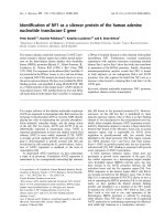
VEGF
165
degradation
The efficiency describes the quotient of VEGF
165
val-
ues scored in our test setting and initially applied
VEGF
165
. Only 10% of initially applied 0.8 μg were
finally detected in the present study. Ninety per cent
were lost during production, transport or storage. Of
the applied 10 μg and 80 μg, 96% respectively 97% were
lost (Fig. 3).
Figure 1 Half-life of VEGF.
Kleinheinz et al. Head & Face Medicine 2010, 6:17
/>Page 4 of 7
Light microscopy
In light microscopy the VEGF
165
collagen complex
appears homogenously, presents a reticular structure and
shows no signs of structural defaults caused by fixation or
coupling with VEGF
165
. Only in the periphery single
agglutinated fibres are detected; these are artefacts
caused by the production process (Fig. 4).
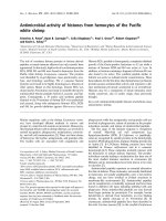
SEM
In scanning electron microscopy the VEGF
165
collagen
complexes feature more agglutinated parts, even in cen-
tral areas, in contrast to the collagen matrix without
cytokine (Fig. 5a and 5b).
During the five days of degradation process the ultra
structure of the VEGF
165
collagen complexes changes
considerably. On day 0, the collagen matrix is coated by a
VEGF
165
layer that varnishes the single collagen fibrils.
After 3 days of simulated circulation the collagen fibres
are clearly detectable; this effect is more obvious on day
five. The collagen matrix appears porose and knotty (Fig.
6a and 6b).
With immuno-gold-labelling the VEGF
165
molecules
are visible. A homogenous distribution of VEGF
165
in the
collagen scaffold can be proved (Fig. 7).
TEM
In transmission electron microscopy the gold particles
present themselves as black round structures (Fig. 8). Sin-
gle VEGF antibody complexes can be precisely assigned
to their corresponding collagen fibril. Due to the close
vicinity between fibre and VEGF an adhesion must be
assumed that overcomes the preliminary chemical proce-
dure for TEM (Fig. 9).
Discussion
To restore form and function to an existing bony defect,
vascularisation is the key to success.
Clinical experience shows that avascular bony struc-
tures namely in chronically infected bones tend to atro-
phy and fracture [20].
Circulation and angiogenesis are responsible for a
restored perfusion of impaired bone areas.
Bone cells on the other hand release growth factors to
stimulate angiogenesis. Osteo- and angiogenesis are
clearly linked in a strong co-dependent relation. The high
susceptibility and the low applicable doses of cytokines
Figure 2 Release kinetics of VEGF.
Figure 3 Natural degradation of VEGF.
Figure 4 Collagen matrix, azan staining (100×): representative
central area of pure collagen matrix.
Figure 5 Collagen matrix with (a) and without (b) VEGF, SEM
(100×); the smear layer coffering the surface of the collagen ma-
trix can be seen on the left picture.
A B
Kleinheinz et al. Head & Face Medicine 2010, 6:17
/>Page 5 of 7
make high demands: next to good biocompatibility, an
easy application mode is critical for the successful use of
biomaterials for regenerative medicine strategies [21,22].
VEGF
165
has been exposed as the central angiogenetic
protein in the process of bone regeneration; many in-
vitro studies underlined its potency to stimulate osteo-
genesis physiologically via induction of neo-vascularisa-
tion [23]. Xenogenic collagen is a well established drug
carrier in daily clinical use. As freeze-dried sponge it
comes with excellent biocompatibility and is hence the
ideal carrier for cytokine application.
In the present study the combination of a xenogenic
collagen carrier and recombinant human VEGF
165
is anal-
ysed pharmacologically and morphologically. This kind
of research is crucial for forthcoming in-vivo studies
where biological factors will overlie and falsify the thera-
peutical effects of the VEGF
165
collagen complex. To be
able to interpret these results properly drug release kinet-
ics has to be established before. In cell cultures the
VEGF
165
specific half-maximum growth stimulation has
been determined. The effect of applied cytokines is sup-
posed to range above this score [24].
Our data accounts for VEGF
165
release from the colla-
gen over 48 hours; considering the 90 minutes half-life of
free VEGF
165
it is a surprising result. Obviously, a stabili-
sation of VEGF
165
can be achieved by connecting the
cytokine with collagen fibrils. The trial at hand provides
only indirect evidence for this assumption but is observed
in the whole test series.
During the first 50 hours an elevated release rate was
observed as described in the literature before. The
VEGF
165
release is divided in two phases: first, the quick
elusion of VEGF
165
and diffusion into the buffer medium,
and second, the slow sustained disposal when the
VEGF
165
molecules are dissolved from the degrading col-
lagen fibrils in the deeper areas of the matrix.
This pharmacological behaviour corresponds with our
morphological findings in REM: hydrolytic erosion
reveals the single collagen fibrils and facilitates VEGF
165
release.
The fraction of released VEGF
165
varies in our data
from 3% to 10%. Despite ideal test condition the main
Figure 6 VEGF
165
-collagen complex on day 3 (a) and day 5 (b),
SEM (20000×).
A B
Figure 7 VEGF
165
-collagen complex, 10 μg, TEM, (5000×).
Figure 8 VEGF
165
-collagen complex, 10 μg, TEM (3400×).
Figure 9 VEGF
165
-collagen complex, 10 μg, TEM, (21500×); a
VEGF-antibody complex in relation to its collagen fibre.
Kleinheinz et al. Head & Face Medicine 2010, 6:17
/>Page 6 of 7
section of VEGF
165
is lost during production, transport
and storage.
The decreasing efficacy of the higher concentrated
VEGF
165
carriers argues for a saturation effect, higher
doses of VEGF
165
in the collagen scaffold do not lead to
higher VEGF
165
release [6].
To sum up: The biphasic release kinetic allows a hyper-
physiological stimulation caused by the applied VEGF
165
over 50 hours. It is more efficient than free VEGF
165
.
Higher doses of VEGF
165
do not lead to better effects for
there is no proportional connection between the dose in
the collagen carrier and the emitted total quantity.
The next steps to elucidate the biological behaviour of
the cytokine collagen complex are in-vivo trials to elimi-
nate the shortcomings of our setting
- PBS as an inadequate model for blood flow in human
tissues
- disregard of enzymatic degradation processes
- insufficient verification of biologically active cytokine
areas
The interfacing of VEGF
165
to a collagen scaffold is not
the only way of cytokine application: its transport in
micro spheres was described; cytokine mRNA was cou-
pled with a viral vector and cytokine plasmid DNA was
directly transferred into the tissue [25-27].
Conclusions
The restitution of bony defaults with a technique that
provides biologic functionality, easy mechanical handling
and reliable outcome is a significant challenge in maxillo-
facial surgery.
Our idea was to combine an osteoconductive scaffold
with osteoinductive proteins and hence to stimulate and
support natural healing and regenerating processes.
Our in-vitro trial substantiates the position of cytokine
collagen complexes as innovative and effective treatment
tools in regenerative medicine and paves the way for fur-
ther clinical research.
Competing interests
The authors declare that they have no competing interests.
Authors' contributions
CF established the circulation model.
JK carried out the immunoassays.
SJ and KW participated in the design of the study and performed the statistical
analysis.
UJ, JK and CF conceived of the study, and participated in its design and coordi-
nation and helped to draft the manuscript.
CF and UJ were involved in revising the article.
All authors read and approved the final manuscript.
Author Details
1
Department of Cranio-Maxillofacial Surgery, Research Unit "Vascular Biology
of Oral, Structures (VABOS)", University Hospital Muenster, Waldeyerstrasse 30,
D-48149, Muenster, Germany and
2
Private practice, Duelmen, Germany
References
1. Reddi A: Bone and cartilage differentiation. Curr Opin Gen Develop 1994,
4:737-744.
2. Caplan AI: Cartilage begets bone versus endochondral myeloporests.
Clin Orthop 1990, 261:257-267.
3. Kübler N: Osteoinduktion und -reparation. Mund Kiefer GesichtsChir
1997, 1:2-25.
4. Schmidt K, Swoboda H: Die Bedeutung matrixgebundener Zytokine für
die Osteoinduktion und Osteogenese. Implantologie 1995, 2:127-148.
5. Sauter E, Nesbit M, Watson J, Klein-Szanto A, Litwin S, Herlyn M: Vascular
endothelial growth factor is a marker of tumor invasion and metastasis
in squamous cell carcinomas of the head and neck. Clin Cancer Res
1999, 5:775-782.
6. Schliephake H, Jamil M, Knebel J: Experimental reconstruction of the
mandible using polylactic acid tubes and basic fibroblast growth
factor in alloplastic scaffolds. J Oral Maxillofac Surg 1998, 56:616-626.
7. Ferrara N, Carver-Moore K, Chen H, Dowd M, Lu L, O'Shea K, Powell-
Braxton L, Hillan K, Moore M: Heterozygous embryonic lethality induced
by targeted inactivation of the VEGF gene. Nature 1996, 380:439-443.
8. Kleinheinz J, Joos U: Serum concentration of VEGF and bFGF in patients
with sagittal split ramus osteotomy. Int J Oral Maxillofac Surg 1999,
28:539.
9. Hollinger J, Wong M: The integrated process of hard tissue regeneration
with special emphasis on fracture healing. Oral Surg Oral Med Oral
Pathol Oral Radiol Endod 1996, 82:594-606.
10. Mattei MG, Borg JP, Rosnet O, Marmé D, Birnbaum D: Assignment of
vascular endothelial growth factor (VEGF) and placenta growth factor
(PLGF) genes to human chromosome 6p12-p21 and 14q24-q31
regions, respectively. Genomics 1996, 32:168-9.
11. Drake CJ, Little CD: Exogenous vascular endothelial growth factor
induces malformed and hyperfused vessels during embryonic
neovascularization. Proc Natl Acad Sci USA 1995, 92:7657-61.
12. Keck PJ, Hauser SD, Krivi G, Sanzo K, Warren T, Feder J, Connolly DT:
Vascular permeability factor, an endothelial cell mitogen related to
PDGF. Science 1989, 246:1309-12.
13. Plate KH, Breier G, Risau W: Molecular mechanisms of developmental
and tumor angiogenesis. Brain Pathol 1994, 4:207-18.
14. Senger DR, Van de Water L, Brown LF, Nagy JA, Yeo KT, Yeo TK, Berse B,
Jackman RW, Dvorak AM, Dvorak HF: Vascular permeability factor (VPF,
VEGF) in tumor biology. Cancer Metastasis Rev 1993, 12:303-24.
15. Hutmacher D, Kirsch A, Ackermann K, Hürzeler M: Matrix and carrier for
bone growth factors: state of the art and future perspectives. Berlin
Heidelberg, Springer; 1998.
16. Wang D, Yamazaki K, Nohtomi K, Shizume K, Ohsumi K, Shibuya M, Sato K:
Increase of vascular endothelial growth factor mRNA expression by
1,25-dihydroxyvitamin D3 in human osteoblast-like cells. J Bone Miner
Res 1996, 11:472-479.
17. Ramselaar M, Driessens F, Kalk W, De Wijn J, Van Mullem P:
Biodegradation of four calcium phosphate ceramics; in vivo rates and
tissue interactions. J Mat Sci 1991, 2:63-70.
18. Hemprich A, Lehmann R, Khoury F, Schulte A, Hidding J: Filling cysts with
type 1 bone collagen. Dtsch Zahnarztl Z 1989, 44:590-592.
19. Basle M, Lesourd M, Grizon F, Pascaretti C, Chappard D: Typ-I-Kollagen im
xenogenen Knochenmaterial reguliert Anbindung und Verbreitung
von Osteoblasten über die β1-Integrin-Untereinheit. Orthopäde 1998,
27:136-142.
20. Burchardt H: Biology of bone transplantation. Orthop Clin North Am
1987, 18:187-196.
21. Crotts G, Park T: Protein delivery from poly(lactic-co-glycolic acid)
biodegradable microspheres: release kinetics and stability issues. J
Microencapsulation 1998, 15:699-713.
22. Arnold F, West D: Angiogenesis in wound healing. Pharm Ther 1991,
52:407-422.
23. Iruela-Arispe M, Dvorak H: Angiogenesis: a dynamic balance of
stimulators and inhibitors. Thrombosis and Haemostasis 1997,
78:672-677.
Received: 2 June 2010 Accepted: 19 July 2010
Published: 19 July 2010
This article is available from: 2010 Kleinheinz et al; licensee BioMed Central Ltd. This is an Open Access article distributed under the terms of the Creative Commons Attribution License ( which permits unrestricted use, distribution, and reproduction in any medium, provided the original work is properly cited.Head & Face Medicine 2010, 6:17
Kleinheinz et al. Head & Face Medicine 2010, 6:17
/>Page 7 of 7
24. Kremer C, Breier G, Risau W, Plate KH: Up-regulation of flk-1/vascular
endothelial growth factor receptor 2 by its ligand in a cerebral slice
culture system. Cancer Res 1997, 57:3852-9.
25. Safi J, DiPaula A, Riccioni T, Kajstura J, Ambrosio G, Becker L, Anversa P,
Capogrossi M: Adenovirus-mediated acidic fibroblast growth factor
gene transfer induces angiogenesis in the nonischemic rabbit heart.
Microvasc res 1999, 58:238-249.
26. Franceschi RT: Biological approaches to bone regeneration by gene
therapy. J Dent Res 2005, 84:1093-103.
27. Isner J, Pieczek A, Schainfeld R, Blair R, Haley L, Asahara T, Rosenfield K,
Razvi S, Wash K, Symes J: Clinical evidence of angiogenesis after gene
transfer of phVEGF165 in patient with ischemic limb. Lancet 1996,
348:370-374.
doi: 10.1186/1746-160X-6-17
Cite this article as: Kleinheinz et al., Release kinetics of VEGF165 from a colla-
gen matrix and structural matrix changes in a circulation model Head & Face
Medicine 2010, 6:17