báo cáo khoa học: " Embryonic stem cells in scaffold-free threedimensional cell culture: osteogenic differentiation and bone generation" ppt
Bạn đang xem bản rút gọn của tài liệu. Xem và tải ngay bản đầy đủ của tài liệu tại đây (2.97 MB, 6 trang )
RESEA R C H Open Access
Embryonic stem cells in scaffold-free three-
dimensional cell culture: osteogenic
differentiation and bone generation
Jörg Handschel
1
, Christian Naujoks
1*
, Rita Depprich
1
, Lydia Lammers
2
, Norbert Kübler
1
, Ulrich Meyer
1
and
Hans-Peter Wiesmann
3
Abstract
Extracorporeal formation of mineralized bone-like tissue is still an unsolved challenge in tissue engineering.
Embryonic stem cells may open up new therapeutic options for the future and should be an interesting model for
the analysis of fetal organogenesi s. Here we describe a technique for culturing embryonic stem cells (ESCs) in the
absence of artificial scaffolds which generated mineralized miromasses. Embryonic stem cells were harvested and
osteogenic differentiation was stimulated by the addition of dexamethasone, ascorbic acid, and ß-
glycerolphosphate (DAG). After three days of cultivation microspheres were formed. These spherical three-
dimensional cell units showed a peripheral zone consisting of densely packed cell layers surrounded by minerals
that were embedded in the extracellular matrix. Alizarine red staining confirmed evidence of mineralization after 10
days of DAG stimulation in the stimulated but not in the control group. Transmission electron microscopy
demonstrated scorching crystallites and collagenous fibrils as early indication of bone formation. These extracellular
structures resembled hydroxyl apatite-like crystals as demonstrated by distinct diffraction patterns using electron
diffraction analysis. The micromass culture technique is an appropriate model to form three-dimensional bone-like
micro-units without the need for an underlying scaffold. Further studies will have to show whether the technique
is applicable also to pluripotent stem cells of different origin.
Keywords: Embryonal stem cell, osteogenic tissue engineering, three-dim ensional culture technique, scaffold free
tissue, hydroxyl apatite
Introduction
Bony defects have various causes and of ten turn out to
be a major therapeutic challenge. Until today, the recon-
struction of bone using autologous grafts has been
recog nized as the gold standard because it provides bio-
logical active cells with osteoinductive properties and
avoids any immunological reactions [1]. Unfortunately,
the harvesting of these grafts causes donor-side defects
and shows a quantitative limitation [2-4]. Artificial
materials and extracorporeal tissue formation are alter-
native approaches for the reconstruction of bone defects,
because they neither cause donor-site lesions nor is their
availabilty restricted.
Bone is a highly specialized tissue of the organism
which is generated by mineral ization of the extracellular
matrix called osteoid. Osteoblasts and osteoclasts contri-
bute to the formation and remodelling of bone tissue.
However, there are further cell types e.g. endothelia
cells, which are also essential for bone formation [5].
The complex cell-driven process of bone formation
starts early in the embryo and results in bone tissue
with unique features that combines stiffness and elasti-
city with the ability to regenerate itself [6]. A key feature
of bone tissue is the presence of biological active apatite
crystals. These crystals were formatted by the minerali-
zation of the extracellular matrix (osteoid) with calcium
and p hosphate ions. The process of mineralization can
be monitored histologically by special stainings like ali-
zarin red or ultrastructurally by transmission (TEM) and
scanning electron microscopy (SEM).
* Correspondence:
1
Department for Cranio- and Maxillofacial Surgery, Heinrich-Heine-Universität,
Moorenstr. 5, D- 40225 Düsseldorf, Germany
Full list of author information is available at the end of the article
Handschel et al. Head & Face Medicine 2011, 7:12
/>HEAD & FACE MEDICINE
© 2011 Handschel et al; lic ensee BioMed Central Ltd. This is an Open Access article distributed under the terms of the Creative
Commons Attribution License (http://cre ativecommons.org/licenses/by/2.0), which permits unrestricted u se, distribution, and
reproduction in any mediu m, provided the original w ork is properly cited.
Common approaches for en gineering bone ex vivo are
usually based on a combination of cells and scaffolds
[7-9]. Even the ex vivo de novo bone building starts
with the secretion of collagen via matrix vesicles fol-
lowed b y the mineralisation of the extracellular matrix
molecules [7]. It has been reported that cells in three-
dimensional cultures exert higher proliferation rates
than cells cultured in monolayers, suggesting that their
differentiation resembles more closely that seen in situ
[10-12]. Furthermore, it is assumed that cells are more
flexible to change their shape and behaviour upon speci-
fic cell signals when they are cultured in three-dimen-
sional as compared to two-dimensional cultures [13,14].
Whereas a multitude of extracorporeal bone tissue
engineering approaches have been undertaken to fabri-
cate bone tissue ex vivo, up to now cell culture-based
methods for synthesizing bone-like tissue on a structural
level are still limited due to tec hnical restrictions [15].
Here we describe that mineralized bone-like matrix is
produced by osteoinduced totipot ent embryonic st em
cells cultured in three-dimensional micromass technique
in the absence of any scaffold. The osteogenic differen-
tiation of the cells was induced by the addition of dexa-
methasone, ascorbic acid, and ß-glycerolphosphate
(DAG) to the medium [16,17]. The features of ossifica-
tion mimic in-vivo bone formation, thus enabling
matured mineralized bone matrix to be generated.
Materials and methods
Cell culture
A cell culture method for producing mineralized bioma-
terial-free, three-dimensional cell units up to 0.4 mm in
diameter was established. Feeder-independent murine
embry onic stem cells (ESCs) were kindly provide d by K .
Pfeffer (Institute f or Microbiology, Heinrich Heine Uni-
versity of Düsseldorf, Germany). The cells were derived
from the inner cell mass of blastocysts extracted from
C57BL/6 mice and tested posit ive for the stem cell mar-
kers Pouf1 (alias Oct4) and Foxd3 [18]. Cells were cul-
tured in Dulbecco’ s modified Eagle medium (DMEM,
Gibco) supplemented with penicillin (100 U/ml, Grü-
nenthal), streptomycin (100 U/ml, Hefa-pharma), 2-mer-
captoethanol (500 mM, Gibco), ultraglutamine (2 mM;
Cambrex), leukemia inhibitor factor (1000 U/ml; Chemi-
con) and 15% fetal calf serum. The cells were split every
second day and the medium was changed every day by
detaching the cells with 0.25% trypsin (Pan Biotech).
ESCs were detached from the plate, centrifuged and
resuspended in normal growth medium (1 × 10⁶ cells/
ml).
To prevent adherence of the cells leading to the for-
mation of monolayers, the microsphere assembly bior-
eactor was prepared by filling 60 μlofasolution
consisting of 2% agarose in DMEM (without any
supplements) into 96-well plates. After curing of the
agarose solution to each well, 180 μlofcellsuspension
was a dded and the cells were incubated overnight. The
old medium was replaced by equal volumes (160 μl) of
control medium and control medium containing 100
nM dexamethasone, 50 μM ascorbic acid, and 10 mM
b-glyerolphosphate (all from Sigma), respectivey. Thus,
half of the culture chambers were incubated in the pre-
sence of dexamethasone, ascorbic acid, and DAG, (DAG
(+)) to induce the osteogenic differentiation, w hile the
other half used as a control was cultivated in medium
without these stimuli (DAG (-)). Both cell populations
were kept in culture for three weeks in an incubator
under a humified a tmosphere (37°C, 90% humidity, 5%
CO
2
). The medium was changed every day. After 3, 7,
10, and 21 days one quarter of the cultivated wells with
microspheres of the + and - DAG group was harvested
and transferred into Petri dishes for a washing step with
phosphate-buffered saline (PBS). Subsequently the pre-
paration of the spheres for the different analysis was
performed.
Histological analysis
For histologiacal analysis, micromasses were fixed in for-
malin (4%) until further procession. Formaline-fixed
microspheres were dehydrated in increasing ethanol
concentrations (50%, 75%, 90% and 100%) and
embedded in paraffin (Paraplast plus). Sections (4 μm)
were mounted on Superfrost slides, deparaffinized with
xylol and rehydrated in decreasing ethanol concentra-
tions. Samples were stained with alizarine red solution
(2%) to detect calcium and counterstained with toluidine
blue, as mentioned in the literature. Briefly, af ter stain-
ing with toludine blue the slides were counterstained
with alizarin red (mixture of 0.5 g alizarin red and 0.5
ml 0.28% NH3 with 45 ml distilled water (pH: 6.4)).
Before the slides were finally covered with entellan, they
were incubated in xylene. A descriptive analysis was
performed.
Scanning electron microscopy
For scanning electron microscopy, micromasses were
fixedinglutaraldehyde(4%)followedbyawashingstep
with 0.1 M PBS. Microspheres were dehydrated in
increasing isopropanol concentrations (30%, 50%, 70%,
90%, 96%, and 100%; 30 minutes for each concentra-
tion). The critical pont drying was performed following
the instructors protocol. In this procedure isopropanol
was substituted for CO2 by five wash ing steps. Af ter
drying, the specimens were directly put on a carbon pad
of a SEM-holder (Cambridge). For morphological stu-
dies, probes were sputtered with platinum, whereas for
EDX analysis, samples were coated with carbon using
standard techniques. Scanning electron microscopy was
Handschel et al. Head & Face Medicine 2011, 7:12
/>Page 2 of 6
performed with a DSM 960 (Zeiss) microscope using an
acceleration voltage of 5-15 kV.
Transmission electron microscopy
For TEM, specimens were fixated i n glutaraldehyde
(2.5%) and embedded in araldite. For morphological
analysis, a fixation with osmium tetroxide and glutaral-
dehyde was carried out. Specimens were washed three
times with 0.1 M PBS for 10 minutes each. Micro-
spheres were dehydrated in increasing iso propanol con-
centrations (50%, 70%, 90%, 96% and 100%; 30 minutes
for each concentration) and followed by a transfer into
propylene oxid. Afterwa rds the spheres were transferred
to pure araldite by using intermediate r atios of mixtures
(100% propylene oxide, 2/1 propylene oxide/araldite, 1/
1, 1/2, 100% araldite). To harden the araldite the speci-
men were kept at 42°C for 24 hours and afterwards
were sectioned with a microtome (Ultracut S, Reichert).
For morphological studies ultrathin sections were
stained with osmium tetroxide (OsO
4
). For ultrastr uc-
tural assessment of the mineral substance no staining
was performed and the water contact during prepara-
tion, particulary during sectioning, was reduced to a
minimum in order to avoid dissolution or redistribution.
The ultrathin slides were applied to copper grids and
contrasted with uranyl acetate. Analyses were performed
with an acceleration voltage of 80 kV with EM902
(Zeiss). Electron spectroscop ic diffraction analysis w as
performed with the specimens used for the TEM. Con-
tact time of the slides with water on the microtome was
limited to a few seconds to avoid redistribution of the
crystallites. Analyses were performed with an EM902
(Zeiss ) microscopy using 80 kV acceleration voltage and
a camera length of 650 mm. D-values for the 002 dif-
fraction patterns were calculated according to Arnold et
al. and Plate et al. [19,20].
Results
After three days all cell cultures formed spheroid, th ree-
dimensional cell units in high density (5 × 10
6
cells/ml),
which appeared as oval micromasses. At that time,
neither in specimens from the DAGstimulated group
nor in the non-stimu lated group signs of mineralization
were detectable. After 10 days of cultivation t he first
indications of mineralization were visible in the DAG-
treated cells, while they were absent in non-stimulated
cells. Mineralization proceeded in the cen tre of the sti-
mulated specimens and became more clearly visible
after 3 weeks of cultivation in the presence of osteoin-
ductive stimuli. Numerous living cells were detected in
the mineralized centre of the spheres by means of tolui-
dine blue staining (Figure 1). Generally, the m ineraliza-
tion was most p rominent i n the centre of the sphere, as
demonstrated in histological sections stained with
alizarin red (Figure 1). The SEM analysis confirmed the
differences regarding the distribution pattern of the
formed mineral and the quantitative differences. The +
DAG group showed an intense mineralization in the
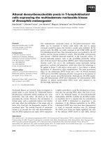
centre of the spheres (Figure 2). Transmission electr on
Figure 1 Micromasses consisting of embryonic stem cells were
cultured with or without DAG and stained with toluidine blue
followed by counterstaining with alizarin red. Shown is evidence
for the mineralization in the centre of the micromasses, which were
stained in red.
Figure 2 SEM image of the minerali zation in the cent re of a
+DAG spheres after 21 days.
Handschel et al. Head & Face Medicine 2011, 7:12
/>Page 3 of 6
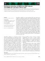
microscopy (TEM) confirmed the presence of scorching
crystallites in the mineralized area, which appeared after
21 days of cultivation (Figure 3a). Theses crystals were
typically embedded in an extracellular matrix containing
numerous collagenous fibrils (Figure 3b). The spherical
cell units had a peripheral zone consisting of densely
packed cell layers, which surrounded the minerals. To
demonstrate that the mineralized matrix in the DAG-
treated group is composed of hydroxyl apatite crystals,
electron spectroscopic diffraction analysis was per-
formed (Figure 4). In accordance with Arnol d et al. and
Plate et al., the d-value for the diffraction ring 002 was
calculated (0.344 nm) (Figure 3 and 4).
Discussion
Thede-novoformationofboneintermsoftissueengi-
neering requires cells, matrix and growth factors. For
crea ting larger tissue constructs for surgical use, natural
or artificial biomaterials are additionally needed as scaf-
folds. However, there is controversy about the use of
biomaterials as a scaffold because the physicochemical
properties of the biomateri als influence the proliferation
and gene expression of the cells [9,21,22]. Even protein
coating of the scaffold has impact on the attachment of
the cells [23-25]. It is generally accepted that no existing
artificial or natural scaffold can meet all the require-
ments f or ruling out undesired effects. The micromass
culture technique may be an alternative for substituting
artificial scaffolds. In contrast to monolayers, cell cul-
ture-based techniques in three-dimensional space appear
to more closely resemble in-vivo conditions [11]. It is
well known that many functions of the cells, e.g. differ-
ent iation and prolifera tion, rely on intact cell-cell inter -
actions and a tight attachment to extracellular matrix
components. In micromasses, the cells can interact with
each other and maintain these interactions [26]. Former
studies have shown that in micromass culture techni-
ques a cartilaginous differentiation of ESCs is feasible
[27,28]. In the presented study we show that stimulated
ESCs cultured in micromass techniqu e form minealiz ed
microspheres during cultivation.
Aggregation of cells is the pivotal stage in the develop-
ment of skeletal tissues and the primary resource from
which the skeleton is built and through which the skele-
ton is modified ontogenetically [29] . Mineralized bony
units formed ex vivo seem to be an ideal biomaterial
because they combine the structural features of bone.
Currently, the b est treatment option for bone defects
utilises the enhanced regeneration potential of embryo-
nic stem cells [30]. In this respect, fusion of multiple
bony units may allow the reconst ruction of larger skele-
tal elements. Through the ability of embryonic stem
cells to differentiate along the whole osteogenic path-
way, embryonic stem cell transplantation may play a
future role in the treatment of generalized bone dis-
eases. Furthermore, we show that osteoinductive stimuli
including DAG support the mineralization of the extra-
cellular matrix and that stimulated micromasses produce
more mineralized extracel lular matrix than micromasses
cultured in the absence of these stimuli. To verify that
the matrix consists of hydroxyl apatite, we performed
transmission e lectron microscopy and revealed a time-
dependent occurrence of scorching crystallites in the
interior of the microspheres. Using electron spectro-
scopic diffraction we confirmed that the crystallites con-
sisted of hydroxyl apatite. Furthermore, we detected
Figure 3 ESC micromass cultured for 21 days in the presence
of medium containing dexamethasone, ascorbic acid, and ß-
glycerolphosphate (DAG). Transmission electron microscopy
demonstrated scorching crystallites (a) and collagen fibrils (b) in the
mineralized area.
Figure 4 Electron spectroscopic diffraction in the centre of
DAG-treated ESC microspheres showed typical patterns for
hydroxyl apatite formation (day 21).
Handschel et al. Head & Face Medicine 2011, 7:12
/>Page 4 of 6
collagen fibrils that were morphologically very similar to
collagenous fibril s within bone tissue. Coll agen I fibrils
are known to be a major extracellular matrix component
of bone tissue [5,31]. Plate and co-workers described the
formation of hydroxyl apatite in bone and dentin as a
multistage process resulting in the depositi on of a
mineralized matrix. These calcium-phosphate crystals
coordinate longitudinally and accumulate a s scorching
crystallites [20]. In samples from stimulated ESCs we
detected crystal-like structures in the i nterior of the
microspheres. These ESC microspheres resemble aggre-
gates consisting of preosteoblasts.
Our finding of a mineralization in microspheres of
DAG-treated ESCs seems to share similarity to the for-
mation of bone and dentin in vivo. Thus, it appears that
osteologous differentiation of ESC micromasses may be
a f easible approach to advance the bony reconstruction
of large defects. However, the size of the microspheres
is limited possibly due to restricted diffusion of nutrients
and we are currently unable to format larger tissue con-
structs without support by artificial matrices. The use of
bioreactors may be an adequate technique to gain larger
tissue constructs without the need for a scaffold by sim-
ply transferring osteologously differentiated ECS micro-
masses [32].
Nevertheless, the micromass culture technique may be
an appropriate model to analyse the formation of the
skeleton during embryonic or fetal organogenesis.
Aggregation of cells to a critical size is a fundamental
step in initiating organogenesis of vertebrates [33]. Hall
and Miyake assume that the condensation of cells is a
precondition for skeleton formation that promotes the
differentiation of cells to osteoblasts and chondroblasts
[29,34]. Furthermore, the three-dimensional micromass
culture technique may be a useful method for identify-
ing substances that enhance mineralization.
The use of embryonic stem cells will probably play a
major role in tissue engineering in the future be cause of
the remakable potential and differentiation capacity of
ESCs. Prior to clinical application, many challenges need
to be faced in future studies, particularly with respect to
immune tolerance and the f ormation of malignant
tumors in the host organism. However, the studies by
Burt and coworkers are promising with regard to
immuntolerance. They grafted ESCs into MHC-mis-
matched mice and found no clinical or histological evi-
dence for a graft-versus-host or host-versus-graft
reaction [35].
Furthermore, Zavazava has demonstrated that ESCs
have the potential to induce immune tolerance [36] and
revealed evidence for a suppression of the M HC gene
expression [37]. Trounson and colleagues show ed that
transplanted undifferentiated ESCs may induce teratoma
and teratocarcinoma [38]. Even if many other authors
could not find any indication of malignant transforma-
tion in their studies [39], the eventuality of cancer
induction is still an argument for the restricted use of
these cells. Lastly, there are legal and ethical restrictions
for the use of human ESCs.
Despite the above mentioned doubts about the use of
ESCs, they may open up new therapeutic options for
future application and may turn out to be interesting
models for the study of fetal organogenesis. Further-
more, the results may be transferred to other pluripo-
tent stem cells, such as umbilical somatic stem cells,
which have not so many restrictions.
Author details
1
Department for Cranio- and Maxillofacial Surgery, Heinrich-Heine-Universität,
Moorenstr. 5, D- 40225 Düsseldorf, Germany.
2
Department for Cranio- and
Maxillofacial Surgery, Westfälische-Wilhelms-Universität, Waldeyerstr. 30, D-
48149 Münster, Germany.
3
Department for Material Science, Technical
University of Dresden, Helmholtzstr. 7, D-01062 Dresden, Germany.
Authors’ contributions
All authors have read and approved the final manuscript.
Competing interests
The authors declare that they have no competing interests.
Received: 10 February 2011 Accepted: 14 July 2011
Published: 14 July 2011
References
1. Pretorius JA, Melsen B, Nel JC, Germishuys PJ: A histomorphometric
evaluation of factors influencing the healing of bony defects
surrounding implants. Int J Oral Maxillofac Implants 2005, 20:387-398.
2. Nkenke E, Schultze-Mosgau S, Radespiel-Troger M, Kloss F, Neukam FW:
Morbidity of harvesting of chin grafts: a prospective study. Clin Oral
Implants Res 2001, 12:495-502.
3. Nkenke E, Weisbach V, Winckler E, Kessler P, Schultze-Mosgau S, Wiltfang J,
Neukam FW: Morbidity of harvesting of bone grafts from the iliac crest
for preprosthetic augmentation procedures: a prospective study. Int J
Oral Maxillofac Surg 2004, 33:157-163.
4. Sasso RC, LeHuec JC, Shaffrey C: Iliac crest bone graft donor site pain
after anterior lumbar interbody fusion: a prospective patient satisfaction
outcome assessment. J Spinal Disord Tech 2005, 18(Suppl):S77-81.
5. Löffler G: Basiswissen Biochemie. 4 Auflage edition. Berlin, Heidelberg, New
York: Springer Verlag; 2000.
6. Weiner S, Traub W, Wagner HD: Lamellar bone: structure-function
relations. J Struct Biol 1999, 126:241-255.
7. Boskey AL: Musculoskeletal disorders and orthopedic conditions. Jama
2001, 285:619-623.
8. Meyer U, Wiesmann HP: Bone and cartilage tissue engineering Heidelberg,
Berlin, Tokyo, New York: Springer; 2005.
9. Handschel J, Berr K, Depprich R, Naujoks C, Kubler NR, Meyer U,
Ommerborn M, Lammers L: Compatibility of Embryonic Stem Cells with
Biomaterials. J Biomater Appl 2008.
10. Abbott A: Cell culture: biology’s new dimension. Nature 2003,
424:870-872.
11. Handschel JG, Depprich RA, Kubler NR, Wiesmann HP, Ommerborn M,
Meyer U: Prospects of micromass culture technology in tissue
engineering. Head Face Med 2007, 3:4.
12. Cukierman E, Pankov R, Stevens DR, Yamada KM: Taking cell-matrix
adhesions to the third dimension. Science 2001, 294:1708-1712.
13. Weaver VM, Petersen OW, Wang F, Larabell CA, Briand P, Damsky C,
Bissell MJ: Reversion of the malignant phenotype of human breast cells
in three-dimensional culture and in vivo by integrin blocking antibodies.
J Cell Biol 1997, 137:231-245.
Handschel et al. Head & Face Medicine 2011, 7:12
/>Page 5 of 6
14. Sivaraman A, Leach JK, Townsend S, Iida T, Hogan BJ, Stolz DB, Fry R,
Samson LD, Tannenbaum SR, Griffith LG: A microscale in vitro
physiological model of the liver: predictive screens for drug metabolism
and enzyme induction. Curr Drug Metab 2005, 6:569-591.
15. Green D, Walsh D, Mann S, Oreffo RO: The potential of biomimesis in
bone tissue engineering: lessons from the design and synthesis of
invertebrate skeletons. Bone 2002, 30:810-815.
16. Bielby RC, Boccaccini AR, Polak JM, Buttery LD: In vitro differentiation and
in vivo mineralization of osteogenic cells derived from human
embryonic stem cells. Tissue Eng 2004, 10:1518-1525.
17. Chaudhry GR, Yao D, Smith A, Hussain A: Osteogenic Cells Derived From
Embryonic Stem Cells Produced Bone Nodules in Three-Dimensional
Scaffolds. J Biomed Biotechnol 2004, 2004:203-210.
18. Baharvand H, Ashtiani SK, Taee A, Massumi M, Valojerdi MR, Yazdi PE,
Moradi SZ, Farrokhi A: Generation of new human embryonic stem cell
lines with diploid and triploid karyotypes. Dev Growth Differ 2006,
48:117-128.
19. Arnold S, Plate U, Wiesmann HP, Stratmann U, Kohl H, Hohling HJ:
Quantitative electron spectroscopic diffraction analyses of the crystal
formation in dentine. J Microsc 1999, 195(Pt 1):58-63.
20. Plate U, Arnold S, Stratmann U, Wiesmann HP, Hohling HJ: General
principle of ordered apatitic crystal formation in enamel and collagen
rich hard tissues. Connect Tissue Res 1998, 38:149-157, discussion 201-145.
21. Meyer U, Joos U, Wiesmann HP: Biological and biophysical principles in
extracorporal bone tissue engineering. Part I. Int J Oral Maxillofac Surg
2004, 33:325-332.
22. Kasaj A, Reichert C, Gotz H, Rohrig B, Smeets R, Willershausen B: In vitro
evaluation of various bioabsorbable and nonresorbable barrier
membranes for guided tissue regeneration. Head Face Med 2008, 4:22.
23. Dennis JE, Haynesworth SE, Young RG, Caplan AI: Osteogenesis in
marrowderived mesenchymal cell porous ceramic composites
transplanted subcutaneously: effect of fibronectin and laminin on cell
retention and rate of osteogenic expression. Cell Transplant 1992, 1:23-32.
24. Meyer U, Meyer T, Jones DB: Attachment kinetics, proliferation rates and
vinculin assembly of bovine osteoblasts cultured on different pre-coated
artificial substrates. J Mater Sci Mater Med 1998, 9:301-307.
25. Petrovic L, Schlegel AK, Schultze-Mosgau S, Wiltfang J: Different substitute
biomaterials as potential scaffolds in tissue engineering. Int J Oral
Maxillofac Implants 2006, 21:225-231.
26. Boudreau NJ, Jones PL: Extracellular matrix and integrin signalling: the
shape of things to come. Biochem J 1999, 339(Pt 3):481-488.
27. Naito J, Kaji H, Sowa H, Hendy GN, Sugimoto T, Chihara K: Menin
suppresses osteoblast differentiation by antagonizing the AP-1 factor,
JunD. J Biol Chem
2005, 280:4785-4791.
28. Naujoks C, Meyer U, Wiesmann HP, Jasche-Meyer J, Hohoff A, Depprich R,
Handschel J: Principles of cartilage tissue engineering in TMJ
reconstruction. Head Face Med 2008, 4:3.
29. Hall BK, Miyake T: All for one and one for all: condensations and the
initiation of skeletal development. Bioessays 2000, 22:138-147.
30. Rando TA: Stem cells, ageing and the quest for immortality. Nature 2006,
441:1080-1086.
31. Gerber I, ap Gwynn I: Influence of cell isolation, cell culture density, and
cell nutrition on differentiation of rat calvarial osteoblast-like cells in
vitro. Eur Cell Mater 2001, 2:10-20.
32. Depprich R, Handschel J, Wiesmann HP, Jasche-Meyer J, Meyer U: Use of
bioreactors in maxillofacial tissue engineering. Br J Oral Maxillofac Surg
2008, 46:349-354.
33. Atchley WR, Hall BK: A model for development and evolution of complex
morphological structures. Biol Rev Camb Philos Soc 1991, 66:101-157.
34. Hall BK, Miyake T: The membranous skeleton: the role of cell
condensations in vertebrate skeletogenesis. Anat Embryol (Berl) 1992,
186:107-124.
35. Burt RK, Verda L, Kim DA, Oyama Y, Luo K, Link C: Embryonic stem cells as
an alternate marrow donor source: engraftment without graft-versus-
host disease. J Exp Med 2004, 199:895-904.
36. Zavazava N: Embryonic stem cells and potency to induce transplantation
tolerance. Expert Opin Biol Ther 2003, 3:5-13.
37. Heng BC, Cao T, Stanton LW, Robson P, Olsen B: Strategies for directing
the differentiation of stem cells into the osteogenic lineage in vitro. J
Bone Miner Res 2004, 19:1379-1394.
38. Trounson A: Human embryonic stem cells: mother of all cell and tissue
types. Reprod Biomed Online 2002, 4(Suppl 1):58-63.
39. Zhang SC, Wernig M, Duncan ID, Brustle O, Thomson JA: In vitro
differentiation of transplantable neural precursors from human
embryonic stem cells. Nat Biotechnol 2001, 19:1129-1133.
doi:10.1186/1746-160X-7-12
Cite this article as: Handschel et al.: Embryonic stem cells in scaffold-
free three-dimensional cell culture: osteogenic differentiation and bone
generation. Head & Face Medicine 2011 7:12.
Submit your next manuscript to BioMed Central
and take full advantage of:
• Convenient online submission
• Thorough peer review
• No space constraints or color figure charges
• Immediate publication on acceptance
• Inclusion in PubMed, CAS, Scopus and Google Scholar
• Research which is freely available for redistribution
Submit your manuscript at
www.biomedcentral.com/submit
Handschel et al. Head & Face Medicine 2011, 7:12
/>Page 6 of 6