báo cáo khoa học: " Autologous bone marrow stem cell intralesional transplantation repairing bisphosphonate related osteonecrosis of the jaw" docx
Bạn đang xem bản rút gọn của tài liệu. Xem và tải ngay bản đầy đủ của tài liệu tại đây (1.37 MB, 6 trang )
CASE REP O R T Open Access
Autologous bone marrow stem cell intralesional
transplantation repairing bisphosphonate related
osteonecrosis of the jaw
Luigi Cella
1
, Aldo Oppici
1
, Mariacristina Arbasi
2
, Mauro Moretto
2
, Massimo Piepoli
3
, Daniele Vallisa
4
,
Adriano Zangrandi
5
, Camilla Di Nunzio
4
and Luigi Cavanna
4*
Abstract
Purpose: Bisphosphonate - related osteonecrosis of the JAW (BRONJ) is a well known side effect of
bisphosphonate therapies in oncologic and non oncologic patients. Since to date no definitive consensus has
been reached on the treatment of BRONJ, novel strategies for the prevention, risk reduction and treatment need
to be developed. We report a 75 year old woman with stage 3 BRONJ secondary to alendronate and pamidronate
treatment of osteoporosis. Th e patient was unresponsive to recommended treatment of the disease, and her
BRONJ was worsening. Since bone marrow stem cells are know as being multipotent and exhibit the potential for
differentiation into different cells/tissue lineages, including cartilage, bone and other tissue, we performed
autologous bone marrow stem cell transplantation into the BRONJ lesion of the patient.
Methods: Under local anesthesia a volume of 75 ml of bone marrow were harvested from the posterior superior
iliac crest by aspiration into heparinized siringes. The cell suspension was concentrated, using Ficoll - Hypaque
®
centrifugation procedures, in a final volume of 6 ml. Before the injection of stem cells into the osteonecrosis, the
patient underwent surgical toilet, local anesthesia was done and spongostan was applied as a carrier of stem cells
suspension in the bone cavity, then 4 ml of stem cells suspension and 1 ml of patient’s activated platelet-rich
plasma were injected in the lesion of BRONJ.
Results: A week later the residual spongostan was removed and two weeks late r resolution of symptoms was
obtained. Then the lesion improved with progressive superficialization of the mucosal layer and CT scan,
performed 15 months later, shows improvement also of bone via concentric ossification: so complete healing of
BRONJ (stage 0) was obtained in our patient, and 30 months later the patient is well and without signs of BRONJ.
Conclusion: To our knowledge this is the first case of BRONJ successfully treated with autologous stem cells
transplantation with a compl ete response.
Keywords: Osteonecrosis of the Jaw, bisphoshonate, stem cell transplantation, organ repair
Background
Bisphosphonates are widely used in the management o f
bone diseases including osteoporosis, Paget’ sdisease,
hyp ercalcemia related to malignancy and in the preven-
tion of skeletal complication from bone metastasis.
Bisphosphonates are incorporated into skeleton and
suppress bone resorption, without being degraded [1,2].
Bisphosphonates have shown direct anti-tumor effects,
possibly related to growth factors release reduction and
inhibition of cell adhesion molecules [2,3]. Bisphospho-
nates - related osteonecrosis of the Jaw (BRONJ) has
been characterized as a main s ide effect of bisphospho-
nate therapy [4-6]. The most frequent clinical sign of
BRONJ is bone exposure, associated with pain, swelling
and purulent secretion that does not heal over a period
of 6-8 weeks [7,8]. To date no definitive consensus has
been reached on the treatment of BRONJ and several
studies reported relatively conflicting results fo llowing
* Correspondence:
4
Department of Oncology and Hematology, Hospital of Piacenza, Via
Taverna, 49. 29100. Italy
Full list of author information is available at the end of the article
Cella et al. Head & Face Medicine 2011, 7:16
/>HEAD & FACE MEDICINE
© 2011 Cella et al; licensee BioMed Central Ltd. This is an Op en Access article distributed under the terms of the Creative Com mons
Attribution License ( which permits unrestricted u se, distribution, and reproductio n in
any medium, provided the original work is properly cited.
surgery, antibiotics, laser or hyperbaric oxygen adminis-
tration [9-16]. For this reason, new strategies for the
prevention, risk reduction and treatment for BRONJ
need to be developed [16,14,17-20].
Bone marrow harvested stem cells and progenitor cells
(BMSC) may be capab le of solid-organ repair [21], and
it has been demonstrated that adult human mesenchy-
mal stem cells (MSC) from bone marrow can represent
a promising source for skeletal regeneration [22].
Based on these data, we report here a patient with
BRONJ, unresponsive to the recommended procedures,
that showed clinical and radiographic improvement after
autologous bone marrow stem cells intralesional
transplantation.
Case report
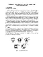
In January 2008 a 75 year old woman was referred to
us for a stage III BRONJ of the mandible (Figure 1);
she was previously treated for a severe osteoporosis
with alendronate 70 mg every four weeks for 9
months, then pamidronate 60 mg every four weeks for
two years. In the same period the patient was treated
with Eritropoietin beta (EPO) for three years (10.000
U/weeks) for a mild renal failure related anemia.
BRONJ was defined in accordance w ith the American
Association of Oral and Maxillofacial Surgeons Posi-
tion Paper on bisphosphonates - related osteonecrosis
of the Jaws [23,17] and all the three characteristics
were present in the patient:
1. Current or previous treatment with a
bisphosphonate;
2. Exposed bone in the maxillofacial region that has
persisted fore more 8 weeks;
3. No history of radiation therapy to the jaws.
Conservative, non - surgical treatment was initially
performed, as recommended [17-20], such as oral
hygiene (brushing and mouth rinses), topical and sys-
temic antibiotics active against common oral/dental bac-
terial infection; su bsequen tly both debridement, toilet of
exposed bone and Er:YAG laser treatment were uneffec-
tive [15,16] and patient’s conditions deteriorated with
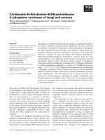
pain and worsening of BRONJ progressively. Computed
Tomography (CT) scan showed bone destruction (Figure
2). Resection and immediate reconstruction was pro-
posed to the patient, however she refused resection, as
recommended [17,20].
The concept that bone marrow stem cells upon tra ns-
plantation into adult recipients transdifferentiate and
contribute to the rigeneration of a variety of non -hema-
topoietic lineages in multiple organs, has provoked great
interest for its potential clinical applications [24,25] as
recently reported also by our group [ 26]. So we offered
the opportunity to our patient of injection of autologous
bone marrow stem cells into the osteonecro sis site
lesion.
Methods
In september 2008 the patient (who is the mother of
one of us) was informed about the procedure and gave
written informed consent.
Under local a nesthesia an average of 75 ml of bone
marrow were harvested from the posterior superior iliac
crest by aspiration into heparinized syringe s as pre-
viously reported by our group [26]. Progenitor cells
were isolated and enriched using Ficoll - Hypaque
®
cen-
trifugation procedures. This procedure allowed the
depletion of mature myeloid and erythroid cell from the
Figure 1 Clinical onset and appearance of BRONJ stage III.
Figure 2 Clinical Onset: computed tomography scan shows
bone destruction.
Cella et al. Head & Face Medicine 2011, 7:16
/>Page 2 of 6
harvest. The cell suspension consisted of an heteroge-
neous cell population including hematopoietic,
mesenchymal, endothelial and other progenitor cells as
well as mononuclear cells. The cells were suspended
into an oppo rtune quantity of PBS-EDTA buffer con-
taining 5% of human albumin.
The cell fraction was concentrated in a final volume of
6 ml. Finally, bef ore intralesional transplantation, the
cells were subjected to quality control procedures (i.e.
sterility test for aerobic and anaerobic bacteria, Elisa test
forHCV,HBV,HIVviruses)inordertoexcludeany
contamination as previously reported [26]. In addition,
full blood count and immune-phenotype analyses of the
cell suspension were performed, including absolute
CD34 and CD45 positive cell c ount and five colour
MoAb panel for the identification of the cellular subpo-
pulation (Table 1).
Before the injection of s tem cells into the osteo necro-
sis the patient underwent a surgical toilet in local
anesthesia of the bone lesion.
The bone cavity was fullfilled with fibrine sponge
(Spongostan
®
) as a carrier, then 4 ml of stem cells sus-
pension and 1 ml of patient’ s activated platele t-rich
plasma were injected in the lesion of BRONJ.
Results
The procedure was well tolerated, and a week later the
dehiscence of the surgical wound was observed, the resi-
dual carrier was removed. The n a soft, uniform layer of
whitishmucosadressingthebonecavitywasobserved.
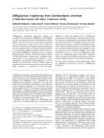
Two weeks later, resolution of symptoms was ob tained
and the lesion improved (Figure 3) w ith the pink
coloured new layer. Subsequently the patient was seen
at our out patients dental-maxillofacial service every two
weeks for six months, then every four weeks and
showed a progressive improvement. Clinical controls
showed progressive improvement of the mucosal layer
(Figure 4). CT scan performed 15 months later showed
improvement of bone and concentric ossification (Figure
5). A complete healing of BRONJ (stage 0) was obtain ed
and the patient is well without sings of BRONJ 30
months later. To our knowled ge this is the first case of
BRONJ treated with autologous stem cells injection.
Discussion
Since the first description by Marx , 2003 [27]and Wang
et al 2003 [28], cases with BRONJ are being increasingly
reported, first of all, in oncologic patients in line with
the increased use of bisphospho nates (mainly zolendro-
nate and pamidronate) as the main pathogenetic factor
of BRONJ.
A review of the literature through march 2006 per-
formed by our group [9] identified more than 250
reported case on BRONJ, and more recently over 6.000
cases have been reported to the US Food and Drug
Table 1 Multiparameter flow cytometric analysis of the injected BMC
Cellular subset harvest (ml 75) final (ml 6) injected (ml 4)
nuclear cells/ul (total E
6
) 16.800/ul (1.260) 71.000/ul (426)
WBC (CD45
+
)(E
6
) * 875,7 252 168
CD 34+ cells/ul 95,2 800 800
CD 34+ cells (E
6
) 7,14 4,8 3,2
CD34+/CD117+ cells (E
6
) 5,64 4,22 2,8
CD34+/CD133+ cells (E
6
) 3,42 1,15 0,76
CD45-/CD31+ cells (E
6
) 2,64 0,97 0,64
CD133+/CD117+ cells (E
6
) 4,1 1,95 1,3
CD133+ cells (E
6
) 5,92 2,25 1,5
CD117+ cells (E
6
) 28 20,2 13,5
CD45-/CD105+/CD71- cells (E
6
) 302,2 255,6 170
Ficoll mediated myeloid depletion % *
BMC: Bone marrow cells
Figure 3 Two weeks later after bone marrow cells
transplantation: pink coloured new layer shows progressive
improvement of the mucosa.
Cella et al. Head & Face Medicine 2011, 7:16
/>Page 3 of 6
Administration [29]. The t reatment goals for pat ients
with an established diagnosis of BRONJ are, as recently
reported [16-20], to eliminate pain, to control infection
of the soft and hard tissue and to minimize the occur-
rence or the progression of bone necrosis.
However the response to treatments of the patient s
with BRONJ is less predictable than the established sur-
gical treatment modalities for osteomyelitis or osteora-
dionecrosis, and new treatment procedures need to be
developed [16-20].
From the hystopathological point of view the BRONJ
is characterized by an avascular necrosis. In osteonecro-
sis is found a lack of osteogenic precursor cells that
derive from mesenchymal stem c ells (MSCs), but also a
lack in va scular support that deriv es from endothelial
progenitor cells (EPCs).
MSCs are known as being multipotent and exhibit the
potential for differentia tion into different cell/tiss ue
lineages, including cartilage, bone, adipose tissue, tendon
and ligament [30]. These pluripotent mesenchymal pro-
genitor cell are denoted as stromal or mesenchymal
stem cells.
In vivo osteogenesis occurs only if the density of
implanted cells at the treated site is sufficiently high. To
achieve this goal, either large amounts of concentrated
bone marrow stem cells or bone marrow stem cells in
combination with growth factors can be used [31,32].
This situation has been reproduced by in vitro studies
which confirmed that composite implantation of
mesenchymal stem cells with endothelial progenitor
cells enhances tissue-engineered bone formation [33].
The homing m echanisms of MSCs are poorly under-
stood; it is known that, based on chemokine/chemo-
kine-receptor interactions and adhesion molecules,
MSCs are potentially capable on finding the site of
injury and when, given intravenously, o f restoring
damaged tissue on site due to their plasticity and/or
paracrine properties [30]. How ever, it mu st be emp ha-
sized that a direct approach, bringing direct into the
osteonecrotic site a significant amount of bone marrow
enriched in mononuclear cells, could allow a better
osteogenesis of the damaged bone based on evidence
data of the presence in this cell-fraction of osteoid and
angiogenic precursors [34-36].
Bone marrow contains three main cell lines: hemato-
poietic cells, mesenchymal and proendotelial cells
[34,35]. Recently we reported in a randomized con-
trolled trial the effects of intracoronary transfer of au to-
logous bone marrow stem cells in patients with acute
anterior myocardial infarction and we demonstrated that
this procedure improves cardiac, autonomic, and func-
tional indexes in this setting of treated patients [26].
These positive effects may be mediated by a direct
transdifferentiation of transplanted stem cells to cardio-
myocytes [37], but also indirectly by parackrine secre-
tion of cytokines and growth factors with resulting
stimulation of survivors cardiac stem cells and/or an gio-
genesis, improving microvascular function [38].
Recently, a stabilizing effect of the injection cells via
changes in the connettive tissue has been hypothesized
[39].
Stem cells are easily obtained from the bone marrow
with a minimally invasive approach and can be easily
transplanted into the osteonecrotic lesion as demon-
strated in our patient. This simple, cheap procedure
allowed a clinical improvement of symptoms, and
induced novel ossification as demonstrated by CT scan
Figure 4 Four months later: the lesion of the mucosa is
ulteriorly improved.
Figure 5 Computed Tomography scan, 15 months later, shows
a concentric ossification of the bone lesion.
Cella et al. Head & Face Medicine 2011, 7:16
/>Page 4 of 6
15 months after the treatment, and it must be emphasized
that the patient is in complete remission from a stage 3
BRONJ after 30 months. In addition, our patient showed a
particularly rich bone marrow, not only in red cells pre-
cursors (as we expected since the patient was treated with
EPO), but also in total stem cells subset (table 1). Recently,
Kikuiri et al, [40] infused m esenchymal stem cells in
BRONJ-like mice. They demonstrated that systemic infu-
sion with MSCs prevents and cures BRONJ-like disease
possibly via introduction of peripheral tolerance, shown as
an inhibition of T-helper-producing i nterlukin 17 cells
(th17)and increase in T regulatory cells (Tregs). Handschel
and Meyer [41] suggest that stem cells might be a promis-
ing treatment option for BRONJ and our case demon-
strates thei r hypothesis is right. In our case bone marrow
stem cells were directly infused in the bone lesion of
BRONJ with a complete remission.
We are aware that a case report can be of limited
interest, however it could suggest that this technique
may be studied in patients with BRONJ unrensponsive
to standard treatment and can be tried before major
demolitive surgery procedures for the reconstruction of
defect of the ONJ by bisphosphonates.
Consent
Written informed consent was obtained from the patient
for publication of this case report and accompanying
images. A copy of the written consent is available for
review by the Editor-in-Chief of this journal
Acknowledgements
Authors acknowledge Fondazione di Piacenza e Vigevano (Italy) for the
excellent support and assistence
Author details
1
Departments of Oral and Maxillofacial Surgery, Hospital of Piacenza, Via
Taverna, 49. 29100. Italy.
2
Department of Immunohematology, Hospital of
Piacenza, Via Taverna, 49. 29100. Italy.
3
Department of Cardiology, Hospital of
Piacenza, Via Taverna, 49. 29100. Italy.
4
Department of Oncology and
Hematology, Hospital of Piacenza, Via Taverna, 49. 29100. Italy.
5
Department
of Pathology, Hospital of Piacenza, Via Taverna, 49. 29100. Italy.
Authors’ contributions
All authors read and approved the final manuscript. LC, MA, LC conceived of
the study, and participated in its design and coordination and helped to
draft the manuscript. AO, MM have been involved in drafting the manuscript
and to collect the results from follow-up examinations. MP has been
involved in revising the manuscript critically for important intellectual
content. DV, AZ, CDN have done substantial contributions to conceptions to
conception and design and interpretation of data.
Competing interests
The authors declare that they have no competing interests.
Received: 24 May 2011 Accepted: 17 August 2011
Published: 17 August 2011
References
1. McClug MR: Bisphosphonates. Endocrinol Metab Clin North Am 2003, 32,
253 71.
2. Santini D, Vespasiani Gentilucci U, Vincenzi B, Picardi A, Vasaturo F: The
antineoplastic role of bisphosphonates: from basic research to clinical
evidence. Ann Oncol 2003, 14:1468-76.
3. Boissier S, Magnetto S, Frappart L, Cuzin B, Ebetino FH, Delmas PD:
Bisphosphonates inhibit prostate and breast carcinoma cell adhesion to
unmineralized and mineralized bone extracellular matrices. Cancer Res
1997, 57 :3890-3894.
4. Wysoski DK: Reports of esophageal cancer with oral bisphosphonates
use. N Engl J Med 2009, 360:89-90.
5. Ibrahim T, Barbanti F, Giorgio Marrano G, Mercatali L, Ronconi S, Vicini C:
Osteonecrosis of the Jaw in patients with bone matastases treated with
bisphosphonates: a retrospective study. Oncologist 2008, 13:330-336.
6. Bagan JV, Jimenez Y, Murillo J, Hernandez S, Poveda R, Sanchis JM: Jaw
osteonecrosis associated with bisphosphonates: multiple exposed areas
and its relationship to teeth extractions. Study of 20 cases. Oral oncol
2006, 42 :327-329.
7. Hewitt C, Farah C: Bisphosphonates-related osteonecrosis of the jaws: a
comprehensive review. J Oral Pathol Med 2007, 36:319-328.
8. American Dental Association Council on Scientific Affairs: Dental
Management of patients receiving oral bisphosphonate Therapy: Expert
panel recommendations. J Am Assoc 2006, 137:1144-1150.
9. Cavanna L, Bertè R, Arcari A, Mordenti P, Pagani R, Vallisa D: Osteonecrosis
of the Jaw. A newly emerging sete-specific osseous pathology in
opatients with cancer treated with bisphosphonates. Report of 5 cases
and review of the literature. Eur J Internal Med 2007, 18:417-422.
10. Rizzoli R, Burlet N, Cahall D, Delmas PD, Eriksen EF, Felsenberg D:
Osteonecrosis of the jaw and bisphosphonate treatment for
osteoporosis. Bone 2008, 42:841-847.
11. Marx RE, Sawatari Y, Fortin M, Broumand V: Bisphosphonate-induced
exposed bone (osteonecrosis/osteopetrosis) of the jaws: risk factors,
recognition, prevention and treatment. J Oral Maxillofac Surg 2005,
63:1567-1575.
12. Yarom N, Yahalom R, Shoshani Y, Hamed W, Regev E, Elad S: Osteonecrosis
of the jaw induced by orally administered bisphosphonates: incidence,
clinical features, predisposing factors and teatment outcome. Osteoporos
Int 2007, 18:1363-1370.
13. Elad S, Yarom N, Hamed W, Ayalon S, Yahalom R, Regev E: Osteomyelitis
and necrosis of the jaw in patients treated with bisphosphonates A
comparative study. Clin Lab Haematol 2006, 28:393-398.
14. Ruggiero SL, Fantasia J, Carlson E: Bisphosphonates-related osteonecrosis
of the jaw: background and guidlines for diagnosis, staging and
management. Oral Surg. Oral Med Oral Path Oral Radio Endod 2006,
102:433-41.
15. Vescovi P, Merigo E, Meleti M, Fornaini C, Bonanini M, Rocca EP, De
Moor RJ, Nammour S: Surgical
treatment of maxillary osteonecrosis due
to bisphosphonates using an Er: YAG (2940 nm) laser. Discussion of 17
clinical cases. Rev Belge Med Dent 2009, 64(2):87-95.
16. Rugani P, Stephan A, Truschnegg A, Obermayer-Pietsch B, Jakse N:
Bisphosphonate - associated osteonecrosis of the jaws: surgical
treatment with ErCrYSGG-laser. Case report. OOOOE 2010, 110:e1-e6.
17. Ruggiero SL, Dodson TB, Assale LA, Landesberg R, Marx RE, Mehrotra B:
American Association of Oral and Maxillofacial Surgeons Position Paper
on Bisphosphonate-Relate Osteonecrosis of the Jaws-2009 Update. J Oral
Maxillofac Surg 2009, 67(Suppl):2-12.
18. Madrid C, Bouferrache K, Abarca M, Jaques B, Broome M: Bisphosphonate-
related osteonecrosis of the jaws: how to manage cancer patients. Oral
Oncology 2010, 46:468-470.
19. Lee JJ, Cheng SJ, Jeng JH, Ching CP, Lau HP, Kok SH: Successful treatment
of advanced bisphosphonate-related osteonecrosis of the mandible with
adjunctive teriparatide therapy. Wiley InterScience 2010 [http://www.
interscience.wiley.com].
20. Wilde F, Heufelder M, Winter K, Hendricks J, Frerich B, Schramm A,
Hemprich A: The role of surgical therapy in the management of
intravenous bisphosphonates-related osteonecrosis of the jaw. OOOOE
2010.
21. Korbling M, Estrov Z: Adult stem cells for tissue repair–a new therapeutic
concept? N Engl J Med 2003, 349:570-582.
22. Schneuder RK, Puellen A, Kramann R, Raupach K, Bornemann J, Knuechel R,
Perez-Buoza A, Neuss S: The osteogenic differentiation of adult bone
marrow and perinatal umbilical mesenchymal stem cells and matrix
Cella et al. Head & Face Medicine 2011, 7:16
/>Page 5 of 6
remodelling in three-dimensional collagen scaffolds. Biomaterial 2010,
31:467-480.
23. Ruggiero S, Gralow J, Marx RE, Hoff AO, Schubert MM, Huryn JM, Toth B,
Damato K, Valero V: Practical guidelines for the prevention, diagnosis,
and treatment of osteonecrosis of the jaw in patients with cancer. J
Oncol Prac 2006, 2:7-14.
24. Gussoni E, Soneoka Y, Strickland CD, Buzney EA, Khan MK, Flint AF,
Kunkel LM, Mulligan RC: Dystrophin expression in the mdx mouse
restored by stem cell transplantation. Nature 1999, 401:309-394.
25. Petersen BE, Bowen WC, Patrene KD, Mars WM, Sullivan AK, Murase N,
Boggs SS, Greenberger JS, Goff JP: Bone marrow as a potential source of
hepatic oval cells. Science 1999, 284:1168-1170.
26. Piepoli M, Vallisa D, Arbasi C, Cavanna L, Cerri L, Mori M, Passerini F,
Tommasi L, Rossi A, Capuccci A: Bone marrow cell transplantation
improves cardiac, autonomic, and functional indexes in acute anterior
myocardial infarction patients (Cardiac Study). European Journal of Heart
Failure .
27. Marx RE: Pamidronate (Aredia) and zoledronate (Zometa) induced
avascular necrosis of the jaw: a growing epidemic. J Oral Maxillofac Surg
2003, 61 :1115-1117.
28. Wang J, Goodger NM, Pogrel MA: Osteonecrosis of the jawsmassociated
with cancer chemotherapy. J Oral Maxillofac Surg 2003, 61:1104-1107.
29. Edwards BJ, Gounder M, McKoy JM, Boyd I, Farrugia M, Migliorati C, Marx R,
Ruggiaero S, Dimopoulos M, Raisch DW, Singhal S, Carson K, Obadina E,
Trifilio S, West D, Mehta J, Bennett CL: Bisphosphonate use and
osteonecrosis of the jaw: Pharmacovigilance and reporting of this
serious adverse event. Lancet Oncol 2008, 9:1166.
30. Salamon A, Toldy E: Use of mesenchymal stem cells from adult bone
marrow for injured tissue repair. Orv Hetil 2009, 150(27):1259-1265.
31. Connolly J, Grese R, Lipello L, Dehne R: Development of an osteogenic
bone marrow preparation. J Bone Joint Surg 1989, 71A:684-691.
32. Muschler G, Boehm C, Easley K: Aspiration to obtain osteoblast progenitor
cells from human bone marrow: the influence of aspiration volume. J
Bone Joint Surg 1997, 79A:1699-1707.
33. Usami K, Mizuno H, Okada K, Narita Y, Aoki M, Kondo T, Mizuno D, Mase J,
Nishiguchi H, Kagami H, Minoru U: Composite implantation of
mesenchymal stem cells with endothelial progenitor cells enhances
tissue-engineered bone formation. J Biomed Mater Res A 2009,
90(3):730-41.
34. Bittira B, Shum-Tim D, Al-Khaldi A, Chiu RC: Mobilization and homing of
bone marrow stromal cells in Myocardial infarction. Eur J Cardiotoracic
Surg 2003, 24:393-398.
35. Prockop DJ: Marrow stromal cells as stem cells for nonhematopoietic
tissues. Science 1997, 276:71-74.
36. Asahara T, Takahashi T, Masuda H, Kalka C, Chen D, Iwaguro H, Inai Y,
Silver M, Isner JM: VEGF contributes to postnatal neovascularization by
mobilizing bone marrow-derived endothelial progenitor cells. EMBO J
1999, 18 :3964-3972.
37. Orlic D, Kajstura J, Chimenti S, Bodine DM, Leri A, Anversa P: Bone marrow
cells regenerate infracted myocardium. Nature 2001, 410:701-705.
38. Smart N, Riley PR: The stem cell movement. Circ Res 2008, 102, 1155 1168.
39. Sun J, Li SH, Liu SM, Wu J, Weisel RD, Zhuo YF, Yau TM, Li Rk, Fazel SS:
Improvement in cardiac function after bone marrow cell therapy is
associated with an increase in myocardial inflammation. Am J Physiol
Heart Circ Physiol 2009, 296:H43-H50.
40. Kikuiri T, Kin I, Yamaza T, Akiyama K, Zhang Q, Li Y, Chen C, Chen C,
Wang S, le AD, Shi S: Cell-based immunotherapy with mesenchymal stem
cells cures bisphosphonate-related osteonecrosis of the jaw - like
disease in mice. J Bone Miner Res 2010, 25(7):1668-79.
41. Handschel J, Meyer U: Infection, vascularization, remodelling - are stem
cell the answers for bone disease of the jaw? Head & Face Medicine 2011,
7:5.
doi:10.1186/1746-160X-7-16
Cite this article as: Cella et al.: Autologous bone marrow stem cell
intralesional transplantation repairing bisphosphonate related
osteonecrosis of the jaw. Head & Face Medicine 2011 7:16.
Submit your next manuscript to BioMed Central
and take full advantage of:
• Convenient online submission
• Thorough peer review
• No space constraints or color figure charges
• Immediate publication on acceptance
• Inclusion in PubMed, CAS, Scopus and Google Scholar
• Research which is freely available for redistribution
Submit your manuscript at
www.biomedcentral.com/submit
Cella et al. Head & Face Medicine 2011, 7:16
/>Page 6 of 6