Báo cáo y học: " Production and purification of VP2 protein of porcine parvovirus expressed in an insectbaculovirus cell system" docx
Bạn đang xem bản rút gọn của tài liệu. Xem và tải ngay bản đầy đủ của tài liệu tại đây (509.74 KB, 6 trang )
SHOR T REPOR T Open Access
Production and purification of VP2 protein of
porcine parvovirus expressed in an insect-
baculovirus cell system
Hongchao Zhou
1†
, Guizhe Yao
2†
, Shangjin Cui
2*†
Abstract
The porcine parvovirus (PPV) VP2 protein was expressed in an insect-baculovirus cell system and was purified using
Ni-NTA affinity column chromatography. The recombinant 6-His-tagged VP2 protein with molecular mass (Mr) of
about 64 kDa was detected by anti-his antibody and anti-PPV serum. Electron microscopy showed that the purified
VP2 protein assembled into spherical particles with diameters ranging from 20 to 22 nm. The expressed VP2 was
antigenically similar to the native capsid protein according to HA and a Western blotting assay performed with
polyclonal antibodies collected from an outbreak of PPV in one farm. This study provides a foundation for the
application of VP2 protein in the clinical diagnosis of PPV or in the vaccination against PPV in the future.
1. Introduction
Porcine parvovirus (PPV) causes reproductive failure in
pregnant sows. The infection occurs w ithout clinical
symptoms in adults; however, the virus can cross the pla-
cental barrier during the infection and cause t he death of
the fetuses, stillbirths and return to estrus [1]. More
recently, PPV has gained importance as an agent able to
increas e the effects of porcine circovirus type 2 infection
in the clinical course of post weaning mul tisystemic wast-
ing syndrome (PMWS) [2,3], which is a significant dis-
ease in global swine production [4]. Widespread
vaccination has been proposed as a cost-effective method
to reduce the economical losses due to the endemic and
worldwide prevalence of this virus [5,6].
PPV is a small, non-enveloped, single-stranded, nega-
tive-sense DNA virus. Capsids of PPV are assembled
from three viral proteins (VP1, VP2, and VP 3). The
major structural protein, VP2 is the main target for neu-
tralizing antibodies in PPV [7,8]. When VP2 was
expressed in large amounts using the baculovirus
expression vector system, it assembled into virus-like
particles (VLPs) similar in size and morphology to the
original virions [7]. PPV VP2 VLPs induced antibodies
against PPV in immunised pigs [7] and rabbits [9]. PPV-
cell or ti ssue-tropism determinants, host-range determi-
nants, and determinants that confer hemagglutination
properties have all been shown to be located in the cap-
sid proteins [ 10-12]. It is noted that PPV VP2 was
expressed by Lu et al. (2002) in pFastBac I and by Si et
al. (2006) in pFastBacDUAL using insect cell-baculovirus
systems and both groups demonstrate d a 64 kDa ban d
by Western-blot analysis.
Because VP2 is the main structural protein of PPV
and c onstitutes most of the viral capsid, VP2 produced
in vitro can self a ssemble into virus-li ke particles [1 3].
PPV VP2 VLPs exhibited positive immunoreactivity for
PPV in a commercial ELISA [14]. Rueda et al. (2000)
showed that contaminant baculovirus could be inacti-
vated in preparations of PPV VP2 VLPs while retaining
physical and immunological properties. VP2 VLPs have
been produced and purified using a specific affinity
Immobilized Metal Affinity Chromatography (IMAC)
system for other parvoviruses such as B19 [15] and for
infectious bursal disease virus [16]. T o facilitate the use
of PPV VP2 for diagnosis and vaccination, the current
study a ttempted to identify an improved procedure for
producing VP2 in vitro and for purifying this fusion
protein.
There are many advantages to the use of VLPs in vac-
cines and for diagnosis. Compared t o inactivated virus,
* Correspondence:
† Contributed equally
2
Division of Swine Infectious Disease, State Key Laboratory of Veterinary
Biotechnology, Harbin Veterinary Research Institute of CAAS, Harbin, China
150001
Full list of author information is available at the end of the article
Zhou et al. Virology Journal 2010, 7:366
/>© 2010 Zhou et al; licensee BioMed Central Ltd. This is an Open Access article distributed unde r the terms of the Creative Commons
Attribution License (http://cre ativecommons.o rg/licenses/by/2.0), which permits u nrestricted use, distribution, and reproduction in
any medium, provided the original w ork is properly cited.
which is currently used in vaccines, VLPs do not require
the propagation of infectious virus, there is no risk of
virus transmission or infection, production l evels are
much higher, product ion is cost effective, and VLPs are
generally sta ble. The authors have p reviously expressed
PPV VP2 in E. coli using the plasmid pET-32a (+) [17].
VP2 expressed in bacteria had similar antigenicity to
native PPV VP2, as determined by Western blot analysis
using polyclonal antibodies from pigs vaccinated against
PPV [17]. PPV VP2 expressed in bacteria appears to
have good immunogenicity, this is better for using as
vaccine than the use as diagnosis antigen, for the anti-
body for E. coli in sera effects the ELISA assay. This
provide us a compelling reason for expressing PPV VP2
in a baculovirus system, although baculovirus expression
systems are likely to be more costly than bacterial
expression systems for producing viral proteins and may
present difficulty in purifying expressed proteins from
insect cell and baculovirus constituents. PPV VP2 has
been expressed in insect cell-baculovirus systems by a
number of other groups previously [7,9,14,15]. P PV VP2
expressed by baculovirus in sf9 cells produced VLPs
[7,9,14,15]. PPV VP2 VLPs induced antibodies against
PPV in immunised pigs [7]and r abbits [14]. PPV VP2
VLPs exhibited positive immunoreactivity for PPV in a
commercial ELISA [15]. Ruedaetal.[1]showedthat
contaminant baculovirus could be inactivated in pre-
parations of PPV VP2 VLPs while retaining phy sical and
immuno logical properties. It is noted th at PPV VP2 was
expressed by [15] in pFastBac I and by Si et al. [9] in
pFastBacDUAL using insect cell-baculovirus systems
and both groups demonstrated a 64 kDa band by We s-
tern-blot analysis. VLPs produced in baculovirus-
infected insect cells could then be used as a v accine or
as a diagnostic agent to detect the antibody produced by
PPV infection or vaccination [ 18]. Baculovirus-infected
insect cells have been used to produce VP2 VLPs of
infectious bursal disease virus [19], and parvoviruses
including PPV [14] and B19 [15]. To further facilitate
the use of PPV VP2 for diagnosis and vaccination, the
goal of the current study was to find an improved pro-
cedure for e xpressing PPV VP2 in vitro and to purify
the fusion protein.
2. Materials and methods
2.1. Cells, virus, and reagents
Strain 20-06 of PPV was isolated from a dead fetus
delivered from a sow diagnos ed with reproductive fail-
ure. HRP-labeled anti-pig serum was purchased from
Sigma (St. Louis, Missouri, USA). Ni-NTA His Bind
resin was obtained from Invitrogen (Carlsbad, California,
USA). Prestained protein ladder was purchased from
Fermentas International Inc. (Burlington, Canada).
Swine anti-PPV serum samples, with serum
neutralization titer, were obtained from the Harbin
Veterinary Research Institute, CAAS. The serum sam-
ples were collected from an outbreak of PPV in 2008 in
one farm in Heilongjiang Province.
2.2. Construction of recombinant plasmids and
recombinant bacmid
Genomic DNA was extracted from the cell-cultured
strain 20-06 of PPV by the classical phenol-chloroform
extraction method and was used a s a template to
amplify the VP2 fragment by PCR. The PPV VP2 gene
was amplified with the primers PPV-VP2 FD (TATG-
GATCCGATGAGTCATCATCACCATC ACCATAGT-
GAAAATGTGGAACAAC) and PPV-VP2 RV (GCGT
CGACTATGAGTTAGAGT TTGTATTAG). The under-
lined nucleotides represent BamHI and SalI re striction
sites, respectively. The PCR products wer e digested with
BamHI and SalI and subsequently cloned into the corr e-
sponding restriction sites of the pFastbac1 vector to pro-
duce the recombinant plasmid, pFastPVP2. The insert
of the recombinant plasmid was confirmed by DNA
sequencing.
After the recombinant pFastPVP2 donor plasmid was
determined to be correct, the DNA was transformed
into DH10Bac™ for transposition into the bacmid. The
transposition assay and subsequent transfection steps
were the same for all vectors. White colo nies cont ained
the recombinant bacmid, and therefore were selected for
isolation of recombinant bacmid DNA. Before DNA was
isolated, candidate colonies were streaked to ensure they
were truly white. Bacmid DNA (B-pFastPVP2) was
extracted by the phenol-c hloroform extraction me thod.
The rec ombinant Bacmid (B-pFastPVP2) was then ana-
lyzed by PCR.
2.3. Expression of the VP2 protein in sf9 cells
The recombinant baculoviruses, containing the coding
sequences of VP2 with t he polyhistidine tag at the N-
terminus, were generated by using the Bac-to-BacTM
system (Invitrogen; Luckow et al., 1993). Propagation of
the recombinant virus was performed according to stan-
dard procedures (Summers et al., 2006). For production
of the recombinant VP2 proteins, sf9 (Spodoptera frugi-
perda) cells were grown in 2-l Erlenmeyer flasks on
orbital shakers (120 rpm) to a concentration of abo ut
2×10
6
cells per ml of culture medium ( 30 ml growing
volume) and infected with the recombinant viruses at a
multiplicity of infection (MOI) of 2-3. At 72 h postinfec-
tion (p.i.), cells were collected and processed as
described below. The infected cells were collected by
low-speed centrifugation at 3500 × g (Hermle Labor-
technik, Wehingen, Germany; swing-out rotor 4 × 750
ml) for 15 min at 4°C and solubilized in 30 ml of ice-
cold lysis buffer [20 mM Tris, 0.3 M NaCl, 1.0% (v/v)
Zhou et al. Virology Journal 2010, 7:366
/>Page 2 of 6
Triton X-100, pH 7.4] for 1 5 min with gentle mixing
(about 20 × 10
6
cells/ml). The crude cell lysate was clar-
ified by high-speed centrifu gation at 23,400 × g (Sorvall,
Thermo Fisher Scientific, Waltham, MA; GSA rotor) for
20 min at 4°C. The supernatant fraction was collected,
and t he soluble recombinant protein products purified
by IMAC as described below.
2.4. Purification of PPV VP2 protein
Cells were harvested at different times after infection,
centrifuged at 200 × g for 15 min, and resuspended in
25 mM Na
2
HCO
3
,pH8.3,atadensityof2×10
7
cells/
ml; lysis was allowed to occur for 20 min. Afterward, cell
debris was removed by centrifugation at 10,000 g for
15 min. The recombinant fusion protein VP2 was puri-
fied by IMAC. The clarified lysate was incubated w ith
3 ml of pr e-equilibrated Ni2 +- Str eamline Chelating
GelTM ( Amersham Biosciences, Piscataw ay, NJ) on a
rotating wheel for 16 h at 4°C, and was then placed in a
10-ml chromatography column (PolyPrep 0.8 by 4 cm;
BioRad, Hercules, CA). Weakly bound and contaminating
proteins were washed from the chelating gel by using 10×
the co lumn vol ume (20 mM Tris, 0.3 M NaCl, 20 mM
imidazole, pH 7.4). The recombinant polyhistidine-
tagged protein products were finally eluted from the
packed bed with 3-4× the column vo lume (20 mM Tris,
0.3 MNaCl, 500 mM Mimidazole, pH 7.4). One-ml frac-
tions were collected, and the protein contents were ana-
lyzed using a NanoDrop ND-1000 spectrophotometer
(NanoDrop Technologies, Wilmington, DE).
2.5. SDS-PAGE and Western blotting
Thepurityandtheapparentmolecularweightofthe
recombinant VP2 specific proteins were assessed by
sodium dodecyl sulfate polyacrylamide gel electrophor-
esis (SDS-PAGE) and immunoblot analysis. The purified
proteins were separated by SDS-P AGE and were either
stained with Commassie Brilliant Blue or were trans-
ferred onto nitrocellulose membranes using a wet t rans-
fer cell for Western blotting. The protein expressed 0 to
5 days after insect cells were challenged was obtained
for Western blotting. The membranes were blocked
with 5% skimmed milk in TBS-T (50 mM Tris-HCl, 150
mM NaCl; 0.05% Tween 20, pH 7.5) for 1 h at room
temperature (RT). Swine anti-PPV sera (1:1 000 dilution)
or anti-His monoclonal antibody (1:5000 dilution) was
added to the membranes and shaken overnight at 4°C.
The membranes were then washed three times ( 5 min
each time) with TBS-T. Secondary antibody, either anti-
swine or anti-mouse at 1:5000 dilution, was then ad ded
and incubated for 1 h. After the membranes were
washed, 3,3’-diaminobenzidine (DAB) was added for col-
our development.
2.6. Hemagglutination assay
Following the method of Senda et al. [20], two-fold dilu-
tions of samples of VP2 protein were prepar ed, mixed
with guinea pig red blood cells, and added to the wells
of a 96-well plate. Af ter 60 minutes at 37°C, the wells
were photographed.
3. Results
3.1 Construction of recombinant plasmids pFastpVP2 and
recombinant bacmid
The recombinan t plasmids pFastpVP2 and B-pFastpVP2
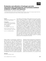
were identified by BamHI and SalI enzyme digestion.
The fragments were about 1800 bp and 4775 bp, respec-
tively, which conformed to the expected sizes (Figure 1).
Recombinant bacmid was identified by PCR with the
primers PPV-VP2 FD and PPV-VP2 FD. The fragment
was about 1840 bp.
3.2 Expression and purification of polyhistidine-tagged
VP2 of PPV
We have previously shown that formation of chimeric
VLPs of PPV fusion constructs is feasible in pET-PPV
with E. coli cells (Qi T et al., 2009 ). The re combinant
baculoviruses encoding the structural proteins VP2 of
PPV N -terminally fused to a polyhistidine tag (6 × his)
were engineered to simplify the overall purification pro-
cess of these the viral antigens. The recombinant bacu-
loviruses were used for infection of Sf9 insect cells
Figure 1 Restriction endonuclease digests of PPV VP2 plasmid
DNA. Products produced by restriction enzyme digestion of the
vector plasmid (lane 1), the PCR product of the VP2 gene (lane 2),
and the recombinant plasmid (lane 3). Lane M shows the DL15000
DNA marker.
Zhou et al. Virology Journal 2010, 7:366
/>Page 3 of 6
according to established procedures, and the infected
insect cells were collected and solubilized by treatment
with a non-ionic detergent. The cell lysates were clari-
fied by centrifugation, and the viral pro teins were finally
extracted from the cytoplasmic extracts by IMAC
(Ni2+). After isolation, the recombinant VP2 specific
fusion proteins were analyzed by SDS-PAGE, Western-
blotting, and hemagglutination assay (HA).
3.3 SDS-PAGE and Western blotting
The VP2 protein was identified with the SDS-PAGE and
Western b lotting. To confirm the identity of his-tagged
VP2, the purified fusion protein was subjected to Wes-
tern blot assay using PPV-positive pig sera. The polyco-
lonal antibodies recognized his-tagged VP2, and the
band had the appropriate molecular weight. Immuno-
blot of these membranes using anti-PPV antibodies
showed that the fusion protein had epitopes derived
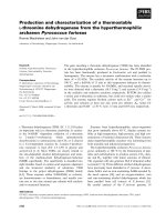
from PPV. The Western blots of purified protein
obtained 0 to 5 days after insect cells were challenged
indicated that the protein was expressed on day 4 and 5
(Figure 2).
To purify the fusion protein (his-tagged VP2), Ni-
NTA agarose were used to finish the SDS-PAGE a nd
Western blotting. The results showed that the target
protein could be conjugated to the resin. A single band
was detected by SDS-PAGE Western blotting (Figure 3).
To confirm the identity of VP2 minus the his-tag tail,
the purified VP2 without a His-Tag was subjected to
Western blotting assay using PPV-positiv e pig sera. The
polyclonal antibodies recognized VP2 w ithout a His-
Tag, and the band had a molecular weight of 64 KDa.
The a ssay therefore provided evidence that the protein
could be used as an efficient immunological reagent.
This conclusion was supported by the HA (see next two
sections).
3.4 HA
Sample VP2 protein caused hemagglutination when
diluted up to 1:8,192 (Figure 4). Therefore, the HA titer
of this protein stock was 8,192.
4. Discussion
Currently, vaccines against PPV are produced by chemi-
cally inactivating isolated virus particles grown in primary
cell cultures of porcine origin. The method is both labor
intensive and costly, with the additional hazard of requir-
ing the handling of large quantities of infectious virus
[21]. Economic and safety considerations, as well as prac-
tical limitations associated with low yields of PPV parti-
cles from in vitro cultures, led us to the investigate
recombinant sub-unit vaccines for PPV. The VP2 protein
of PPV had been previously shown to self-assemble into
virus-like particles when expressed in insect cells by
baculovirus infection [7]. In addition, the virus-like parti-
cles of PPV were found to be highly immu nogenic, and
breeding sows were protected against reproductive failure
in PPV challenge experiments [22]. Nonetheless, baculo-
virus-based systems for the production of recombinant
proteins are still technically demanding, requiring sterile
bioreactors that may be prohibitively costly for the pro-
duction of vaccines for farm animals. Given that PPV
causes serious economic losses for swine producers,
Figure 2 Western blot analysis of 6-His-tagged recombinant
PPV VP2 protein expressed at different times after insect cells
were infection with baculovirus. lane 1, the protein expressed in
insect cells challenged with negative baculovirus; lanes 2 to 6, the
protein expressed 1 to 5 days after insect cells were challenged
with recombinant baculovirus; M, prestained protein ladder. The first
antibody is PPV-positive pig sera, the second antibody is horseradish
peroxidase-conjugated rabbit anti-pig antibody.
Figure 3 Western blot analysis of expressed VP2.M:prestained
protein ladder, 1: the purified VP2 without the His-Tag, 1: the
control of VP2 before induction. The first antibody is PPV-positive
pig sera, the second antibody is horseradish peroxidase-conjugated
rabbit anti-pig antibody.
Zhou et al. Virology Journal 2010, 7:366
/>Page 4 of 6
development of safe, effective, and inexpensive methods
for producing va ccines a nd diagnosi ng the dise ase is
warranted.
This paper describes a meth od for producing the VP2
protein of PPV in an insect-baculovirus cell system.
After expression was optimized, a his-tagged VP2 was
obtained. The paper also describes an alternative
method (IMAC) for efficient recovery of PPV VP2 and
for purifying the protein using a Ni-NTA affinity col-
umn chromatography. This purification method avoids
time-consuming ultracentrifugation steps such as
sucrose gradient or cesium chloride gradient centrifuga-
tion. When larger amounts of recombinant proteins are
needed, the purification process could be easily scaled-
up by using an expanded-bed adsorption column techni-
que. The virus-like particles formed by the fusion pro-
tein had high HA titer, could be useful as antigens for
detecting PPV and could be useful for the development
of a vaccine against PPV that is effective but less expen-
sive than current vaccines [23].
The author s have previously expressed PPV VP2 in E.
coli using the plasmid pET-32a (+) [ 17]. VP2 expressed
in bacteria had similar antigenicity to native PPV VP2,
as determined by Western blot analysi s using polyclonal
antibodies from pigs vaccinated against PPV [17].
Although PPV VP2 expressed in bacteria appears t o
have good immunogenicity, it is better for using as vac-
cine than the use as diagnosis antigen, for the a ntibod y
for E. coli in sera bothers the ELISA assay. This provide
us a compelling reason for expressing PPV VP2 i n a
baculovirus system. The authors note a number of other
studies where recombi nant PPV VP2 expressed in bacu-
lovirus has been engineered with other viral antigens or
immunogenic epitopes to produce multivalent vaccine
candidates. The current diagnostic tests and vaccines for
PPV are well establis hed and considered to be adequate
for most practical purposes; therefore the authors would
like to demonstrate that any new diagnostic technology
using recombinant PPV VP2 has advantages over cur-
rent diagnostic tests and that any new vaccines against
PPV using VP2 are superior to current vaccines in the
future.
Acknowledgements
The study was supported in part by funding from the National High-tech
R&D Program (863 Program-2007AA100606).
Author details
1
College of Veterinary Medicine, Northwest A&F University, Yangling, Shaanxi,
712100, China.
2
Division of Swine Infectious Disease, State Key Laboratory of
Veterinary Biotechnology, Harbin Veterinary Research Institute of CAAS,
Harbin, China 150001.
Authors’ contributions
GY and HZ carried out the molecular studies, and drafted the manuscript. SC
participated in the design of the study and conceived of the study, and
participated in its design and coordination. All authors read and approved
the final manuscript.
Competing interests
The authors declare that they have no competing interests.
Received: 24 June 2010 Accepted: 10 December 2010
Published: 10 December 2010
References
1. Rueda P, Fominaya J, Langeveld JP, Bruschke C, Vela C, Casal JI: Effect of
different baculovirus inactivation procedures on the integrity and
immunogenicity of porcine parvovirus-like particles. Vaccine 2000,
19:726-734.
2. Allan GM, Kennedy S, McNeilly F, Foster JC, Ellis JA, Krakowka SJ,
Meehan BM, Adair BM: Experimental reproduction of severe wasting
disease by co-infection of pigs with porcine circovirus and porcine
parvovirus. Pathol 1999, 1:1-11.
3. Krakowka S, Ellis JA, Meehan B, Kennedy S, McNeilly F, Allan G: Viral
Wasting Syndrome of Swine: Experimental reproduction of postweaning
multisystemic wasting syndrome in gnotobiotic swine by coinfection
with Porcine Circovirus 2 and Porcine Parvovirus. Vet Pathol 2000,
37:254-263.
Figure 4 Hemagglutination assay with the VP2 protein (two
columns on right), with a sample known to be negative for
PPV (two columns on the left), or with a sample known to be
positive for PPV (two center columns).
Zhou et al. Virology Journal 2010, 7:366
/>Page 5 of 6
4. Segales J, Allan GM, Domingo M: Porcine circovirus diseases. Anim Health
Res Rev 2005, 6:119-42.
5. Gardner IA, Carpenter TE, Leontides L, Parsons TD: Financial evaluation of
vaccination and testing alternatives for control of parvovirus-induced
reproductive failure in swine. J Am Vet Med Assoc 1996, 208:863-869.
6. Parke CR, Burgess GW: An economic assessment of porcine parvovirus
vaccination. Aust Vet J 1993, 70:177-180.
7. Martinez C, Dalsgaard K, de Turiso JAL, Cortes E, Velac , Casal JI: Production
of porcine parvovirus empty capsids with high immunogenic activity.
Vaccine 1992, 10(10):684-90.
8. Kamstrup S, Langeveld J, Botner A, et al: Mapping the antigenic structure
of porcine parvovirus at the level of peptides. Virus Res 1998, 53:163-73.
9. Si Y-h, Fang M-g, Wang H-z: [Expression of porcine parvovirus vp2 gene
and construction of virus like particles (Chinese)]. Virologica Sinica 2006,
21:148-152.
10. Bergeron J, Hebert B, Tijssen P: Genome organization of the Kresse strain
of porcine parvovirus: identification of the allotropic determinant and
comparison with those of NADL-2 and field isolates. J Virol 1996,
70(4):2508-15.
11. Li J, Carroll J, Ellar DJ: Crystal Structure of Insecticidal d-Endotoxin from
Bacillus thuringiensis at 2.5 Å resolutions. Nature 1991, 353(6347):815-821.
12. Truyen U, Parrish CR: Canine and feline host ranges of canine parvovirus
and feline panleukopenia virus: distinct host cell tropisms of each virus
in vitro and in vivo. J Virol 1992, 66(9):5399-408.
13. Pan Q, He K, Huang K: Development of recombinant porcine parvovirus-
like particles as an antigen carrier formed by the hybrid VP2 protein
carrying immunoreactive epitope of porcine circovirus type 2. Vaccine
2008, 26(17):2119-26.
14. Maranga L, Brazao TF, Carrondo MJ: Virus-like particle production at low
multiplicities of infection with the baculovirus insect cell system.
Biotechnology and Bioengineering 2003, 84:245-253.
15. Luckow VA, Luckow VA, Lee SC, Barry GF, Olins PO: Efficient generation of
infectious recombinant baculoviruses by site-specific transposon-
mediated insertion of foreign genes into a baculovirus genome
propagated in Escherichia coli. J Virol 1993, 67(8):4566-79.
16. Michel PO, Mäkelä AR, Korhonen E, Toivola J, Hedman L, Söderlund-
Venermo M, Hedman K, Oker-Blom CJ: Purification and analysis of
polyhistidine-tagged human parvovirus B19 VP1 and VP2 expressed in
insect cells. J Virol Methods 2008, 152(1-2):1-5.
17. Qi T, Cui S: Expression of porcine parvovirus VP2 gene requires codon
optimized E. coli cells Virus Genes 2009.
18. Lo-Man R, Rueda P, Sedlik C, Deriaud E, Casal I, Leclerc C:
A recombinant
virus-like particle system derived from parvovirus as an efficient antigen
carrier to elicit a polarized Th1 immune response without adjuvant. Eur J
Immunol 1998, 28(4):1401-7.
19. Hu YC, Bentley WE, Edwards GH, Vakharia VN: Chimeric infectious bursal
disease virus-like particles expressed in insect cells and purified by
immobilized metal affinity chromatography. Biotechnol Bioeng 1999,
63(6):721-9.
20. Senda M, Hirayama N, Yamamoto H, Kurata K: An improved
hemagglutination test for study of canine parvovirus Vet. Microbio 1986,
12(1):1-6.
21. Casal JI: Use of parvovirus-like particles for vaccination andinduction of
multiple immune responses. Biotechnol Appl Biochem 1999, 29:141-150.
22. Casal JI: Parvovirus diagnostic and vaccine production in insect cells.
Cytotechnology 1996, 20:261-270.
23. Antonis AFG, Bruschke CJM, Paloma R, Maranga L, Casal JI, Vela C,
Hilgers LAT, Belt PBGM, Weerdmeester K, Carrondo MJT, Langeveld JPM: A
novel recombinant virus-like particle vaccine for prevention of porcine
parvovirus-induced reproductive failure. Vaccine 2006, 24:5481-5490.
doi:10.1186/1743-422X-7-366
Cite this article as: Zhou et al.: Production and purification of VP2
protein of porcine parvovirus expressed in an insect-baculovirus cell
system. Virology Journal 2010 7:366.
Submit your next manuscript to BioMed Central
and take full advantage of:
• Convenient online submission
• Thorough peer review
• No space constraints or color figure charges
• Immediate publication on acceptance
• Inclusion in PubMed, CAS, Scopus and Google Scholar
• Research which is freely available for redistribution
Submit your manuscript at
www.biomedcentral.com/submit
Zhou et al. Virology Journal 2010, 7:366
/>Page 6 of 6