Báo cáo y học: " Rapid isolation of mycoviral double-stranded RNA from Botrytis cinerea and Saccharomyces cerevisiae" pot
Bạn đang xem bản rút gọn của tài liệu. Xem và tải ngay bản đầy đủ của tài liệu tại đây (675.22 KB, 7 trang )
MET H O D O LO G Y Open Access
Rapid isolation of mycoviral double-stranded RNA
from Botrytis cinerea and Saccharomyces cerevisiae
Antonio Castillo
*
, Luis Cottet, Miguel Castro, Felipe Sepúlveda
Abstract
Background: In most of the infected fungi, the mycoviruses are latent or crypti c, the infected fungus does not
show disease symptoms, and it is phenotypically identical to a non-infected strain of the same species. Because of
these properties, the initial stage in the search for fungi infected with mycoviruses is the detection of their viral
genome, which in most of the describe d cases corresponds to double-stranded RNA (dsRNA). So to analyze a large
number of fungal isolates it is necessary to have a simp le and rapid method to detect dsRNA.
Results: A rapid method to isolate dsRNA from a virus-infected filamentous fungus, Botrytis cinerea, and from a
killer strain of Sacchar omyces cerevisiae using commercial minicolumns packed with CF11 cellulose was developed.
In addition to being a rapid method, it allows to use small quantities of yeasts or mycelium as starting material,
being obtained sufficient dsRNA quantity that can later be analyzed by agarose gel electrophoresis, treated with
enzymes for its partial characterization, amplified by RT-PCR and cloned in appropriate vectors for further
sequencing.
Conclusions: The method yields high qualit y dsRNA, free from DNA and ssRNA. The use of nucleases to degrade
the DNA or the ssRNA is not required, and it can be used to isolate dsRNA from any type of fungi or any
biological sample that contains dsRNA.
Background
Mycoviruses or fungal viruses have properties that differ-
entiate them from viruses that infect animals, plants and
bacteria [1-4]; they do not infect intact cells and are
transmitted vertically by intracellular routes (meiosis and
mitosis) and horizontally by anastomosis of compatible
hyphae or through sexual mating of yea st cells. Mycov-
iruses may also be latent and/or cryptic, since in most
cases the infected fungus does not show disease symp-
toms and is phenotypically identical to a non-infected
strain of the same species. Due to these peculiarities, the
initial stage in the search for infected fungi with m ycov-
iruses is the detection of their viral genome, which in
most of the described cases corresponds to dsRNA [1-4].
Although the number of ssRNA viruses described so far,
such as the F and X viruses of Botrytis cinerea [4-6], has
incr eased considerably, dsRNA continues to be the more
predominant mycoviral genome. Therefore, to analyze a
large number of fungal isolates it is necessary to have a
rapid method that allows the isolation and partial charac-
terizat ion of viral dsRNA us ing small amounts of mycelia
or yeast cells as starting material.
Some of the main and general met hods described until
now to isolate dsRNA molecules are: total nucleic acid
isolation and further enzymatic digestion of the DNA
and ssRNA [7]; phenol acid extraction (pH 4.5) in the
presence of ammonium sulphate [8]; boiling of the fungal
sample in the presence of a high salt concentration buffer
[9], and use of CF11 cellulose, a chromatographic resin
tha t allows the selective separation of dsRNA from DNA
and ssRNA, using 16% ethanol in the elution buffer
[10-13]. All of the former methods require a considerable
quantity of initial sample t o obtain sufficient dsRNA for
its later characterization, so it is very difficult to analyze a
large number of fungal isolates with these techniques.
Of the previous methodologies, the most widely used
one is c hromatographic separation on CF11-cellulose,
since it allows getting dsRNA free of ssRNA, rRNA or
tRNA, without further treatment.
* Correspondence:
Laboratorio de Virología de Hongos, Departamento de Biología, Facultad de
Química y Biología, Universidad de Santiago de Chile. Avenida Libertador
Bernardo O’Higgins 3363, Estación Central, Santiago, Chile
Castillo et al. Virology Journal 2011, 8:38
/>© 2011 Castillo et al; licensee BioMed Cent ral Ltd. This is an Open Access article d istributed under the terms of the Creative Commons
Attribution Lice nse (http://c reativecommons.org/licenses/by/2.0), which permits unrestricted use, distribution, and reprodu ction in
any medium, provided the original work is properly cited.
In this paper we describe a rapid method for isolating
dsRNA from a filamentous fungus, Botrytis cinerea,and
ayeast,Saccharomyces cerevisiae. Besides being a rapid
method, i t allows the use of small amounts of yeasts or
micelia a s initial material to obtain a sufficient quantity
of dsRNA that can later be analyzed by electrophoresis
in agaro se gel, quantified by densitometric analysis, and
treated with enzymes for their partial characterization.
ThemethodallowsgettinghighqualitydsRNA,freeof
DNA and ssRNA, and it can be applied to isolate
dsRNA from a ny type of fungus or any biological sam-
ple that contains dsRNA.
Methods
Fungal strains and culture conditions
Botrytis cinerea strains CCg378, THg324, and SUg275
were grown at 20°C for 7-10 days in 50 mL of liquid cul-
ture medium containing 1.5% (w/v) malt extract and
0.75% (w/v) yeast extract (Merck, Darmstadt, Germany).
S. cerevisiae 1743 (kindly provided by Dr. Reed B. Wickner)
[14] was grown for 16-20 hours in 50 mL of liquid YPD
medium containing 1.0% (w/v) yeast extract, 2.0% (w/v)
peptone and 2.0% (w/v) glucose. In both cases the culture
mediaweresterilizedbyautoclavingat121°Cfor20min.
dsRNA purification
For the three B. cinerea strains the mycelia were manu-
ally separated from culture media with forceps and
excess moisture was removed by pressing between paper
towels. S. cerevisia e 1743 yeast cells were sedimented by
centrifugation and excess moisture was removed by
incubation at 60°C for 10-15 min. The following steps
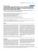
are the same for both fungi and are represented sche-
matically in Figure 1.
(1) Both the fungal mycelium and yeast cell pellet (3-
5 g wet weight) were frozen in liquid nitrogen and
ground to a fine powder with m ortar and pestle. The
powder was resuspended in 5 mL of STE 1X buffer (25
mM Tris-HCl pH 7.5, 50 mM NaCl and 0.5 M EDTA)
containing 50 μLofb-mercaptoethanol. Then one
volume of phenol:chloroform:isoamyl alcohol solution
(25:24:1) was added. The mixture was stirred for 10
minutes on ice and was then centrifuged at 10,000 × g
for 15 minutes. The aqueous phase was transferred to a
sterile tube, e thanol was added to a final concentration
of 16% (v/v) and the mixture was centrifuged for 15
minutes at 10,000 × g. T he supernatant co ntaining the
total nucleic acids was recovered, discarding the pellet
containing only a fraction of DNA and proteins.
(2 & 3) CF11 cellulose Whatman (0.2 g) prewashed
with STE buffer containing 16% (v/v) ethanol and 2%
(v/v) b-mercaptoethanol was added to a previously used
commercial minicolumn without its original resin (Pro-
mega Wizard Plus Midipreps or Qiagen QIAprep empty
Minicolumns). The minicolumn was coupled to a 5 mL
syringe and mounted in a vacuum system.
(4)The supernatant (recovered in step 1), containing
the total nucleic acids, was loaded in the minicolumn
and eluted under vacuum, then 5 mL of STE buffer con-
taining 16% (v/v) ethanol were immediately added to
completely elute the DNA and ssRNA.
(5) The minicolumn was coupled to a 1.5 mL micro-
centrifuge tube and centrifuged for 30 s at 10,000 × g to
eliminate the residual washed buffer.
(6 & 7) In order to recover dsRNAs bound to the
resin, the minicolumn was coupled to a new microcen-
trifugetubeand200μL of STE buffer without ethanol
were added over the CF11 cellulose contained in the
minicolumn. The dsRNA was eluted by centrifugation
for 2 min at 10,000 × g. Each recovered sample was
added to 0. 2 g o f CF11 cellulose prewashed wit h STE
buffer containing 16% (v/v) ethanol and 2% (v/v) b-
mercaptoethanol, and steps 3 to 10 were repeated.
(8) Double-stranded RNA was precipitated overnight
with 2 volumes of absolute ethanol at -20°C.
(9 & 10) After centrifugation for 15 min at 10,000
× g, the pellet containing the dsRN A was dried and
resuspended in 10 μL of sterile triple-distilled water for
its further analysis. The electrophoretic characterization
of dsRNA was performed in 0.8% (w/v) agarose gel
using TAE as running buffer (2 μL of dsRNA sample is
sufficient to visualize bands of regular intensity in gel).
The gel was subsequently stained by incubation in 0.5
μg/mL of ethidium bromide.
Nucleic acid analysis
The electrophoretic and RNase A treatment conditions
were as described by Castro et al. [13].
Densitometric analysis
With the images of the gels, densitometry c urves of the
bands were processed with the aid of specific MediaCy-
bernetics, Gel-Pro Analyzer Version 6.0 software.
Molecular cloning of the 2.2 kpb dsRNA from B. cinerea
CCg378
The dsRNAs of B. cinerea CCg378 were purified by CF11
cellulose chromatography and separated by agarose gel
electrophoresis. Then, the 2.2 kbp band was cut-out of the
gel and the dsRNA molecules were eluted putting the
agarose piece in a eppendorf tube with triple-distilled
water and incubating it to 4°C overnight. The agarose-free
dsRNAs molecules were concentrated by ethanol precipi-
tation. The obtaining of the cDNA by reverse transcrip-
tion, the cDNA amplifying by PCR and cloning of the
cDNA fragments in pGEM-T easy vector (Promega) were
done essentially as described by Darissa et al. [15], using
the single-primer amplification technique (SPAT). For the
Castillo et al. Virology Journal 2011, 8:38
/>Page 2 of 7
cDNA amplification we used the Go TaqDNApolymer-
ase with the colorless buffer (Promega).
Results and Disc ussion
Electrophoretic analysis of nucleic acids obtained from
Botrytis cinerea and Saccharomyces cerevisiae
The electrophoretic analysis of the CF11 cellulose col-
umn fractions, obtained after elution of the total nucleic
acids using STE buffer containing 16% ethanol, revealed
thepresenceofbandscorrespondingmainlytoDNA
and ssRNA (not shown). The dsRNAs retained by the
CF11 cellulose resin were eluted with STE buffer with-
out ethanol (Figure 2) and their chemical nature was
demonstrated by their resistance to digestion with
RNase A in a high ionic strength buffer. The dsRNAs
obtained from different Botrytis cinerea strains are
Figure 1 Schematic representation of the steps that should be carried out to purify dsRNA. For details of the technique, see dsRNA
purification in Methods.
Castillo et al. Virology Journal 2011, 8:38
/>Page 3 of 7
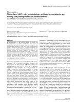
shown in Figure 2. The electrophoretic profile of the
CCg378 strain revealed the presence of four dsRNA
bands with approximate molecular sizes of 2.2, 1.95,
1.75 and 1.4 kiloba se pairs (kbp) (Figure 2, lane 1),
whereas the THg324 and SUg275 strains contained only
one dsRNA molecule of about 12.0 and 7.5 kbp, respec-
tively (Figure 2, lanes 2 and 3).
According to their molecular size, the 2.2-kbp dsRNAs
of B. cinerea CCg378 may correspond to or be part of
the genome of a partitivirus [16], whereas those o f the
THg324 and SUg275 strains may correspond to the gen-
ome of members of the Hypoviridae and Totiviridae
families, respectively [17,18].
The purified dsRNAs of S. cerevisiae 1743 are shown
in Figure 3A. The 4.6 kbp L-dsRNA of the L-A virus
and its satellite 1.8-kbp M-dsRNA are clearly noticeable
(Figure 3A, lane 1).
Binding capacity of dsRNA molecules by CF11 cellulose
In order to determine the b inding capacity of dsRNA by
the CF11 cellulose resin, different amounts of total nucleic
acids were loaded in the minicolumns until the binding
sites of the resin were saturated with dsRNA molecules.
To achieve this we worked with three parallel experiments
using the total nucleic acid preparation of S. cerevisiae
1743 [14]. Two, four and six milligrams of total nucleic
acids were loaded into separate columns, each containing
0.2 g of CF11 cellulose. The electrophoretic profile of
nucleic acids eluted with STE buffer containing 16% (v/v)
ethanol (see stage 4 of methods) revealed that when the
amount of total nucleic acids loaded on the column was
approximately 4 mg, dsRNA bands were also seen in addi-
tion to the bands corresponding to DNA and ssRNAs
(Figure 3B, lane 5). Three sharp bands that correspond to
Figure 2 Agarose gel electrophoresis of dsRNA from Botrytis
cinerea wild-type strains. Lane St
1
,O’GeneRuler™ 1 kb DNA
Ladder, Fermentas; lanes 1, 2 and 3, dsRNA from B. cinerea CCg378,
THg324 and SUg275 wild-type strains; lane St
2
, Lambda DNA/EcoRI
+ HindIII marker. The numbers on the left and right side indicate
molecular sizes expressed in kilobase pairs (kbp).
Figure 3 Agarose gel electrophoresis of dsRNA from Saccharomyces cerevisiae 1743. (A) Lane St, Lambda DNA/EcoRI + HindIII marker; lane
1, dsRNAs from Saccharomyces cerevisiae 1743. The numbers on the left side indicate molecular sizes expressed in kilobase pairs (kbp). (B) Lane
St, Lambda DNA/EcoRI + HindIII marker; lanes 1, 2 and 3, different samples of dsRNA from Saccharomyces cerevisiae 1743 (for details see Binding
capacity of dsRNA molecules by CF11 cellulose in Results and Discussion); lane 4, empty; lane 5, nucleic acids of S. cerevisiae 1743 eluted with STE
buffer containing 16% (v/v) ethanol. The column was loaded with 4 mg of total nucleic acids. The numbers on the left side indicate molecular
sizes expressed in kilobase pairs (kbp).
Castillo et al. Virology Journal 2011, 8:38
/>Page 4 of 7
the genomic DNA, 25S rRNA and 18S rRNA from S. cere-
visiae 1743 were seen clearly in the gel (Figure 3B, lane 5).
Furthermore, it was possible to visualize the band corre-
sponding to the L-dsRNA, but it was not possible t o see
the band of the M-dsRNA, since it migrates at the same
speed that 25S rRNA and therefore both bands overlap in
the gel (Figure 3B, lane 5). These results indicate that a
portion of the dsRNA molecules contained in the total
nucleic acids was not binding to the resin, which was satu-
rated without leaving binding sites available. Also, it was
possible to see clearly that the a mounts of dsRNA
obtained from the minicolumn loaded with four and six
milligrams of total nucleic acids are equivalent (Figure 3B,
lanes 2 and 3), confirming that the resin was saturated
with the dsRNAs contained in 4 mg of total nucleic acids.
Considering this, for this particular experiment 4 mg o f
total nucleic acids would be a sufficient initial amount to
ensure an adequate yield in relation to the amount of
CF11 cellullose (0.2 g of resin) packed in the minicolumn.
The initial 5 g cell pellet contained approxi mately
14 mg of total nucleic acids. Therefore, roughly 1.5 to
2.0 g wet weight of cells would be enough as starting
material for a column. Alternatively, 3 columns can be
loaded with the total nucleic acids obtained from the 5
g wet weight of fungal cells.
Under these conditions, the amount of total dsRNA
recovered from a minicolumn was approximately 4 μg,
since of t he 10 μLobtained,only2μLwereloadedin
each lane of the gel. This amount was enough to cor-
rectly visualize the bands in the gel after staining with
ethidium bromide (Figure 3B, lanes 1, 2 and 3), since in
the three lanes the bands corresponding to the L and M
dsRNA from S. cerevisiae 1743, were clearly visualized.
According to the densitometric analysis, the amount of
dsRNA corresponding to each band in the gel was
approximately 600 and 200 ng for the L and M dsRNA,
respectively (Figure 3B, lane 2).
Molecular cloning of the 2.2 kbp dsRNA from B. cinerea
CCg378
To test the integrity and applicability of the dsRNA
molecules purified by CF11 cellulose chromatography,
the 2.2 kbp dsRNA band of B. cinerea CCg378 was
eluted from the agarose gel, and after ligation of a syn-
thetic oligonucleotide in their 3’ ends, the cDNA was
obtained by reverse transcription and amplified by PCR
[15]. The obtained PCR fragment of about 2.2 kbp is
shown in Figure 4A, lanes 1 and 2. Later, the cDNA was
cloned in pGEM-T easy vector and the recombinant
plasmid was characterized with restric tion enzymes
(Figure 4B). In F igu re 4B, lane 1, the recombinant plas-
mid of about 5.2 kbp linearized with NcoI is shown.
Treatment with NcoI and SpeI generated two ba nds, a
corresponding to t he cDNA of about 2.2 kbp and the
other to the linear pGEM-T easy vector of about 3.0
kbp (Figure 4B, lane 2). Therefore, the obtention of the
full-length cDNA corresponding to the 2.2 kbp dsRNA
from B. cinerea CCg378, is a confirmation that the
dsRNA molecules isolated using the methodology
described in this work are obtained chemically intact
and can be used for other applications, such as cDNA
preparation, PCR, and molecular cloning.
The dsRNA purification technique presented in this
paper is very fast and very e asy to p erform. The res ults
show clearly that from small samples (mycelia or yeast
cells) it was possible to obtain dsRNAs free from DNA
and ssRNA, and in sufficient amount for their prelimin-
ary characterization. The refore, using this methodology
it should be possible to analyze simultaneously a large
number of fungal strains to detect the presence of
mycoviral dsRNA. These da ta show that neither the ori-
gin, the source, the size, or the number of dsRNA seg-
ments are imp ediments to obtain dsRNA free from
DNA and ssRNA.
A similar methodology has been described to isolate
dsRNA from the fungus Paecilomyces, but it requires
treatment with DNase I to remove the DNA present in
the dsRNA preparations [19]. Another technique that
uses guanidinium thiocyanate as the main reagent to
isolate dsRNA of Uncinula necator, requires that t he
dsRNA samples be treated with RNase A to eliminate
ssRNAs that are visualized as smears in the lanes where
the obtained dsRNA samples had been loaded [20].
More recently a technique that uses polyvinylpolypyrro-
lidone instead of phenol-chloroform has been described
[21]. This procedure is very simil ar to that described in
this paper, and the results are equivalent in terms of the
electrophoretic quality of the dsRNA. However, it i s not
possible to make a quantitative comparison of both
methods, since the authors of that paper do not quantify
the dsRNA obtained.
Two aspects that have been improved in the metho-
dology described in the present paper are the required
time and the amounts of reagents used. The original
technique of CF11cellulose chromatography [10-13]
requires that the sample be incubated overnight with
the resin, followed by two chromatographic cycles to
obtain high purity dsRNA after three days of work. In
the case of the minicolumns described in this paper, the
sample is loaded directly in the column, eliminating the
incubation time with the resin, and only about 20 min-
utes of elution are needed to obtain high purity dsRNA.
It is worth noting that in the original method of chro-
matography on cellulose CF11, 15 to 20 grams of myce-
lium or yeast cells are needed, and the total nucleic acid
extract obtained is used wholly to make a column o f
about 20 mL, while in the case of a minicolumn only
2 mL of total nucleic acid extraction are required, thus
Castillo et al. Virology Journal 2011, 8:38
/>Page 5 of 7
allowing running two chromatographic experiments in
parallel, using only about 2 g of mycelia or yeast cells as
starting material.
The most critical aspects of the technique are related
to cellular breaking and the degree of hydration of the
chromatographic resin during the whole procedure. In
order for the breaking of the mycelia or yeast cells to be
more efficient it is advisable to withdraw most of the
water from this s tarting material, as described in meth-
ods section. With the addition of liquid nitrogen, aqu-
eous crystals would be f ormed if the samples have too
much moisture. Under these conditions the cell break-
ing would be very difficult and the final yield would
decrease significantly. In the whole purification process
of dsRNA the resin (CF11 cellulose) must remain
hydrated. If for some reason the resin becomes dehy-
drated, the yield will decrease remarkably and in
extreme cases may result in no production of dsRNAs.
Conclusions
We developed a very simple and rapid method to isolate
dsRNA from fungi, using commercial minicolumns
packed with CF11 cellulose. From small quantities of
fungal cells as initial samples it was possible to obtain
sufficient amount of chemically intact dsRNA for its
part ial characterization. The dsRNA obtained is electro-
phoretically free of other nucleic acids, such as DNA
and ssRNA, and no additional treatment with nucleases
was required. We co nsider that t his methodology may
be used to isolate dsRNA from any type of fungi and
also can be applied to isolate genomic RNA from
dsRNA viruses that infect cells of higher eukary otes,
since the phenol-chloroform extraction would help to
eliminate capsid proteins. We think that this method
may be used to isolate dsRNA from any biological sam-
plethatcontainsdsRNAandthatthedsRNAobtained
can be used for further downstream applications.
Acknowledgements
The authors wish to thank Dr. Reed B. Wickner for kindly providing the killer
strain, Saccharomyces cerevisiae 1743.
This work was supported by DICYT-USACH project.
Authors’ contributions
AC and MC designed the study, participated in the characterization of the
dsRNA, performed the densitometric analysis, performed the molecular
cloning of the 2.2 kbp dsRNA from B. cinerea CCg378 and co-wrote the
manuscript; LC and FS carried out the isolation, purification and
Figure 4 Agarose gel electrophoresis of the cDNA obtained by the single-primer amplification technique. (A) Lane St, MassRuler™ DNA
Ladder 80-10,000 bp (Fermentas); lanes 1 and 2, PCR fragment corresponding to cDNA obtained from 2.2 kbp dsRNA from B. cinerea CCg378.
The numbers on the left side indicate molecular sizes expressed in kilobase pairs (kbp). (B) Analysis of the digestion patterns with restriction
enzymes of the recombinant plasmid (pGEM-T easy + cDNA) containing as insert the cDNA shown in (A). Lane St, Lambda DNA/EcoRI + HindIII
marker; lane 1, recombinant plasmid treated with NcoI; lane 2, recombinant plasmid treated with NcoI and SpeI; lane 3, recombinant plasmid
without treatment. The numbers on the left side indicate molecular sizes expressed in kilobase pairs (kbp).
Castillo et al. Virology Journal 2011, 8:38
/>Page 6 of 7
characterization of dsRNA from Saccharomyces cerevisiae and Botrytis cinerea.
All the authors read and approved the final manuscript.
Competing interests
The authors declare that they have no competing interests.
Received: 9 September 2010 Accepted: 25 January 2011
Published: 25 January 2011
References
1. Buck KW: Fungal virology - an overview. In Fungal Virology. Edited by:
Buck KW. Boca Raton: CRC Press; 1986:1-84.
2. Koltin Y, M Leibowitz M: Viruses of Fungi and Simple Eukaryotes. New
York: Marcel Dekker; 1988.
3. Ghabrial SA: Origin, adaptation and evolutionary pathways of fungal
viruses. Virus Genes 1998, 16:119-131.
4. Pearson MN, Beever RE, Boine B, Arthur K: Mycoviruses of filamentous
fungi and their relevance to plant pathology. Mol Plant Pathol 2009,
10:115-128.
5. Howitt RL, Beaver RE, Pearson MN, Forster RL: Genome characterization of
Botrytis virus F, a flexuous rod-shaped mycovirus resembling plant
‘potex-like’ viruses. J Gen Virol 2001, 82:67-78.
6. Howitt RL, Beaver RE, Pearson MN, Forster RL: Genome characterization of
a flexuous rod-shaped mycovirus, Botrytis virus X, reveals high amino
acid identity to genes from plant ‘potex-like’ viruses. Arch Virol 2006,
151:563-579.
7. Castillo A, Cifuentes V: Presence of double-stranded RNA in Phaffia
rhodozyma. Curr Genet 1994, 26:364-368.
8. Flegr J: A rapid method for isolation of double stranded RNA. Prep
Biochem 1987, 17:423-433.
9. Seroussi E, Peery T, Ginzberg I, Koltin Y: Detection of killer-independent
dsRNA plasmids in Ustilago maydis by a simple and rapid method of
extraction of dsRNA. Plasmid 1989, 21:216-225.
10. Franklin RM: Purification and properties of the replicative intermediate of
the RNA bacteriophage R17. Proc Natl Acad Sci USA 1966, 12:1504-1511.
11. Valverde RA, Nameth ST, Jordan RL: Analysis of double-stranded RNA for
plant virus diagnosis. Plant Dis 1990, 74:255-258.
12. Castillo A, Cifuentes V: Purification and Characterization of
Extrachromosomal Genetic Elements of Double-Stranded RNA (dsRNA)
of Xanthophyllomyces dendrorhous. In Non-Conventional Yeasts in Genetics,
Biochemistry and Biotechnology. Edited by: Wolf K, Breunig KD, Barth G.
Berlin Heidelberg: Springer-Verlag; 2003:329-334.
13. Castro M, Kramer K, Valdivia L, Ortiz S, Castillo A:
A double-stranded RNA
mycovirus confers hypovirulence-associated traits to Botrytis cinerea.
FEMS Microbiol Lett 2003, 228:87-91.
14. Ball SG, Tirtiaux C, Wickner RB: Genetic Control of L-a and L-(Bc) DsRNA
Copy Number in Killer Systems of Saccharomyces cerevisiae. Genetics
1984, 107:199-217.
15. Darissa O, Willingmann P, Adam G: Optimized approaches for the
sequence determination of double-stranded RNA templates. J Virol Meth
2010, 169:397-403.
16. Ghabrial SA, Buck KW, Hillman BI, Milne RG: Family Partitiviridae. In Virus
Taxonomy: Eighth Report of the International Committee for the Taxonomy of
Viruses. Edited by: Fauquet CM, Mayo MA, Maniloff J, Desselberger U, Ball
AL. San Diego: Elsevier/Academic Press; 2005:581-590.
17. Nuss DL, Hillman BI, Rigling D, Suzuki N: Family Hypoviridae. In Virus
Taxonomy: Eighth Report of the International Committee for the Taxonomy of
Viruses. Edited by: Fauquet CM, Mayo MA, Maniloff J, Desselberger U, Ball
AL. San Diego: Elsevier/Academic Press; 2005:597-601.
18. Wickner RB, Wang CC, Patterson JL: Family Totiviridae. In Virus Taxonomy:
Eighth Report of the International Committee for the Taxonomy of Viruses.
Edited by: Fauquet CM, Mayo MA, Maniloff J, Desselberger U, Ball AL. San
Diego: Elsevier/Academic Press; 2005:571-580.
19. Inglis PW, Valadares-Inglis MC: Rapid isolation of double-stranded RNAs
from entomopathogenic species of the fungus Paecilomyces using a
commercial minicolumn system. J Virol Methods 1997, 67:113-116.
20. Délye C, Corio-Costet MF: Rapid isolation of both double-stranded RNA
and PCR-suitable DNA from the obligate biotrophic phytopathogenic
fungus Uncinula necator using a commercially available reagent. J Virol
Methods 1998, 74:149-153.
21. Balijja A, Kvarnheden A, Turchetti T: A non-phenol-chloroform extraction
of double-stranded RNA from plant and fungal tissues. J Virol Methods
2008, 152:32-37.
doi:10.1186/1743-422X-8-38
Cite this article as: Castillo et al.: Rapid isolation of mycoviral double-
stranded RNA from Botrytis cinerea and Saccharomyc es cerevisiae.
Virology Journal 2011 8:38.
Submit your next manuscript to BioMed Central
and take full advantage of:
• Convenient online submission
• Thorough peer review
• No space constraints or color figure charges
• Immediate publication on acceptance
• Inclusion in PubMed, CAS, Scopus and Google Scholar
• Research which is freely available for redistribution
Submit your manuscript at
www.biomedcentral.com/submit
Castillo et al. Virology Journal 2011, 8:38
/>Page 7 of 7