Báo cáo y học: " Recombinant luciferase-expressing human cytomegalovirus (CMV) for evaluation of CMV inhibitors" pps
Bạn đang xem bản rút gọn của tài liệu. Xem và tải ngay bản đầy đủ của tài liệu tại đây (683.25 KB, 7 trang )
RESEARCH Open Access
Recombinant luciferase-expressing human
cytomegalovirus (CMV) for evaluation of CMV
inhibitors
Ran He
1
, Gordon Sandford
2
, Gary S Hayward
2
, William H Burns
2
, Gary H Posner
3
, Michael Forman
4
, Ravit Arav-Boger
1*
Abstract
Recombinant Towne CMV expressing luciferase under the control of CMV-DNA polymerase (POL) or the late pp28
(UL99) promoters were evaluated for potential application in high-throughput screening of anti-viral compounds.
POL-and pp28-luciferase displayed maximal expression 48 and 72 hours post infection, respectively. The pp28-
luciferase virus achieved a wider dynamic range of luciferase expression (6-7 logs) and was selected for testing of
inhibition by five anti-viral compounds. Luciferase expression highly correlated with plaque reduction and real-time
PCR. The pp28-luciferase reporter system is rapid, reproducible, and highly sensitive. It may be applied to screening
of novel anti-CMV compounds.
Background
Infection with Cytomega lovirus (CMV) continues to be
a major threat in organ transplant recipients and conge-
nitally-infected children [1,2]. Although existing sys-
temic therapies are effective in suppressing virus
replication, serious side effects and the emergence of
resistant viral strains pose significant treatment dilem-
mas [3]. The need to identify and develop new anti-
CMV compounds coincides with the advancement o f
rapid, sensitive, and high-throughput methods for
screening of lead compounds. While the plaque reduc-
tion assay remains the gold-standard for screening of
anti-viral compounds, new techniques based on recom-
binant DNA technology and highly sensitive molecular
assays have recently been suggested as alternatives [4-6].
Real-time PCR, the standa rd of care in the management
of CMV disease in high- risk patient populations, may
also provide a sensitive tool for drug screening [7-12]
In e arlier studies, a series of chloramphenic ol acetyl
transferase (CAT) recombinants expressing CAT under
the control of UL54 (DNA polymerase, POL)orUL99
(pp28) promoters were constructed. The expression of
CAT in in fected cells highly mimicked the expression
pattern of the endogenous UL54 and UL99 [4, 13]. Thus,
these two gene promoters were selected to construct
luciferase-recombinant CMV for quatification of CMV
replication in a rapid and repr oducuble way. We report
on the evaluation of two luciferase recombinant viruses
(pp28 and POL) and the correl ation of the pp28-lucifer-
ase system with plaque reduction and real-time PCR in
evaluation of CMV inhibition by anti-CMV compounds.
Methods
Construction of luciferase viruses
Recombinant CMV based on the laboratory-adapted
strain, Towne, was constructed by homologous recombi-
nation in transfected-infected cells. A b- galactosidase (b
-gal)-expressing Towne virus was first constructed using
an intergenic insertion site between US9 and US10.
Prior studies in which a b-glucuronidase expression cas-
sette was inserted in this intergenic region of the labora-
tory-adapted AD169 virus re vealed no alteration in
expected transcription from this region [4,14,15]. The
recombinant was genetically stable and exhibited normal
in-vitro growth characteristics. The transfer vector, pT,
was constructed from pRL120 which contains the
Towne virus HindIII T fragment [16] . A 2.0 kb BamHI-
ApaI subfragment containing US9 was ligated into
pGEM11z (Promeg a, Madison, WI) and the adjacent 1.3
kb ApaI-ApaI fragment c ontaining US10 was isolated
from agarose gels and ligated int o the ApaI site. DNA
sequencing confirmed the correct orientation of this
* Correspondence:
1
Department of Pediatrics, Johns Hopkins University School of Medicine,
Baltimore, MD, USA
Full list of author information is available at the end of the article
He et al. Virology Journal 2011, 8:40
/>© 2011 He et al; licensee BioMed Central Ltd. This is an Open Access article distributed under the te rms of the Creative Commons
Attribution License (http: //creativecomm ons.org/licenses/by/2.0), which permits unrestricted use, distribution, and reproduction in
any medium, provided the original work is properly cited.
fragment. T he BstEII site, whi ch lies midway between
the US9 and US10 genes, was used as the insertion site
for the b-gal expression cassette containing an SV40
promoter and polyA signal (pSVb from Clontech,
Mountain View, CA). DNA extracted from human fore-
skin fibroblasts (HFF) infected with Towne virus and
lineari zed transfer vector containing the expression cas-
sette were coprecipitated onto subconfluent HFF cul-
tures b y t he calcium phosphate method [17], followed
by a 2 min shock with 20% Di methyl sulfoxide (DMSO)
in Minimum Essential Medium (MEM) 4 t o 6 hrs later .
Virus from cultures developing cytopathic effects was
passed onto fresh HFF cultures, and examined for
b -galactosidase activity. Recombinant virus, designated
T242, was isolated from positive cultures by limiting
dilution in 96 well plates of HFF and selection of b-gal
positive wells at the highest dilutions.
To produce a recombinant virus expressing the luci-
ferase reporter gene under the control of either the pro-
moter of an early gene (POL, UL54) or a late gene
(pp28, UL99), the expression cassette of luciferase was
substituted for the b-gal cassette using the same transfer
vector (pT). Expression cas settes of l uciferase under the
control of POL- or pp28-promoter were constructed by
cloning the PCR products of the upstream 500 bp of
DNA polymerase or 350 bp of pp28 genes and ligating
them into the 5’ position of the luciferase coding regio n.
These expression cassettes were then ligated into the
blunted BstEII site of the pT transfer vector, linearized
and used in coprecipitation experiments with the DNA
of HFF cells infected with T242. Successful replacement
of the b-gal expression cassette by the luciferase expres-
sion cassettes with loss of b-gal expression and acquisi-
tion of luciferase expre ssion as phenotypic markers
facilitated isolation of the desired recombinants. Several
PCR sequencing reactions confirmed the correct posi-
tion and orientation of the luciferase reporter gene. The
following primers were used: pr imer 1- US09 forward
5’ -ACCTTGAAATGGGTCGCGCTCCGCT-3 ’,primer
2- luciferase forward-5’-ACAAGGATATGGGCTCACT-
GAGACT-3’,primer3:luciferasereverse5’-AGTCT-
CAGTGAGCCCATATCCTTGT-3’, and primer 4- US10
reverse- 5’-GCTATCGTCGCCGGAAGGAAACCGA -3’.
Cell Culture and virus infection
HFF a nd human lung fibroblasts (HEL) (ATCC, CRL-
2088 and CCL-137, respectively) were propagated in
Dulbecco’s Modified Eagle Medium (DMEM) containing
10% fetal bovine serum (FBS) and used for infection s
with the luciferase viruses. For assays other than plaq ue
reduction, 4 × 10
4
HFF cells were seeded in each well of
24-well plate one day prior to infection. Luciferase
viruses were used for infections with multiplicity of
infection (MOI) of 1.0 as previously described [18].
After 90 minutes adsorption, virus was removed, and
0.5 ml of media containing specified concentrations of
antiviral compounds was added. Infected non-treated
cells were used as positive controls; non-infected cell
lysates were used as negative controls.
Luciferase Assay
HFF cells were collected and lysed with Wizard
®
SV
Lysis Buffer (Promega, Madison, WI). The lysates were
assayed for luciferase and cell viability using an auto-
mated luminescent assay (Promega, Madison, WI), and
CellTiter-Glo luminescent cell viability assay kit, respec-
tively, on GloMax
®
-Multi+ Det ect ion Syst em (Promega,
Madison, WI) according to manufacturer’s instructions.
Plaque reduction assay
HEL cells were seeded at 3 × 10
5
cells per well in
twelve-well plates and were infected 24 hours later with
the pp28-luciferase CMV at 60 PFU/well. Following
90 minutes adsorption, the medium was aspirated from
the wells, and fresh medium containing selected drug
dilutions of g anciclovir (GCV), Foscarnet (FOS), Cyclo-
heximide (CHX), artesunate (ART), dimer sulfone carba-
mate [19] and 0.5% of carboxymethyl-cellulose were
added into triplicate wells. Af ter incubation at 37°C for
8 days, the overlay was removed, and the monolayer was
stai ned with crys tal viol et. Plaques were counted micro-
scopically under low power (40×). Drug effects were
calculated as the percentage of reduction in number o f
plaques in the presence of each drug concentration to
the number observed in the absence of drug.
Virus yield reduction assay
HFF were infected with the original Towne virus or
pp28/POL– luciferase virus a t an MOI of 0.1. Culture
supernatants were collected every two days unt il day 10
post infection and frozen at -80°C. Collected samples
were thawed and used for titration of infectious virus by
the plaque assay.
Real-time PCR
The quantitative CMV real-time PCR assay is based on
detection o f a 151bp region from the highly conserved
US17 gene [20]. The limit of detection of the assay i s
100 copie s/mL (3.0 copies/reaction), and the me asure-
able range is 2.4-8.0 log
10
copies/mL. The PCR was per-
formed using a total r eaction volume 50 μL. This
included 25 μL of TaqMan 2X Universal PCR Master
Mix (Applied Biosystems, Foster City, CA), 1.5 μL each
of 10 μM primers, 1 μLof10μM FAM-la beled probe,
11 μLofdH
2
0, and 10 μl of template. Amplification was
performed on a 7500 Real-Ti me PCR System (Applied
Biosystems, Foster City, CA). PCR conditions were: 50°C
for 2 min, 95°C for 10 min, 40 cycles of 95°C for 1 5 s
He et al. Virology Journal 2011, 8:40
/>Page 2 of 7
and 60°C for 60 s. Quantification standards were pre-
pared by cloning the US17 amplicon in the pCR
®
2.1-
TOPO
®
plasmid vector (Invitrogen, Carlsbad, CA).
Serial 10-fold dilutions o f plasmid from 7.0 to 1.0 log
10
copies/reaction were included with each assay and used
to establish a standard curve. Assay controls included
quantified CMV AD169 DNA (Advanced Biotechnolo-
gies Inc.) and quantified Towne CMV at 3.0 and 5.0
log
10
copies/mL. Quantitative CMV data were expressed
as viral DNA copies per milliliter.
Antiviral compounds
GCV, sod ium phosphonoformate (FOS) and cyclohexi-
mide (CHX) were obtained from Sigma-Aldrich
(St. Louis, MO). Artemisinin derivativ es, monomeric
trioxane artesunate (ART) and trioxane dimer sulfone
carbamate were synthesiz ed at Johns Hopkins University
(GHP), an d their structural details have been provided
elsewhere [18].
Results
Luciferase constructs
Two luciferase expressing viruses were constructed with
the Tow ne CMV strain (Figure 1A). A recombina nt b-
galactosidase (b-gal) CMV strain was first prepared as a
backbone for luciferase CMV. Recombinant b-gal virus
was isolated from positive cultures. This virus was used
in a second-round DNA recombination to generate two
luciferase-reporter CMV viruses: the luciferase gene
being under the control of either UL54 (POL)orUL99
(pp28) promoters. Successful recombinants were isolated
by loss of b-gal activity and the expression of luciferase
protein. The loss of the b-gal gene and acquisition of
theluciferasegeneintheexpectedlocationwascon-
firmed by DNA sequencing (Genebank submission ID:
1420040, seque nces are also available in Additional
file 1). Insertion at the specific sites was verified by PCR
sequencing (Figure 1B).
Comparison of luciferase expression by the two viral
constructs
The recombinant viruses were expected to express luci-
ferase at different stages of virus replication. The early
gene UL54 (POL) is expressed within the first 24 hours
post infection (hpi), usually later than 12 hpi [21];
whereas the true late UL99 (pp28) gene is expressed
only a t or afte r 48 hpi. Luciferase expression by POL-
and pp28-luciferase was quantified in cell lysates at 12,
24, 36, 48, 72 hpi, and at 36, 48, 72 and 96 hpi, respec-
tively (Figure 2). Using the same cell conditions, infec-
tivity, and luciferase assay system, peak luciferase
activities measured with pp28-luciferase were 20 fold
higher than those measured with POL-luciferase. The
peak activity of pp28-luciferase was reached at 72 hpi,
followed by a plateau towards 96 hpi. POL-luciferase
reached its maximum expression at 48 hours post infe c-
tion. The dynamic range of the luciferase assay using
pp28-luciferase and POL-luciferase was 50 - 5 × 10
6
,
and 50 - 6 × 10
4
respectively; therefore the pp28-lucifer-
ase virus was used in subsequent experiments.
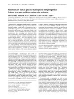
Figure 1 Const ruction of luciferase-recombinant CMV viruses
and confirmation of luciferase orientation by PCR. 1(A):
Construction of luciferase-recombinant Towne, insertion of
promoter and luciferase reporter between US9 and US10.
Appropriate restriction sites, the primers used for verification and
the expected size of PCR products are depicted. 1(B): PCR of pp28-
and POL-luciferase constructs. Lane 1-4: primers 1+ 4, lane 5-8:
primers 1+2, lane 9-12: primer 3+4.
He et al. Virology Journal 2011, 8:40
/>Page 3 of 7
Growth Characteristics of pp28-luciferase and the parent
Towne virus
We evaluated whether insertion of the recombination
cassette affected the growth kinetics and production of
infectious progeny. The parent Towne virus, pp28- and
pol-luciferase Towne viruses were grown in HFF and
the production of infectious progeny was determined
every two days during 10 day course post infection. The
growth characteristics of the viruses were similar
(Figure 3). A marked increase in virus productio n was
observed starting 2 days post infection, and g rowth
kinetics was similar to previous reports [22]
Correlation of plaque reduction and luciferase expression
Parallel experi ments were conducted using the same
MOI of pp28-luciferase CMV with and without anti-
CMVcompounds(GCV,FOS,ART,dimersulfonecar-
bamate, CHX). The relative number of plaques co unted
10 days post infection was compared to re lative lucifer-
ase activities assayed 72 hpi (Figure 4 Table 1). The
drug concentration inhibiting 50% virus replication
(EC
50
) by plaque reduction and luciferase expression
was determined for each compound. For all five com-
pounds a high correlation was observed between plaque
reduction and luciferase expression (Figure 4). Data
obtained with the plaque reduction assay were similar to
previous reports (Table 1).
Inhibition of luciferase expression and DNA replication by
dimer sulfone carbamate and GCV
The supernatants from infected-trea ted and infected-
non treated cells were used for real-time PCR at day 3.
However, the test was no t s ensitive enough to detect
differences between the treatment conditions (data not
shown). Therefore, luciferase activity was compared with
real-time PCR from supernatants of infected cells 6 days
post infection. A high correlation was found between
luciferase expression, and DNA copy number (Figure 5).
Discussion
We report on a highly sensitive and objective luciferase
reporter assay for determination of CMV inhibition by
anti-viral agents. The assa y, based on pp28-luciferase
recombinant CMV, can be performed 72 hpi and drug
treatment, has a large dynami c range of 6-7 logs, and is
highly reproducible. Our work also reveals a high degree
of correlation between late gene (luciferase) expression
and plaque enumeration further confir ming the poten-
tial use of this assay in screening of anti-viral activities.
The su scept ibility of CMV strains, laboratory-adapted
and clinical i solates, to anti-CMV compounds has t radi-
tionally been evaluated by the classic plaque a ssay [23].
Although this assay best reflects viral infectivity, or the
biological behavior of CMV, it suffers from several
drawbacks. The assay is time consuming; r esults are
usually available 8-21 days after infection depending on
the virus strain used, and counting of plaques is labor
intensive. Another disadvantage of the plaque assay is
that the amount of viral replication within a single cell
cannot always be determined. Not infrequently, the end-
point of the test shows enlarg ed cells (CPE) without
spread of the virus to adjacent cells (plaque).
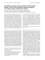
Figure 2 Timing and expression pattern of pp28-and POL-
luciferase CMV. Luciferase expression was determined in cell-
lysates at indicated time points following infection with pp28- or
POL-luciferase with and without treatment with GCV (30 μM). Y axis-
log scale of luciferase read out; X axis- time points in hours.
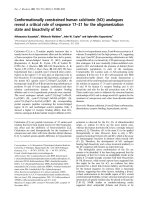
Figure 3 Growth characteristics of Towne, pp28-and POL-
luciferase Towne viruses. The production of virus progeny was
determined in HFF infected with the original Towne virus, and
recombinant pp28- or POL-luciferase virus at an MOI of 0.1. Culture
supernatants were collected at the indicated days and used for
titration of infectious virus by the plaque assay. Y-axis on the left
indicates growth of progeny viruses in log scale, Y-axis on the right
indicated relative virus kinetics of the recombinant viruses as
compared to the parent Towne strain.
He et al. Virology Journal 2011, 8:40
/>Page 4 of 7
Recombinant viruses carrying different reporter genes
have been dev eloped as alternativ e methods to overcome
some of the limitations of the plaque assay. A recombinant
CMV expressing b-galactosidase under the control of the
major immediate early promoter was used i n a 96-well
assay [24]. Although the assay was sensitive and rapid,
background b-galactosidase activity was observed second-
ary to its expression under the control of an immediate
early gene during the initial infection. A secreted alkaline
phosphatase (SEAP) reporter gene driven by the CMV
major immediate early promoter was inserted at the US6
gene [25]. Reduction in SEAP activity under drug treat-
ment was used to de termine drug sensit ivity. Results of
transferring specific mutations in UL97 or POL were com-
pared with results obtained using traditional phenotyping
assays. The assay was validated for approved CMV drugs
(GCV, FOS, and CDV) that target the CMV DNA poly-
merase. The open reading frame between US9 and US10
has been used to construct several recombinant CMV
strains [4,5,26]. For example, a GFP- reporter system
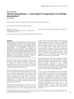
Figure 4 Correlation of plaque reduction and luciferase expression. CMV-infected HFF were treated with GCV, FOS, CHX, ART, dimer sulfone
carbamate with the indicated drug concentrations. Luciferase expression was quantified in cell lysates 72 hpi. Plaque reduction was performed
10 days post infection. The correlation coefficient is provided for each experiment.
Table 1 Inhibition of pp28-luciferase by anti-CMV compounds using plaque reduction or luciferase assay
Compound Plaque Reduction EC
50
(μM) Luciferase
EC
50
(μM)
Reference
FOS 328 +/- 28 268 +/- 20 [28]
Dimer Sulfone Carbamate 0.067 +/- 0.011 0.066 +/- 0.004 [18]
ART 8.03 +/- 0.55 6.74 +/- 0.38 [29]
GCV 4.39 +/- 0.39 4.23 +/- 0.27 [30]
CHX 0.262 +/- 0.067 0.299 +/- 0.036 NA
EC
50
was determined by plaque reduction assay or luciferase expression in pp28-luciferase CMV infected HFF cells. Reported values represent the means ±
standard deviations (SD) of data derived from at least three independent experiments performed in duplicate. Historical controls are provided for EC
50
values
(reference column).
He et al. Virology Journal 2011, 8:40
/>Page 5 of 7
generated with the laboratory-adapted strai n AD169 was
applied successfully to both qua litative and semiquantita-
tive applications [5]. Compared to the GFP-CMV system,
the luciferase-CMV offers a highly accurate and quantita-
tive assay which is simpl e and easy to perform. A limited
evaluation of pp28 -luciferase CMV activity in the pre-
sence of GCV, a cyclovir and papaverine, suggested its
potential application for anti-viral screen [26].
In addition to recombinant viruses, reporter cell lines
have been generate d to screen for anti -CMV com-
pounds [6,27]. In one suc h approach, using a luciferase
reporter cell line, the promoter was activated by
immediate early proteins; therefore compounds that
inhibit CMV at later stages of infection cannot be evalu-
ated with this system [6]. Since the pp28-luciferase virus
is driven by the promoter of a true late CMV gene,
which can only occur after DNA replication and the
onset of transcription of late genes, it can be applie d for
screening of compounds that target steps prior to and
during DNA replication. The pp28-luciferase system
therefore has a much wider appli cation for drug screen-
ing compared to the reported luciferase cell line [6].
Quantification of viral genomes by real-time PCR is gen-
erally proportional to production of virus particles [7].
Application of real-time PCR for in-vitro screening of anti-
viral compounds is attractive because the assay is rapid and
highly-sensitive. However, compared to the luciferase assay,
real-time PCR is more labor-intensive. DNA copy number
measured in supernatants collected at 6 days post infection
with Towne virus correlated with luciferase activity in cell
lysates at 3 and 6 days post infection. For a clinical isolate,
generally 10 days were required for quantification of DNA
in cell lysates [18]. Recently, a real-time PCR assay of a con-
served region in UL54 was performed in cell lysates four
days following infection and treatment with compounds
and showed a high correlation with plaque reduction assay
[12]. Additional studies are need ed to determine th e best
timing and compartment for performance of the real-time
PCR assay.
Our study reveal s late CMV protein exp ression highly
correlates with the production of infectious progeny
(plaque assay) and DNA replication. Advantages of the
luciferase assay over the real-time PCR include: faster
turn-around tim e after infection, and lower cost (20
times less than real-time PCR). The luciferase assay
yielded similar data to the plaque assay, but its perfor-
mance (accuracy and rapidity) was superior. In conclu-
sion, the recombinant pp28-lucifarese fulfills important
characteristics that are require d for high-throughput
screening o f anti-viral co mpounds: rapidity, reproduci-
bility, low cost, and high sensitivity.
Additional material
Additional file 1: Sequences of the pp28, POL promoters and
luciferase in the region between US9 and US10. Several regions can
be distinguished- bold sequences are of CMV Towne, underlined
sequences are POL (sequence #1) and pp28 (sequence #2) promoters,
and the italic regions are the sequence of firefly luciferase gene.
Abbreviations
CMV: Cytomegalovirus; PCR: polymerase chain reaction; EC
50
: effective
concentration 50; HEL: human embryonic lung fibroblasts; HFF: human
foreskin fibroblasts; MOI: multiplicity of infection; US: unique short; POL:
polymerase.
Figure 5 Luciferase expression and real-time PCR. HFF were infected with pp28-luciferase and treated with either GCV or dimer sulfone
carbamate. Luciferase activity was determined in cell lysates of infected-treated cells and infected non-treated cells. DNA copy number was
determined by real-time PCR in supernatants of infected-treated cells and infected non-treated cells 6 days post infection.
He et al. Virology Journal 2011, 8:40
/>Page 6 of 7
Acknowledgements
Supported by NIH KO8 AI074907 to RAB.
Author details
1
Department of Pediatrics, Johns Hopkins University School of Medicine,
Baltimore, MD, USA.
2
The Sidney Kimmel Comprehensive Cancer Center,
Johns Hopkins University School of Medicine, Baltimore, MD, USA.
3
Department of Chemistry, School of Arts and Sciences, The Johns Hopkins
University, Baltimore, MD, USA.
4
Department of Pathology, Johns Hopkins
Medical Institutions, Baltimore, MD, USA.
Authors’ contributions
RH carried out the plaque/luciferase assays and verification of viral
constructs. He participated in drafting the manuscript. GS, GSH and WHB
designed and constructed the luciferase viruses, GHP synthesized and
provided artemisinin derivatives, MF carried out the real-time PCR assays,
RAB directed the study, analyzed and interpreted the data, drafted and
revised the manuscript. All authors read and approved the manuscript.
Competing interests
The authors declare that they have no competing interests.
Received: 21 December 2010 Accepted: 26 January 2011
Published: 26 January 2011
References
1. Fishman JA, Emery V, Freeman R, Pascual M, Rostaing L, Schlitt HJ,
Sgarabotto D, Torre-Cisneros J, Uknis ME: Cytomegalovirus in
transplantation - challenging the status quo. Clin Transplant 2007,
21:149-158.
2. Kenneson A, Cannon MJ: Review and meta-analysis of the epidemiology
of congenital cytomegalovirus (CMV) infection. Rev Med Virol 2007,
17:253-276.
3. Chou S: Cytomegalovirus UL97 mutations in the era of ganciclovir and
maribavir. Rev Med Virol 2008, 18:233-246.
4. Kohler CP, Kerry JA, Carter M, Muzithras VP, Jones TR, Stenberg RM: Use
of recombinant virus to assess human cytomega lovirus ea rly and late
promoters in the context of the viral genome. JVirol1994,
68:6589-6597.
5. Marschall M, Freitag M, Weiler S, Sorg G, Stamminger T: Recombinant
green fluorescent protein-expressing human cytomegalovirus as a tool
for screening antiviral agents. Antimicrob Agents Chemother 2000,
44:1588-1597.
6. Fukui Y, Shindoh K, Yamamoto Y, Koyano S, Kosugi I, Yamaguchi T,
Kurane I, Inoue N: Establishment of a cell-based assay for screening of
compounds inhibiting very early events in the cytomegalovirus
replication cycle and characterization of a compound identified using
the assay. Antimicrob Agents Chemother 2008, 52:2420-2427.
7. Boeckh M, Boivin G: Quantitation of cytomegalovirus: methodologic
aspects and clinical applications. Clin Microbiol Rev 1998, 11:533-554.
8. Boeckh M, Huang M, Ferrenberg J, Stevens-Ayers T, Stensland L,
Nichols WG, Corey L: Optimization of quantitative detection of
cytomegalovirus DNA in plasma by real-time PCR. J Clin Microbiol 2004,
42:1142-1148.
9. Hadaya K, Wunderli W, Deffernez C, Martin PY, Mentha G, Binet I, Perrin L,
Kaiser L: Monitoring of cytomegalovirus infection in solid-organ
transplant recipients by an ultrasensitive plasma PCR assay. J Clin
Microbiol 2003, 41:3757-3764.
10. Gerna G, Furione M, Baldanti F, Percivalle E, Comoli P, Locatelli F:
Quantitation of human cytomegalovirus DNA in bone marrow transplant
recipients. Br J Haematol 1995, 91:674-683.
11. Lilleri D, Baldanti F, Gatti M, Rovida F, Dossena L, De Grazia S, Torsellini M,
Gerna G: Clinically-based determination of safe DNAemia cutoff levels
for preemptive therapy or human cytomegalovirus infections in solid
organ and hematopoietic stem cell transplant recipients. J Med Virol
2004, 73:412-418.
12. Schnepf N, Boiteau N, Petit F, Alain S, Sanson-Le Pors MJ, Mazeron MC:
Rapid determination of antiviral drug susceptibility of human
cytomegalovirus by real-time PCR. Antiviral Res 2009, 81:64-67.
13. Kerry JA, Priddy MA, Kohler CP, Staley TL, Weber D, Jones TR, Stenberg RM:
Translational regulation of the human cytomegalovirus pp28 (UL99) late
gene. J Virol 1997, 71:981-987.
14. Jones TR, Muzithras VP, Gluzman Y: Replacement mutagenesis of the
human cytomegalovirus genome: US10 and US11 gene products are
nonessential. J Virol
1991, 65:5860-5872.
15. Jones TR, Muzithras VP: Fine mapping of transcripts expressed from the
US6 gene family of human cytomegalovirus strain AD169. J Virol 1991,
65:2024-2036.
16. Lafemina RL, Hayward GS: Replicative forms of human cytomegalovirus
DNA with joined termini are found in permissively infected human cells
but not in non-permissive Balb/c-3T3 mouse cells. J Gen Virol 1983, 64(Pt
2):373-389.
17. Graham FL, van der Eb AJ: A new technique for the assay of infectivity of
human adenovirus 5 DNA. Virology 1973, 52:456-467.
18. Arav-Boger R, He R, Chiou CJ, Liu J, Woodard L, Rosenthal A, Jones-
Brando L, Forman M, Posner G: Artemisinin-derived dimers have greatly
improved anti-cytomegalovirus activity compared to artemisinin
monomers. PLoS One 2010, 5:e10370.
19. Rosenthal AS, Chen X, Liu JO, West DC, Hergenrother PJ, Shapiro TA,
Posner GH: Malaria-infected mice are cured by a single oral dose of new
dimeric trioxane sulfones which are also selectively and powerfully
cytotoxic to cancer cells. J Med Chem 2009, 2:1198-1203.
20. Tanaka Y, Kanda Y, Kami M, Mori S, Hamaki T, Kusumi E, Miyakoshi S,
Nannya Y, Chiba S, Arai Y, Mitani K, Hirai H, Mutou Y: Monitoring
cytomegalovirus infection by antigenemia assay and two distinct plasma
real-time PCR methods after hematopoietic stem cell transplantation.
Bone Marrow Transplant 2002, 30:315-319.
21. Stinski MF: Sequence of protein synthesis in cells infected by human
cytomegalovirus: early and late virus-induced polypeptides. J Virol 1978,
26:686-701.
22. Vieira J, Schall TJ, Corey L, Geballe AP: Functional analysis of the human
cytomegalovirus US28 gene by insertion mutagenesis with the green
fluorescent protein gene. J Virol 1998, 72:8158-8165.
23. Wentworth BB, French L: Plaque assay of cytomegalovirus strains of
human origin. Proc Soc Exp Biol Med 1970, 135:253-258.
24. Hippenmeyer PJ, Dilworth VM: A rapid assay for determination of antiviral
activity against human cytomegalovirus. Antiviral Res 1996, 32:35-42.
25. Chou S, Van Wechel LC, Lichy HM, Marousek GI: Phenotyping of
cytomegalovirus drug resistance mutations by using recombinant
viruses incorporating a reporter gene. Antimicrob Agents Chemother 2005,
49:2710-2715.
26. Song BH, Lee GC, Lee CH: Measurement of antiviral activities using
recombinant human cytomegalovirus. The Journal of Microbiology 2000,
38:255-259.
27. Gilbert C, Boivin G: New reporter cell line to evaluate the sequential
emergence of multiple human cytomegalovirus mutations during in
vitro drug exposure. Antimicrob Agents Chemother 2005, 49:4860-4866.
28. Freitas VR, Fraser-Smith EB, Matthews TR: Increased efficacy of ganciclovir
in combination with foscarnet against cytomegalovirus and herpes
simplex virus type 2 in vitro and in vivo. Antiviral Res 1989, 12:205-212.
29. Efferth T, Marschall M, Wang X, Huong SM, Hauber I, Olbrich A,
Kronschnabl M, Stamminger T, Huang ES: Antiviral activity of artesunate
towards wild-type, recombinant, and ganciclovir-resistant human
cytomegaloviruses. J Mol Med 2002, 80:233-242.
30. Mercorelli B, Muratore G, Sinigalia E, Tabarrini O, Biasolo MA, Cecchetti V,
Palu G, Loregian A: A 6-aminoquinolone compound, WC5, with potent
and selective anti-human cytomegalovirus activity. Antimicrob Agents
Chemother 2009, 53:312-315.
doi:10.1186/1743-422X-8-40
Cite this article as: He et al.: Recombinant luciferase-expressing human
cytomegalovirus (CMV) for evaluation of CMV inhibitors. Virology Journal
2011 8:40.
He et al. Virology Journal 2011, 8:40
/>Page 7 of 7