Báo cáo y học: " Spontaneous acute subdural hematoma as an initial presentation of choriocarcinoma: A case report" potx
Bạn đang xem bản rút gọn của tài liệu. Xem và tải ngay bản đầy đủ của tài liệu tại đây (396.14 KB, 4 trang )
BioMed Central
Page 1 of 4
(page number not for citation purposes)
Journal of Medical Case Reports
Open Access
Case report
Spontaneous acute subdural hematoma as an initial presentation of
choriocarcinoma: A case report
Brandon G Rocque* and Mustafa K Bas¸kaya*
Address: Department of Neurological Surgery, University of Wisconsin, Madison, WI, USA
Email: Brandon G Rocque* - ; Mustafa K Bas¸kaya* -
* Corresponding authors
Abstract
Introduction: Diverse sequelae of central nervous system metastasis of choriocarcinoma have
been reported, including infarction, intra or extra axial hemorrhages, aneurysm formation and
carotid-cavernous fistula. Here we report a case of subdural hematoma as the first presentation of
choriocarcinoma.
Case presentation: The patient is a 34-year-old woman whose initial presentation of widely
metastatic choriocarcinoma was an acute subdural hematoma, requiring decompressive
craniectomy. Histopathologic examination of the tissue showed no evidence of choriocarcinoma,
but the patient was found to have diffuse metastatic disease and cerebrospinal fluid indices highly
suggestive of intracranial metastasis.
Conclusion: Choriocarcinoma frequently metastasizes intracranially. We review the diverse
possible manifestations of this process. In addition, the cerebrospinal fluid:serum beta-human
chorionic gonadotropin ratio is an important factor in diagnosing these cases. Finally, the role of
the neurosurgeon is discussed.
Introduction
Choriocarcinoma is a rare gestational trophoblastic dis-
ease that complicates approximately 1 in 50,000 term
pregnancies and 1 in 30 hydatidiform moles[1]. Among
confirmed cases of choriocarcinoma, 45% occur after
molar pregnancy, 24% after normal term pregnancy, 25%
after spontaneous abortion, and 5% after ectopic preg-
nancy[2]. Prognosis of this disease is generally good, 80–
90% long-term survival with chemotherapy, radiother-
apy, and surgical excision in appropriate cases[3]. One of
the indicators of a poor prognosis is intracranial metas-
tases, which complicate between 3 and 28% of gestational
choriocarcinoma[1]. Here we report a case of subdural
hematoma as the first presentation of choriocarcinoma
and present a review of the literature pertaining to sub-
dural hematoma in this setting.
Case Presentation
The patient is a 34-year-old woman who had an acute epi-
sode of excruciating headache and was later found
obtunded. She had a history of a normal pregnancy three
years prior to presentation. She then had an abnormal
pregnancy requiring dilation and evacuation at 10–12
weeks that was found to be a molar pregnancy. She
became pregnant again 9 months after the dilation and
evacuation of the molar pregnancy. This ended in a spon-
taneous, uncomplicated delivery 5 months prior to her
presentation. There was no history of trauma, recent or
remote.
Published: 19 June 2008
Journal of Medical Case Reports 2008, 2:211 doi:10.1186/1752-1947-2-211
Received: 30 November 2007
Accepted: 19 June 2008
This article is available from: />© 2008 Rocque and Bas¸kaya; licensee BioMed Central Ltd.
This is an Open Access article distributed under the terms of the Creative Commons Attribution License ( />),
which permits unrestricted use, distribution, and reproduction in any medium, provided the original work is properly cited.
Journal of Medical Case Reports 2008, 2:211 />Page 2 of 4
(page number not for citation purposes)
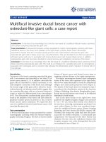
Upon arrival to Emergency Department, she had fixed,
dilated pupils and displayed extensor posturing. Compu-
terized tomography of the head without contrast (Figure
1) showed a 10-mm left hemispheric subdural hematoma
causing significant midline shift and uncal herniation.
The patient was then taken to the operating room for
emergency decompression via frontotemporal craniec-
tomy. A thick, clotted subdural hematoma was removed.
Fresh bleeding from one of the cortical arteries was
encountered and controlled with bipolar coagulation.
Inspection under microscope magnification revealed no
obvious vascular or neoplastic lesion. The coagulated part
of the small cortical artery was divided and sent for his-
topathologic examination along with the evacuated
hematoma.
Examination of the tissue showed no evidence of vascular
malformation or neoplasm, and cytokeratin immunola-
beling showed no signs of choriocarcinoma.
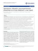
Following neurological and hemodynamic stabilization,
CT angiogram showed no evidence of aneurysm or vascu-
lar pathology. Magnetic resonance imaging (Figure 2)
showed changes associated with herniation injury, but no
appreciable tumor or intracranial mass. After full obstetric
history was obtained, beta-human chorionic gonadotro-
pin (HCG) level was found to be 55,000 mIU/mL (nor-
mal < 5 in non-pregnant patients). CSF examination
showed 675 nucleated cells, 20300 red blood cells, pro-
tein of 291 mg/dL, glucose of 91 mg/dL, and beta-HCG of
2141 mIU/mL, a serum:CSF ratio of 25:1 (normal > 60:1).
CT scan of the chest, abdomen, and pelvis showed lesions
in her liver, spleen, kidneys, and lungs. Her neurological
status continued to improve. On discharge to the Gyneco-
logic Oncology service one month after presentation, she
was extubated and was able to speak slowly, ambulate
with assistance, and had no focal motor deficit. She
underwent whole brain radiation and chemotherapy with
varying regimens of etoposide, cisplatin, bleomycin,
methotrexate, cyclophosphamide, and vincristine. She
initially did well and was able to transfer to inpatient
rehab. However, she developed fibrotic lung disease and
then recurrent pulmonary choriocarcinoma lesions,
which led to her death four months after her initial pres-
entation.
Discussion
Approximately one half of tumor-related hemorrhages are
the first manifestation of the tumor. In addition, there are
numerous reports in the literature of other presentations,
CT scan showing left subdural hematoma with midline shift and right-side subarachnoid hemorrhageFigure 1
CT scan showing left subdural hematoma with midline shift
and right-side subarachnoid hemorrhage.
Coronal MRI T2 FLAIR sequence showing herniation injuryFigure 2
Coronal MRI T2 FLAIR sequence showing herniation injury.
Journal of Medical Case Reports 2008, 2:211 />Page 3 of 4
(page number not for citation purposes)
including intracranial hemorrhage[4], subarachnoid
hemorrhage from rupture of neoplastic aneurysm[5,6],
carotid cavernous fistula[7], and infarct due to tumor
embolus[8].
Here we report a case of choriocarcinoma presenting as
subdural hematoma. This has been reported only twice
before in the literature. In 1986, Toyama et al. reported a
patient who presented with a subdural hematoma due to
ruptured aneurysm of the angular artery following surgi-
cal resection of a choriocarcinoma in the left adnexa[9].
Histological examination of the tissue confirmed chorio-
carcinoma in the aneurysm. Cave reported a case of sud-
den death seven months postpartum due to
choriocarcinoma, metastatic to the wall of a ruptured
occipital artery[10]. The patient presented with an acute
subdural hematoma.
In the female patient of reproductive age, choriocarci-
noma must be considered in the differential for any
intracranial hemorrhage. A lesion may be apparent on CT
scan, but often there is no lesion visible apart from the
hemorrhage. Suresh reports a series of 10 hemorrhages
from confirmed cases of choriocarcinoma in which only
two had visible lesions on CT[11]. The key diagnostic fea-
ture, apart from clinical suspicion, is the elevation of beta-
HCG in the serum and CSF. Elevated HCG in the serum of
a patient with previous abnormal pregnancy strongly sug-
gests choriocarcinoma or retained trophoblastic tissue. If
the ratio of serum to CSF HCG is less than 60, CNS metas-
tasis is strongly suspected[12]. The unique feature of the
case presented here is the lack of histological confirma-
tion of choriocarcinoma. A diagnostic technique not uti-
lized in this case was serial CSF sampling for beta-HCG.
Given the importance of the serum:CSF ratio of beta-HCG
in this patient with no other evidence of intracranial dis-
ease, serial CSF analysis would allow analysis of the trend
as blood is reabsorbed. Presumably, if the decreased
serum:CSF ratio is due to contamination with blood from
hemorrhage, the ratio would normalize on serial studies.
This technique was not utilized in this case, but may be
useful in less clear cases. Given her elevated CSF beta-
HCG, widespread disease elsewhere, and lack of other fac-
tors that could lead to acute subdural hemorrhage, it is
clear that the etiology in this case is metastatic choriocar-
cinoma.
Importantly, CNS metastases are very responsive to chem-
otherapy. There are reports of complete resolution of CNS
disease including intracranial metastases, neoplastic pseu-
doaneurysms, and neoplastic fistulas with chemotherapy
alone [4-7]. Given the good response of this disease to
chemotherapy, in many cases, including resolution of
CNS pathology, it is not necessary to perform surgical
removal of asymptomatic lesions. Surgical treatment
should be reserved for patients with symptomatic intrac-
ranial pathology that represents an immediate threat.
Conclusion
Choriocarcinoma is a relatively uncommon malignancy
associated with pregnancy. The disease may initially
present with intracranial hemorrhage or other CNS mani-
festation in a significant proportion of patients. It is there-
fore critical to have a high level of suspicion regarding
choriocarcinoma in any patient of reproductive age or
with a history of abnormal pregnancy who presents with
intracranial pathology. In the case of hemorrhage, it is
essential to send the evacuated hematoma for histopatho-
logical examination. Increased beta-HCG levels can aid in
the diagnosis, and a low serum:CSF beta-HCG level can be
strongly suggestive of intracranial choriocarcinoma even
in the absence of histopathologically proven disease.
Consent
Written informed consent was obtained from the family
of the patient for publication of this case report and any
accompanying images. A copy of the written consent is
available for review by the Editor-in-Chief of this journal.
Competing interests
The authors declare that they have no competing interests.
Authors' contributions
BGR assembled clinical data and drafted the manuscript,
MKB was the primary surgeon and reviewed and revised
the manuscript. Both authors read and approved the final
manuscript.
References
1. Athanassiou A, Begent RH, Newlands ES, Parker D, Rustin GJ, Bag-
shawe KD: Central nervous system metastases of choriocar-
cinoma. 23 years' experience at Charing Cross Hospital.
Cancer 1983, 52:1728-1735.
2. Redline RW, Abdul-Karim FW: Pathology of gestational tro-
phoblastic disease. Semin Oncol 1995, 22:96-108.
3. Kalafut M, Vinuela F, Saver JL, Martin N, Vespa P, Verity MA: Multiple
cerebral pseudoaneurysms and hemorrhages: the expanding
spectrum of metastatic cerebral choriocarcinoma. J Neuroim-
aging 1998, 8:44-47.
4. Gurwitt LJ, Long JM, Clark RE: Cerebral metastatic choriocarci-
noma: a postpartum cause of "stroke". Obstet Gynecol 1975,
45:583-588.
5. Fujiwara T, Mino S, Nagao S, Ohmoto T: Metastatic choriocarci-
noma with neoplastic aneurysms cured by aneurysm resec-
tion and chemotherapy. Case report. J Neurosurg 1992,
76:148-151.
6. Nakahara T, Nonaka N, Kinoshita K, Matsukado Y: [Subarachnoid
hemmorrhage and aneurysmal change of cerebral arteries
due to metastases of chorioepithelioma (author's transl)].
No Shinkei Geka 1975, 3:777-782.
7. Fadli M, Lmejjati M, Amarti A, El Hassani MR, El Abbadi N, Bellakhdar
F: [Metastatic and hemorrhagic brain arteriovenous fistulae
due to a choriocarcinoma. Case report]. Neurochirurgie 2002,
48:39-43.
8. Nakagawa Y, Tashiro K, Isu T, Tsuru M: Occlusion of cerebral
artery due to metastasis of chorioepithelioma. Case report.
J Neurosurg 1979, 51:247-250.
Journal of Medical Case Reports 2008, 2:211 />Page 4 of 4
(page number not for citation purposes)
9. Toyama K, Tanaka T, Hirota T, Misu N, Mizuno K: [A case report
of neoplastic aneurysm due to metastatic choriocarcinoma].
No Shinkei Geka 1986, 14:385-390.
10. Cave WS: Acute, nontraumatic subdural hematoma of arte-
rial origin. J Forensic Sci 1983, 28:786-789.
11. Suresh TN, Santosh V, Shastry Kolluri VR, Jayakumar PN, Yasha TC,
Mahadevan A, Shankar SK: Intracranial haemorrhage resulting
from unsuspected choriocarcinoma metastasis. Neurol India
2001, 49:231-236.
12. Bagshawe KD, Harland S: Immunodiagnosis and monitoring of
gonadotrophin-producing metastases in the central nervous
system. Cancer 1976, 38:112-118.