Báo cáo y học: "Normothermic treatment in acute clinical encephalitis: a case report" pdf
Bạn đang xem bản rút gọn của tài liệu. Xem và tải ngay bản đầy đủ của tài liệu tại đây (359.73 KB, 5 trang )
BioMed Central
Page 1 of 5
(page number not for citation purposes)
Journal of Medical Case Reports
Open Access
Case report
Normothermic treatment in acute clinical encephalitis: a case
report
Mari Terashima
1
, Hiroshi Kataoka*
1
, Katsuji Hirai
2
and Satoshi Ueno
1
Address:
1
Department of Neurology, Nara Medical University, Kashihara, Nara 634-8522, Japan and
2
Department of Intensive Care Unit, Nara
Medical University, Kashihara, Nara 634-8522, Japan
Email: Mari Terashima - ; Hiroshi Kataoka* - ; Katsuji Hirai - ;
Satoshi Ueno -
* Corresponding author
Abstract
Introduction: Encephalitis is a common infection of the brain, associated with a high risk of
mortality and morbidity despite intensive supportive therapy. This report describes a patient with
acute clinical meningoencephalitis who responded dramatically when her body temperature was
decreased to normothermia (36 to 37°C) in combination with barbiturate therapy.
Case presentation: A 15-year-old, previously healthy girl presented with a 2-day history of
headache and meningeal stiffness and pyrexia. Cranial magnetic resonance imaging showed high-
intensity signals in the splenium of the corpus callosum on T2-weighted and diffusion-weighted
images. On day 4 of admission, the level of consciousness decreased and ataxic respiration and
apnea appeared. After that, fever (body temperature >40°C) developed with remarkable
tachycardia. The body temperature was decreased with the use of a forced-air-cooling blanket and
head cooling. The core temperature, measured in the bladder, was maintained at between 36 and
37°C for 5 days. During the period of normothermia, thiopental sodium was given continuously for
3 days. After normothermia, the level of consciousness increased without the development of
fever, and ventilatory support was withdrawn.
Conclusion: Our experience suggests that normothermic treatment in combination with
barbiturate therapy may be an effective option for the management of brain swelling associated
with acute meningoencephalitis, particularly when accompanied by a persistent high fever.
Introduction
Encephalitis is a common infection of the brain, associ-
ated with a high risk of mortality and morbidity despite
intensive supportive therapy. Hypothermia combined
with barbiturate therapy has been used to treat brain
swelling and intracranial hypertension [1]. Several inves-
tigations have shown that mild hypothermia aimed at
reducing body temperature to 34 to 35°C is an effective
treatment for acute encephalitis and encephalopathy [2]
and has recently been used to treat brain swelling caused
by trauma [3]. Mild hypothermia produces fewer compli-
cations than deep hypothermia, but can cause conditions
such as hypokalemia [2]. On the other hand, using body
surface cooling for 24 hours to achieve a core body tem-
perature between 36 and 37°C was reported to be safe in
patients with acute stroke [4].
Published: 25 July 2008
Journal of Medical Case Reports 2008, 2:246 doi:10.1186/1752-1947-2-246
Received: 3 November 2007
Accepted: 25 July 2008
This article is available from: />© 2008 Terashima et al; licensee BioMed Central Ltd.
This is an Open Access article distributed under the terms of the Creative Commons Attribution License ( />),
which permits unrestricted use, distribution, and reproduction in any medium, provided the original work is properly cited.
Journal of Medical Case Reports 2008, 2:246 />Page 2 of 5
(page number not for citation purposes)
We describe a patient with acute clinical meningoen-
cephalitis who responded dramatically when her body
temperature was decreased to normothermia (36 to
37°C) in combination with barbiturate therapy.
Case presentation
A 15-year-old, previously healthy girl presented with a 2-
day history of headache, fever and vomiting. On admis-
sion to another hospital, she had meningeal stiffness and
pyrexia (body temperature 39°C). Lumbar puncture
showed 137 white blood cells (79% lymphocytes)/mm
3
.
Cranial magnetic resonance imaging (MRI) showed high-
intensity signals in the splenium of the corpus callosum
(SCC) on T2-weighted and diffusion-weighted images
(Figure 1D and 1E). She received intravenous acyclovir
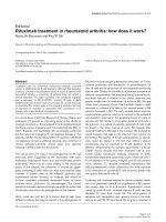
Cranial computed tomography scans obtained before normothermic treatment and during follow-upFigure 1
Cranial computed tomography scans obtained before normothermic treatment and during follow-up. A cranial
computed tomography scan obtained on day 5 (A) before normothermic treatment, showing remarkable meningeal enhance-
ment and brain swelling. Follow-up computed tomography scans obtained on day 12 (B) and day 29 (C), showing reduced
meningeal enhancement and brain swelling after normothermic treatment. T2-weighted magnetic resonance imaging (D) and
diffusion-weighted magnetic resonance imaging (E) scans, showing increased signal intensity of an ovoid lesion in the splenium
of the corpus callosum. A T2-weighted magnetic resonance imaging scan obtained on day 48, showing abnormal increased sig-
nals in the pontine (F).
Journal of Medical Case Reports 2008, 2:246 />Page 3 of 5
(page number not for citation purposes)
and methylprednisolone pulse therapy for a suspected
diagnosis of virus encephalitis. However, her conscious-
ness deteriorated and she was transferred to our hospital.
On the day of admission, she presented with disorienta-
tion and pyrexia (39.5°C) and could not respond to sim-
ple orders. The Glasgow coma score (GCS) was 12; eye
opening, verbal response and motor response were 4, 3
and 5, respectively. The heart rate was 118 beats per
minute with sinus rhythm. Blood pressure was 120/80
mmHg. Blood cell counts and the results of routine bio-
chemical analysis were normal except for hyponatremia
(121 mEq/liter). The osmotic pressure in serum and urine
was 277 and 668 mOsm/liter, respectively. Meningeal
stiffness was present. The deep tendon reflexes were non-
pathological. Lumbar puncture showed 151 white blood
cells (89% lymphocytes)/mm
3
, a protein concentration of
78 mg/dl and a glucose concentration of 49 mg/dl, with
negative bacterial and tuberculosis cultures. On polymer-
ase chain reaction amplification, herpes simplex virus,
varicella-zoster virus, Epstein-Barr virus and cytomegalo-
virus DNA were all negative in the cerebrospinal fluid
(CSF). Infection with various other viruses, such as influ-
enza, parainfluenza, measles and mumps, were excluded
by negative serum or CSF antibody titers (or both). Elec-
troencephalography revealed no epileptic discharges. The
patient received intravenous acyclovir, dexamethasone
and immunoglobulin therapy.
On day 4 after admission, the GCS dropped to 3 (eye
opening, verbal response and motor response were 1, 1
and 1, respectively), and ataxic respiration and apnea
appeared, leading to respiratory failure requiring ventila-
tory support. The patient was given intravenous vidarab-
ine and a continuous infusion of propofol, but fever
(body temperature >40°C) developed with remarkable
tachycardia. The body temperature was decreased with the
use of a forced-air-cooling blanket and head cooling. The
core temperature, measured in the bladder, was main-
tained between 36° and 37°C for 5 days. During the
period of normothermia, thiopental sodium was given
continuously for 3 days. Glycerin and dexamethasone
were also given intravenously. After normothermia, the
level of consciousness increased and the GCS for eye
opening and motor response increased to 4 and 6, respec-
tively, without the development of fever. Verbal response
could not be evaluated because the patient had undergone
a tracheotomy; however, she could respond to simple
orders. Synchronous intermittent mandatory ventilation
was decreased from 16 to 8 breaths per minute.
Thirty-six days after admission, ventilatory support was
withdrawn. Forty-eight days after admission, cranial MRI
showed increased signals in the pontine on T2-weighted
images, suggesting osmotic demyelination (Figure 1F),
and the high intensity in the SCC had disappeared. At that
time, the level of consciousness was normal and the man-
ual muscle test (MMT) scores, based on a 0 to 5 point
scale, were 4 and 1 in the upper and lower extremities,
respectively. The spinal MRI from the Th3 to L2 level
showed no abnormal intensity. Three months after admis-
sion, the patient was discharged, with no mental distur-
bance. The MMT scores were 5 and 1 in the upper and
lower extremities, respectively.
Figure 1 shows the serial changes on computed tomogra-
phy (CT) scans of the brain. A CT scan performed on day
5 (Figure 1A), before normothermia, showed remarkable
meningeal enhancement and brain swelling. In contrast,
CT scans obtained on day 12, during normothermia (Fig-
ure 1B), and on day 29, after normothermia (Figure 1C),
showed reduced meningeal enhancement and brain
swelling. CSF opening pressure decreased from 160 mm/
H
2
O on day 6 (before normothermia) to 130 mm/H
2
O
on day 20.
On transcranial Doppler ultrasonography, systolic flow
velocities in the right and left middle cerebral arteries
before normothermia decreased from 370 to 139 cm per
second and from 265 to 140 cm per second, respectively.
White blood cells, protein concentrations and interleukin
(IL)-6 concentrations in CSF decreased from 101/mm
3
,
74 mg/dl and 69.9 pg/ml on day 6 (before normother-
mia) to 13/mm
3
, 52 mg/dl and 2.3 pg/ml, respectively, on
day 20. The aspartate aminotransferase, alanine ami-
notransferase and serum sodium concentrations changed
from 344 IU/liter, 497 IU/liter and 128 mEq/liter to 159
IU/liter, 320 IU/liter and 132 mEq/liter, respectively, after
normothermia. Other laboratory findings were normal
after normothermia.
Discussion
The patient improved clinically without complications
after normothermic treatment (36 to 37°C) and showed
reduced IL-6 concentrations and leukocyte counts in the
CSF.
Conventional hypothermia (body temperature <30°C)
has been shown to reduce brain metabolic requirements,
which may lessen cerebral edema [5]. Mild hypothermia
(34 to 35°C) was also reported to have a marked protec-
tive effect against ischemic neuronal injury in experimen-
tal models [6], and showed promise for controlling brain
swelling [7]. However, these types of hypothermia often
cannot maintain the core temperature at the target level
for several days and are associated with a risk of complica-
tions, such as cardiovascular instability or infection [8]. A
previous study demonstrated that a decrease in body tem-
perature of 1 to 3°C can minimize or prevent brain energy
failure during hypoxia [8]. Our patient showed a reduc-
Journal of Medical Case Reports 2008, 2:246 />Page 4 of 5
(page number not for citation purposes)
tion in brain edema on cranial CT after body temperature
was decreased by about 3°C. Normothermic treatment
may thus minimize or protect against the brain swelling
associated with meningoencephalitis.
Several cytokines in serum and CSF are elevated in
patients with acute viral encephalitis or encephalopathy
[9,10]. IL-6 levels provide particularly valuable informa-
tion with respect to the diagnosis and severity of encepha-
litis or encephalopathy [10]. In patients with head injury,
moderate hypothermia (32 to 33°C) suppressed
increased arterial IL-6 levels, whereas normothermia (36
to 37°C) did not decrease elevated arterial IL-6 levels after
brain injury [11]. In our patient, IL-6 levels significantly
decreased in response to normothermic treatment plus
immunotherapy. This finding suggests that normother-
mic treatment might suppress the production of cytokines
by brain microglia or astrocytes in response to intense
inflammation in meningoencephalitis. Moreover, hypo-
thermia has been reported to inhibit the production of IL-
6, which may activate neutrophil infiltration [12].
Decreased numbers of leukocytes in the CSF after normo-
thermic treatment also provided evidence that inflamma-
tion-induced production of IL-6 was suppressed.
The patient received other treatments, including immu-
noglobulins, dexamethasone, antiviral agents and anti-
edema therapy, which might have affected outcomes (see
additional file 1). A previous study showed that corticos-
teroid treatment was associated with good outcomes in
patients with herpes simplex virus encephalitis. Pharma-
cologically, the good response was ascribed to mecha-
nisms involving the improvement of brain edema and
regulation of the host immune response associated with
acute encephalitis [13]. In experimental herpes simplex
virus encephalitis, dexamethasone treatment suppressed
not only the expression of inflammatory genes, but also
the expression of viral genes and was associated with neu-
roprotection and survival [14]. IL-6 secretion in smooth
muscle is inhibited by corticosteroids [15].
Recently, the use of intravenous immunoglobulins was
associated with relatively good outcomes in autoimmune
encephalitis [16]. In addition to normothermia and the
effects of barbiturate therapy, immunomodulating, anti-
edema or antiviral treatments might have also contributed
to the reductions in brain edema or CSF cytokine levels in
our patient.
The patient had severe hyponatremia and MRI scans
showed symmetric hyperintensity in the pons, confirming
the diagnosis of osmotic demyelination syndrome [17].
Severe hyponatremia may be caused by a variety of mech-
anisms, including hypovolemia, cerebral salt wasting syn-
drome or inappropriate secretion of antidiuretic hormone
[18]. Although direct evidence is lacking, the
hyponatremia in our patient might have been caused by
hypovolemia due to the persistent high fever or to inap-
propriate secretion of antidiuretic hormone as the
osmotic pressure of serum was less than that of urine. The
total daily correction in our patient was less than 10
mmol/liter/day [17], but the patient presented with para-
plegia as a residual symptom. Among 34 patients with
osmotic demyelination syndrome that were followed up,
11 had a complete recovery but 10 patients had some per-
sistent deficits, similar to our patient [19]. However, the
paraplegia in our patient was not consistent with only
osmotic demyelination at the pons. Although the cause of
paraplegia was unclear on spinal MRI, diseases other than
osmotic demyelination syndrome were suspected.
MRI showed transient high-intensity signals in the SCC,
which have rarely been demonstrated clinically in
encephalitis or encephalopathy [20]. Previous reports
have introduced a concept termed 'intramyelinic edema',
a non-degenerative change characterized by pathological
and neuro-imaging findings of Canavan disease or maple
syrup urine disease, associated with water collection
between the myelinic lamellae and decreased apparent
diffusion coefficient values [21]. A recent pathophysio-
logic study examining various neuro-imaging findings
with techniques such as diffusion tensor MRI or magnetic
resonance spectroscopy demonstrated intramyelinic
(intercellular) edema [22]. Although we did not perform
similar neuro-imaging studies, the reversibility of SCC
lesions on diffusion-weighted imaging in our patient may
also support the presence of intramyelinic (intercellular)
edema. However, confirmation of an association between
acute meningoencephalitis and SCC lesions must await
further studies.
Although the patient had resultant paraplegia, normoth-
ermic treatment in combination with barbiturate therapy
plus immunotherapy prevented a lethal outcome directly
caused by acute encephalitis.
Conclusion
Our experience suggests that normothermic treatment in
combination with barbiturate therapy may be an effective
option for the management of brain swelling associated
with acute meningoencephalitis, particularly when
accompanied by a persistent high fever.
Abbreviations
CSF: cerebrospinal fluid; CT: computed tomography;
GCS: Glasgow coma score; IL: interleukin; MMT: manual
muscle test; MRI: magnetic resonance imaging; SCC: sple-
nium of the corpus callosum.
Publish with BioMed Central and every
scientist can read your work free of charge
"BioMed Central will be the most significant development for
disseminating the results of biomedical research in our lifetime."
Sir Paul Nurse, Cancer Research UK
Your research papers will be:
available free of charge to the entire biomedical community
peer reviewed and published immediately upon acceptance
cited in PubMed and archived on PubMed Central
yours — you keep the copyright
Submit your manuscript here:
/>BioMedcentral
Journal of Medical Case Reports 2008, 2:246 />Page 5 of 5
(page number not for citation purposes)
Competing interests
The authors declare that they have no competing interests.
Authors' contributions
MT, HK and KH reviewed the existing literature and
drafted the manuscript, which was edited by HK and SU.
HK reviewed and selected radiology images. All authors
read and approved the final manuscript.
Consent
Written informed consent was obtained from the patient's
next-of-kin for publication of this case report and any
accompanying images. A copy of the written consent is
available for review by the Editor-in-Chief of this journal.
Additional material
References
1. Shapiro HM, Wyte SR, Loeser J: Barbiturate-augmented hypo-
thermia for reduction of persistent intracranial hyperten-
sion. J Neurosurg 1974, 40:90-100.
2. Munakata M, Kato R, Yokoyama H, Haginoya K, Tanaka Y, Kayaba J,
Kato T, Takayanagi R, Endo H, Hasegawa R, Ejima Y, Hoshi K, Iinuma
K: Combined therapy with hypothermia and anticytokine
agents in influenza A encephalopathy. Brain Dev 2000,
22:373-377.
3. Shiozaki T, Sugimoto H, Taneda M, Yoshida H, Iwai A, Yoshioka T,
Sugimoto T: Effect of mild hypothermia on uncontrollable
intracranial hypertension after severe head injury. J Neurosurg
1993, 79:363-368.
4. Knoll T, Wimmer ML, Gumpinger F, Haberl RL: The low normoth-
ermia concept-maintaining a core body temperature
between 36 and 37 degrees C in acute stroke unit patients. J
Neurosurg Anesthesiol 2002, 14:304-308.
5. Michenfelder JD, Theye RA: The effects of anesthesia and hypo-
thermia on canine cerebral ATP and lactate during anoxia
produced by decapitation. Anesthesiology 1970, 33:430-439.
6. Busto R, Dietrich WD, Globus MY, Valdes I, Scheinberg P, Ginsberg
MD: Small differences in intra-ischemic brain temperature
critically determine the extent of ischemic neuronal injury. J
Cereb Blood Flow Metab 1987, 7:729-738.
7. Boutros A, Hoyt J, Menezes A, Bell W: Management of Reye's
syndrome. A rational approach to a complex problem. Crit
Care Med 1977, 5:234-238.
8. Berntman L, Welsh FA, Harp JR: Cerebral protective effect of
low-grade hypothermia. Anesthesiology 1981, 55:495-498.
9. Ichiyama T, Nishikawa M, Yoshitomi T, Hayashi T, Furukawa S:
Tumor necrosis factor-alpha, interleukin-1 beta, and inter-
leukin-6 in cerebrospinal fluid from children with prolonged
febrile seizures. Comparison with acute encephalitis/
encephalopathy. Neurology 1998, 50:407-411.
10. Aiba H, Mochizuki M, Kimura M, Hojo H: Predictive value of
serum interleukin-6 level in influenza virus-associated
encephalopathy. Neurology 2001, 57:
295-299.
11. Aibiki M, Maekawa S, Ogura S, Kinoshita Y, Kawai N, Yokono S:
Effect of moderate hypothermia on systemic and internal
jugular plasma IL-6 levels after traumatic brain injury in
humans. J Neurotrauma 1999, 16:225-232.
12. Brom J, Konig W: Cytokine-induced (interleukins-3, -6 and -8
and tumour necrosis factor-beta) activation and deactiva-
tion of human neutrophils. Immunology 1992, 75:281-285.
13. Kamei S, Sekizawa T, Shiota H, Mizutani T, Itoyama Y, Takasu T,
Morishima T, Hirayanagi K: Evaluation of combination therapy
using acyclovir and corticosteroid in adult patients with her-
pes simplex virus encephalitis. J Neurol Neurosurg Psychiatry 2005,
76:1544-1549.
14. Sergerie Y, Boivin G, Gosselin D, Rivest S: Delayed but not early
glucocorticoid treatment protects the host during experi-
mental herpes simplex virus encephalitis in mice. J Infect Dis
2007, 195:817-825.
15. Quante T, Ng YC, Ramsay EE, Henness S, Allen JC, Parmentier J, Ge
Q, Ammit AJ: Corticosteroids reduce IL-6 in ASM cells via
upregulation of MKP-1. Am J Respir Cell Mol Biol 2008 in press.
16. Tonomura Y, Kataoka H, Hara Y, Takamure M, Naba I, Kitauti T, Saito
K, Ueno S: Clinical analysis of paraneoplastic encephalitis
associated with ovarian teratoma. J Neurooncol 2007,
84:287-292.
17. Brown WD: Osmotic demyelination disorders: central pon-
tine and extrapontine myelinolysis. Curr Opin Neurol 2000,
13:691-697.
18. Liamis GL, Milionis HJ, Rizos EC, Siamopoulos KC, Elisaf MS: Mech-
anisms of hyponatraemia in alcohol patients. Alcohol Alcohol
2000, 35:612-616.
19. Menger H, Jörg J: Outcome of central pontine and extrapon-
tine myelinolysis (n = 44). J Neurol 1999, 246:700-705.
20. Tada H, Takanashi J, Barkovich AJ, Oba H, Maeda M, Tsukahara H,
Suzuki M, Yamamoto T, Shimono T, Ichiyama T, Taoka T, Sohma O,
Yoshikawa H, Kohno Y: Clinically mild encephalitis/encepha-
lopathy with a reversible splenial lesion. Neurology 2004,
63:1854-1858.
21. Righini A, Ramenghi LA, Parini R, Triulzi F, Mosca F:
Water appar-
ent diffusion coefficient and T2 changes in the acute stage of
maple syrup urine disease: evidence of intramyelinic and
vasogenic-interstitial edema. J Neuroimaging 2003, 13:162-165.
22. Shimizu H, Kataoka H, Yagura H, Hirano M, Taoka T, Ueno S: Exten-
sive neuroimaging of a transient lesion in the splenium of the
corpus callosum. Eur J Neurol 2007, 14:e37-e39.
Additional file 1
Course. Symptoms and treatment during hospitalization period.
Click here for file
[ />1947-2-246-S1.tiff]