Báo cáo y học: " Challenges in the prenatal and post-natal diagnosis of mediastinal cystic hygroma: a case report" docx
Bạn đang xem bản rút gọn của tài liệu. Xem và tải ngay bản đầy đủ của tài liệu tại đây (1.43 MB, 5 trang )
BioMed Central
Page 1 of 5
(page number not for citation purposes)
Journal of Medical Case Reports
Open Access
Case report
Challenges in the prenatal and post-natal diagnosis of mediastinal
cystic hygroma: a case report
Sarfraz Ahmed Nazir*
1
, Syed Arsalan Raza
2
, Sheraz Nazir
2
,
William Sherwood
3
, Colene Bowker
4
and Kokila Lakhoo
3
Address:
1
Department of Radiology, John Radcliffe Hospital, Oxford, OX3 9DU, UK,
2
Department of Cardiology, John Radcliffe Hospital, Oxford,
OX3 9DU, UK,
3
Department of Paediatric Surgery, John Radcliffe Hospital, Oxford, OX3 9DU, UK and
4
Department of Pathology, John Radcliffe
Hospital, Oxford, OX3 9DU, UK
Email: Sarfraz Ahmed Nazir* - ; Syed Arsalan Raza - ;
Sheraz Nazir - ; William Sherwood - ; Colene Bowker - ;
Kokila Lakhoo -
* Corresponding author
Abstract
Introduction: Cystic hygroma is a benign congenital neoplasm that mostly presents as a soft-
tissue mass in the posterior triangle of the neck. Pure mediastinal lesions are uncommon; the vast
majority are asymptomatic and are an incidental finding in adulthood. The diagnosis is often made
intra- or postoperatively. Prenatal identification is exceptional and post-natal diagnosis also proves
challenging.
Case presentation: We report one such case that was mistaken for other entities in both the
prenatal and immediate post-natal period. Initial and follow-up antenatal ultrasound scans
demonstrated a multicystic lesion in the left chest, and the mother was counselled about the
possibility of her baby having a congenital diaphragmatic hernia. Initial post-natal chest radiographs
were reported as normal. An echocardiogram and thoracic computed tomography scan confirmed
a complex multiloculated cystic mediastinal mass. The working diagnoses were of a mediastinal
teratoma or congenital cystic adenomatous malformation. At operation, the lesion was
compressed by the left lung and was found to be close to the left phrenic nerve, which was carefully
identified and preserved. After excision, histopathological examination of the mass confirmed the
diagnosis of cystic hygroma. Postoperative dyspnoea was observed secondary to paradoxical
movement of the left hemidiaphragm and probable left phrenic neuropraxia. This settled
conservatively with excellent recovery.
Conclusion: Despite the fact that isolated intrathoracic cystic hygroma is a rare entity, it needs
to be considered in the differential diagnosis of foetal and neonatal mediastinal masses, particularly
for juxtadiaphragmatic lesions. The phrenic nerve is not identifiable on prenatal ultrasound imaging,
and it is therefore understandable that a mass close to the diaphragm may be mistaken for a
congenital diaphragmatic hernia because of the location, morphology and potential phrenic nerve
compression. Post-natal diagnosis may also be misleading as many mediastinal cystic masses have
similar appearances on imaging. Therefore, as well as cystic architecture, special consideration
needs to be given to the anatomical location and effect on local structures.
Published: 1 August 2008
Journal of Medical Case Reports 2008, 2:256 doi:10.1186/1752-1947-2-256
Received: 20 August 2007
Accepted: 1 August 2008
This article is available from: />© 2008 Nazir et al; licensee BioMed Central Ltd.
This is an Open Access article distributed under the terms of the Creative Commons Attribution License ( />),
which permits unrestricted use, distribution, and reproduction in any medium, provided the original work is properly cited.
Journal of Medical Case Reports 2008, 2:256 />Page 2 of 5
(page number not for citation purposes)
Introduction
Cystic hygromas are slow-growing benign tumours result-
ing from a developmental anomaly of the lymphatic sys-
tem. They are reported to occur in between 1 in 6000 and
1 in 16,000 live births. They can occur anywhere in the
body, but 75% involve the posterior neck, 20% the axilla
and 1% the mediastinum, groin and retroperitoneum [1-
3]. Isolated mediastinal lesions are rare. Prenatal and
post-natal recognition of mediastinal cystic hygromas can
prove equally difficult. We highlight a case where imaging
entertained several diagnoses until the lesion was defini-
tively identified postoperatively. We offer an overview of
the role of imaging and suggest that both the local anat-
omy and the organisation of the cystic structure be borne
in mind in the assessment of these mediastinal masses.
Case presentation
A 27-year-old Sri Lankan woman presented to our hospi-
tal in week 38 of her first pregnancy. An initial antenatal
ultrasound scan (USS) at 37 weeks at a peripheral hospital
demonstrated a multicystic lesion in the left side of the
baby's chest with mediastinal shift. The stomach was
found to be appropriately placed below the diaphragm,
but the lesion had the appearances of a loop of small
bowel. A repeat scan at 38 weeks suggested that the spleen
was intermittently in the left chest. The mother was coun-
selled while at the regional hospital about the possibility
of her baby having a congenital diaphragmatic hernia
(CDH).
The child was born at term by forceps-assisted delivery
and was immediately intubated. He required minimal
ventilation, and an initial chest X-ray was reported as nor-
mal. After extubation, the child exhibited intermittent
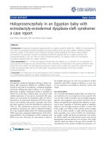
signs of respiratory distress. An echocardiogram revealed
a multiloculated cystic mediastinal mass (Figure 1). A sub-
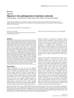
sequent thoracic computed tomography (CT) scan
showed a mainly cystic mass within the anterior mediasti-
num with some solid components in the left lateral
aspect, which was believed to most likely represent a
mediastinal teratoma or a congenital cystic adenomatous
malformation (Figure 2).
The lesion was subsequently completely excised via a
median sternotomy approach and was indeed a multisep-
tated, multicystic mass with solid components. It was
adherent to the thymus and right pleura in the superior
mediastinum, the great vessels and the pericardium, and
traversed the left pleural cavity to the left hemidiaphragm.
The left lung was compressed by the lesion, which was
lying very close to the left phrenic nerve. This was carefully
identified and preserved. Gross and histopathological
examination confirmed the diagnosis of cystic hygroma
(Figure 3).
In the immediate postoperative period, dyspnoea was
observed when feeding. An USS showed paradoxical
movement of the left hemidiaphragm, and a left phrenic
neuropraxia was suspected. This was managed conserva-
tively, and an excellent recovery was made within 3
months. The patient is symptom free at 1 year of age.
Discussion
Redenbacker [4] first described a cystic hygroma in 1828.
It is thought that it results from an early sequestration of
embryonic lymphatic channels. The term 'hygroma'
describes an endothelial-lined mass consisting of small-
to-medium-sized lumina containing lymphatic fluid,
together with a mixture of loose collagen tissue, adipose
tissue and, occasionally, vascular tissue. The cysts may be
unilocular, but more often the structure contains multiple
cysts infiltrating the surrounding structures and distorting
the local anatomy.
Most are found in the cervical region presenting as an
obvious swelling, which can be transilluminated. Medias-
tinal cystic hygromas are usually extensions of cervical
hygromas, as 1% to 2% of cervical cystic hygromas have
mediastinal extensions [5].
Isolated intrathoracic cystic hygroma is a rare finding; less
than 1% of all cystic hygromas are purely mediastinal in
origin. In the majority of cases, these are located in the
anterior mediastinum and reveal themselves after a period
of latency because of their inherent slow growth. Patients
remain completely asymptomatic and so the diagnosis is
Post-natal echocardiogram showing a multiloculated, cystic mass (arrowheads) separately from the heart (arrow)Figure 1
Post-natal echocardiogram showing a multiloculated,
cystic mass (arrowheads) separately from the heart
(arrow).
Journal of Medical Case Reports 2008, 2:256 />Page 3 of 5
(page number not for citation purposes)
not made until adulthood, usually incidentally on routine
plain chest radiography [6]. Larger intrathoracic lesions
tend to envelop neighbouring structures such as the tra-
chea, oesophagus, large blood vessels and heart. In young
infants with larger pure mediastinal cystic hygromas, a
varying degree of respiratory compromise is the most
common presenting symptom, usually secondary to
extrinsic compression of the airway. There may be physi-
cal compression of the lung, as in this case, which may
lead to respiratory distress and possible asphyxia. Rarer
manifestations include dysphagia, superior vena caval
Contrast-enhanced computed tomography scanFigure 2
Contrast-enhanced computed tomography scan. Superiorly, this revealed a mixed attenuation, mainly cystic mass with a
solid component within its left lateral aspect (arrow). More inferiorly, the lesion was of a more fluid density (arrowhead).
These characteristics suggested a teratoma or congenital cystic adenomatoid malformation rather than cystic hygroma as the
working diagnosis.
Gross specimen removed from the patient was confirmed on histopathological testing to be an intrathoracic cystic hygromaFigure 3
Gross specimen removed from the patient was confirmed on histopathological testing to be an intrathoracic
cystic hygroma.
Journal of Medical Case Reports 2008, 2:256 />Page 4 of 5
(page number not for citation purposes)
syndrome, Horner's syndrome, phrenic nerve paresis or
haemoptysis.
A prenatal diagnosis of mediastinal cystic hygromas is dif-
ficult [7]. The usual pathologies considered in the differ-
ential diagnosis of such lesions are listed in Table 1.
Traditionally, ultrasonography has been used as the pri-
mary screening method for prenatal diagnosis, especially
in cervical lesions. CT is avoided because of issues relating
to radiation exposure to mother and foetus. Magnetic res-
onance imaging (MRI) may play a role in prenatal imag-
ing, as amongst other things it provides early
comprehensive information about both the anatomy and
the extension of the tumour [8]. There is, therefore, a case
for advocating an MRI scan for all lesions that are equivo-
cal in prenatal screening.
Our case illustrates the complexities of prenatal diagnosis.
On prenatal sonography, if the stomach is visualized in its
correct anatomical location in the abdomen, any mass in
the left chest is unlikely to be due to a CDH. In the above
case, the stomach was visualized below the diaphragm on
the prenatal ultrasound examination. In what would have
been an atypical presentation, the diagnosis of a CDH was
still entertained because the multicystic morphology of
the foetal chest mass was thought to represent a herniated
intestinal loop through the diaphragm. The mass was
noted to stretch from the superior mediastinum to the
dome of the left hemidiaphragm. The authors appreciate
the fact that the phrenic nerve is not identifiable on pre-
natal ultrasound screening. However, the possibility of
sporadic compression of the left phrenic nerve resulting
from its anatomical location of the lesion, coupled with
the cystic nature of the lesion, would be reason enough for
the lesion to be mistaken for bowel and hence a CDH on
antenatal scanning. On a separate and more general note,
colour Doppler blood flow was not used in prenatal
assessment to evaluate whether the foetal chest mass had
a vascular component. When faced with a mediastinal
mass, we strongly advocate its use to see whether blood
flow is present. It is valuable in narrowing the differential
diagnosis and will help in diagnosing pulmonary seques-
tration or a vascular tumour such as a haemangioma.
Most cases of cystic hygromas are diagnosed post-natally,
and as is borne out in the present case, even this can prove
challenging as many mediastinal cystic masses have simi-
lar appearances on imaging [9]. A diagnostic clue to detec-
tion is mediastinal widening noted on routine chest
radiography, but lesions are better evaluated with a CT
scan [10]. CT is helpful in confirming the cystic nature of
the mass and the anatomical location. It is also particu-
larly useful in planning the potential surgical approach
because of the frequent involvement of local vascular, vis-
ceral and neural structures [9,10]. The most common
characteristic on CT is a well-capsulated, smoothly mar-
ginated and cystic mass, with no evidence of calcification
[11]. However, as this case aptly demonstrates, thoracic
CT may show a complex heterogeneous mass with varied
attenuation values within the lesion [4]. MRI can be used
as a radiation dose-saving modality to demonstrate the
relationship of the mass with surrounding structures or as
an adjunct to other imaging methods. Typically, T1-
weighted magnetic resonance images reveal a mass return-
ing a mainly low-intensity signal, but a faint high-inten-
sity signal may represent mucoid matter within it. T2-
weighted MRI shows a mostly high-intensity signal. How-
ever, a recent series reported various signal characteristics
of such lesions and concluded that the diagnosis of cystic
hygroma on MRI image findings can be difficult [10,12].
We elected not to perform MRI in this case because of the
combination of respiratory distress and the suspicion of a
neoplastic lesion. It was only after the tumour was surgi-
cally excised that the diagnosis of a mediastinal cystic
hygroma was made.
Cervical cystic hygromas have been known to undergo
spontaneous regression because of infection and the asso-
ciated fibrotic process. Similar experience with intratho-
racic lesions has not been observed. The role of non-
surgical therapeutic strategies remains controversial. Aspi-
ration of cysts is fraught with difficulty and incurs a high
risk of recurrence. Sclerotherapy has been used in the
management of cervical cystic hygromas, but there is no
follow-up data documenting long-term success. Notwith-
standing the fact that it is acutely painful, injection of scle-
rosants has not been recommended for the treatment of
isolated mediastinal lesions. Radiotherapy is limited as an
option because of increased risks of thyroid malignancy,
tracheitis, oesophagitis and injury to local neurovascular
tissues. Thus, the only effective treatment of mediastinal
cystic hygroma remains careful surgical excision, which
can be performed in one or more stages. Care should be
taken that the capsule is left intact as rupture predisposes
to incomplete removal and recurrence. Complete excision
may prove hazardous because of adherence of parts of the
Table 1: Differential diagnosis of cystic mediastinal chest masses
Congenital diaphragmatic hernia
Congenital cystic adenomatoid malformation
Cystic hygroma
Pulmonary sequestration
Bronchogenic and neurogenic cysts
Congenital lobar emphysema
Castleman's lymphoma
Thymic cyst or cystic thymoma
Cystic teratoma
Pericardial cyst
Publish with BioMed Central and every
scientist can read your work free of charge
"BioMed Central will be the most significant development for
disseminating the results of biomedical research in our lifetime."
Sir Paul Nurse, Cancer Research UK
Your research papers will be:
available free of charge to the entire biomedical community
peer reviewed and published immediately upon acceptance
cited in PubMed and archived on PubMed Central
yours — you keep the copyright
Submit your manuscript here:
/>BioMedcentral
Journal of Medical Case Reports 2008, 2:256 />Page 5 of 5
(page number not for citation purposes)
tumour to neighbouring vital structures and, therefore, is
not always achievable. In such cases, it is prudent to
deroof the cyst and resect the maximum amount of cyst
wall, rather than risk further harm. The prognosis is
extremely favourable following surgery, although regular
postoperative follow-up is highly recommended. Follow-
up imaging is usually by way of MRI and is performed to
look for rare complications such as infection, local recur-
rence and fistula formation.
Conclusion
In interpreting foetal and neonatal chest masses, medias-
tinal cystic hygroma should be kept in mind as a potential
differential diagnosis. Masses close to the diaphragm may
be problematic to diagnose, particularly if they are cystic.
Even though both prenatal and post-natal imaging is una-
ble to identify the phrenic nerve, the prospect of phrenic
nerve compression should be considered as a possible
consequence of the location of such masses. Therefore,
careful attention should be paid to both the anatomical
site and the organisation of the cystic structure.
Abbreviations
CDH: Congenital diaphragmatic hernia; CT: Computed
tomography; MRI: Magnetic resonance imaging; USS:
Ultrasound scan.
Competing interests
The authors declare that they have no competing interests.
Consent
Written informed consent was obtained from the patient's
next-of-kin for publication of this case report and any
accompanying images. A copy of the written consent is
available for review by the Editor-in-Chief of this journal.
Authors' contributions
SAN liaised with all authors, obtained the radiological
imaging, wrote the paper, rewrote the first draft of the dis-
cussion from the second author and edited the submis-
sion after receiving the reviewers' comments. SAR wrote
the first draft of the discussion. SN obtained the echocar-
diogram image, located the maternal notes from the refer-
ring hospital and edited and proofread the final
submission. WS provided the narrative on the clinical his-
tory and surgical details. CB provided the histopathology
images and reports. KL proofread the first submission and
edited appropriately.
References
1. Bossert T, Gummert JF, Mohr FW: Giant cystic lymphangioma of
the mediastinum. Eur J Cardiothorac Surg 2002, 21:340.
2. Nakazato Y, Ohno Y, Nakata Y, Yamaguchi H, Hazato N, Nagasawa
S, Yokoyama M, Yamada T: Cystic lymphangioma of the medi-
astinum. Am Heart J 1995, 129:406-409.
3. Omell GH, Anderson LS, Bramson RT: Chest wall tumors. Radiol
Clin North Am 1973, 11:197-214.
4. Redenbacker EAH: De Ranula Sublingua, Speciali, cum Casa
Congenita. Monachii, Lindhaue 1828.
5. Glasson MJ, Taylor SF: Cervical, cervicomediastinal and
intrathoracic lymphangioma. Prog Pediatr Surg 1991, 27:62-83.
6. Pike MG, Wood AJ, Corrin S, Warner JO: Intrathoracic extrame-
diastinal cystic hygroma. Arch Dis Child 1984, 59:75-77.
7. Zalel Y, Shalev E, Ben-Ami M, Mogilner G, Weiner E: Ultrasonic
diagnosis of mediastinal cystic hygroma. Prenat Diagn 1992,
12:541-544.
8. Caire JT, Ramus RM, Magee KP, Fullington BK, Ewalt DH, Twickler
DM: MRI of fetal genitourinary anomalies. AJR Am J Roentgenol
2003, 181:1381-1385.
9. Shin MS, Berland LL, Ho KJ: Mediastinal cystic hygromas: CT
characteristics and pathogenetic consideration. J Comput
Assist Tomogr 1985, 9:297-301.
10. Pilla TJ, Wolverson MK, Sundaram M, Heiberg E, Shields JB: CT eval-
uation of cystic lymphangiomas of the mediastinum. Radiology
1982, 144:841-842.
11. Shaffer K, Rosado-de-Christenson ML, Patz EF Jr, Young S, Farver CF:
Thoracic lymphangioma in adults: CT and MR imaging fea-
tures. AJR Am J Roentgenol 1994, 162:283-289.
12. Sigel MJ, Glazer HS, St Aomur TE, Rosenthal DD: Lymphangiomas
in children: MR imaging. Radiology 1989, 170:
467-470.