Báo cáo y học: " Use of a Javid™ shunt in the management of axillary artery injury as a complication of fracture of the surgical neck of the humerus: a case report" ppsx
Bạn đang xem bản rút gọn của tài liệu. Xem và tải ngay bản đầy đủ của tài liệu tại đây (848.47 KB, 4 trang )
BioMed Central
Page 1 of 4
(page number not for citation purposes)
Journal of Medical Case Reports
Open Access
Case report
Use of a Javid™ shunt in the management of axillary artery injury as
a complication of fracture of the surgical neck of the humerus: a
case report
Stuart A Suttie*
1
, Reza Mofidi
2
, Alison Howd
2
and Gareth D Griffiths
2
Address:
1
Department of Surgery and Molecular Oncology, Ninewells Hospital and Medical School, Dundee DD1 9SY, UK and
2
Department of
Vascular Surgery, Ninewells Hospital, Dundee DD1 9SY, UK
Email: Stuart A Suttie* - ; Reza Mofidi - ; Alison Howd - ;
Gareth D Griffiths -
* Corresponding author
Abstract
Introduction: Axillary artery injury is a rare but severe complication of fractures of the surgical
neck of the humerus.
Case presentation: We present a case of axillary artery pseudoaneurysm secondary to such a
fracture, in a 82-year-old white woman, presenting 10 weeks after the initial injury, successfully
treated with subclavian to brachial reversed vein bypass together with simultaneous open
reduction and internal fixation of the fracture. We discuss the use of a Javid™ shunt during
combined upper limb revascularisation and open reduction and internal fixation of the fractured
humerus.
Conclusion: This case highlights the usefulness of a Javid™ shunt, over other forms of vascular
shunts, in prompt restoration of blood flow to effect limb salvage. It can be considered as a
temporary measure whilst awaiting definitive revascularisation which can be performed following
fracture fixation.
Introduction
Proximal humeral fractures are a common injury with an
incidence of approximately 5% of all fractures, with the
majority being secondary to blunt trauma in an elderly
population [1]. Despite the close proximity of the axillary
artery and the surgical neck of humerus, injury to this
artery is a rare complication of proximal humeral frac-
tures. It is, however, associated with significant risks to
both function and viability of the affected upper limb.
Upper limb ischaemia secondary to such a cause requires
prompt intervention to restore blood flow and subse-
quently treat the primary cause. Earlier reports have docu-
mented success in similar settings, using modified
equipment not necessarily designed for use as an intravas-
cular shunt [2,3].
We present a case of delayed presentation of axillary artery
pseudoaneurysm following proximal humeral fracture
and discuss the use of a Javid™ carotid shunt (Bard carotid
shunt, 17F tapered to 10F; Bard
®
Javid™ Carotid Shunts,
Bard Ltd., Forest House, Brighton Rd., Crawley, West Sus-
sex, UK) in maintaining vascular perfusion during open
reduction and internal fixation of the fracture.
Published: 5 August 2008
Journal of Medical Case Reports 2008, 2:259 doi:10.1186/1752-1947-2-259
Received: 29 April 2008
Accepted: 5 August 2008
This article is available from: />© 2008 Suttie et al; licensee BioMed Central Ltd.
This is an Open Access article distributed under the terms of the Creative Commons Attribution License ( />),
which permits unrestricted use, distribution, and reproduction in any medium, provided the original work is properly cited.
Journal of Medical Case Reports 2008, 2:259 />Page 2 of 4
(page number not for citation purposes)
Case presentation
An 82-year-old, white woman with a history of alcohol
abuse, presented to the accident and emergency depart-
ment with a 4-hour history of an acutely ischaemic right
upper limb with motor and sensory deficit. A hard tender,
pulsatile mass was palpable in the right subclavian area
with significant bruising; there was a palpable right sub-
clavian pulse with no pulses distal to this. X-ray revealed
a fracture of the surgical neck of the right humerus with
the humeral head abducted and externally rotated, while
the humeral shaft was displaced medially (Fig. 1).
Ten weeks previously, she had presented with a fracture of
the surgical neck of the right humerus following a fall
whilst under the influence of alcohol. On that occasion,
sensory and motor function of the limb had been
recorded to be fully intact by the medical staff in Accident
and Emergency and there had been a full complement of
pulses. Given she had no neuro-vascular deficit in the
affected limb, the vascular surgeons were not involved ini-
tially. Under guidance of the orthopaedic surgeons, she
had been treated conservatively with a collar and cuff due
to her age and history of current alcohol abuse. She was to
have been followed up fortnightly in the orthopaedic frac-
ture clinic – but failed to attend after her second visit. She
had no neuro-vascular deficit on follow-up. She denied
any further falls or trauma to the right upper limb.
The acute nature of the current presentation together with
neurological compromise prompted classification as cate-
gory-II acute limb ischaemia (Society for Vascular Surgery/
International Society for Cardiovascular Surgery classifica-
tion) [4] and urgent angiography was performed with a
view to revascularisation. This revealed a pseudoaneu-
rysm of the third part of the right axillary artery with com-
plete occlusion of the right brachial artery distal to this
(Fig. 2).
Operative treatment was undertaken with initial exposure
and control of the subclavian artery above the clavicle
(Fig. 3A). Simultaneous exposure of the brachial artery in
the antecubital fossa was performed and a size 3 Fogarty
embolectomy catheter passed distally down the brachial
artery. Both radial and ulnar arteries were found to con-
tain thrombus which was cleared with good back flow.
The proximal brachial and distal subclavian arteries were
ligated in continuity. Two interconnected Javid™ shunts
were inserted to carry blood flow from the subclavian to
the brachial artery in order to maintain perfusion (Fig. 3B)
during open reduction and internal fixation of the frac-
tured humerus, after which a subclavian to brachial
bypass was performed using reversed long saphenous
vein. The fracture was temporarily stabilised using exter-
nal splints to immobilize the limb whilst securing vascu-
lar continuity.
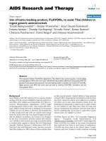
Anteroposterior view of right shoulder 10 weeks after the primary injury, revealing malalignment of fracture ends and attempts at formation of primary callus (arrow)Figure 1
Anteroposterior view of right shoulder 10 weeks
after the primary injury, revealing malalignment of
fracture ends and attempts at formation of primary
callus (arrow).
Catheter angiogram depicting pseudoaneurysm formation of third part of axillary artery with complete occlusion of the distal right brachial arteryFigure 2
Catheter angiogram depicting pseudoaneurysm for-
mation of third part of axillary artery with complete
occlusion of the distal right brachial artery.
Journal of Medical Case Reports 2008, 2:259 />Page 3 of 4
(page number not for citation purposes)
Postoperatively, the patient had strong radial and ulnar
pulses with complete resolution of her motor and sensory
dysfunction within 72 hours. Her postoperative course
was uncomplicated and she was discharged on the 10th
postoperative day. Early postoperative duplex scan per-
formed at 6 weeks revealed satisfactory function of the
vein graft.
Discussion
Despite the fact that a significant proportion of fractures
of the surgical neck of the humerus are displaced, axillary
artery injuries secondary to these fractures are rare [1,4-7].
The majority affect the third part of the artery, due to its
position of relative immobility, being tethered by the sub-
scapular and thoracromial arteries [1,8]. Most of these
injuries lead to thrombosis of the axillary artery and acute
lower limb ischaemia [4,5,9]. Pseudoaneurysm formation
of the axillary artery is rare following blunt and penetrat-
ing trauma to the shoulder, often presenting late as a pul-
satile mass rather than acute limb ischaemia [1,6,7,10].
Endovascular treatment with a covered stent graft has
been reported previously and is the treatment of choice in
patients with pseudoaneurysm of the axillary artery with-
out upper limb ischaemia [7,11]. Due to the presence of
propagating thrombus and displaced fracture requiring
open reduction and internal fixation, endovascular treat-
ment was not an option in this patient. Following proxi-
mal and distal arterial control and thrombectomy, the
limb was revascularised temporarily using a Javid™ shunt,
which allowed safe internal fixation of the fracture before
bypass grafting. The insertion of the Javid™ shunt served
to confirm the viability of the limb and adequacy of distal
thrombo-embolectomy. The use of temporary shunting of
peripheral vasculature in order to maintain distal vascular
perfusion is rarely employed in civilian surgical practice
[2,3], however, it has been gaining popularity in the man-
agement of military trauma [12-14]. Recent reports from
Belfast, whereupon the use of intraluminal shunts has
been advocated for the early restoration of blood flow fol-
lowing complex lower limb vascular injuries, have shown
significant benefits in averting the incidence of fasciot-
omy, contractures, ischaemic nerve palsy and amputa-
tions [15]. This Belfast approach of early shunting allows
for a disciplined surgical approach with adequate time for
wound debridement, safe fracture fixation and optimal
vascular reconstruction. Reports from Operation Iraqi
Freedom suggest that vascular shunts can be used safely to
bypass complex vascular injuries encountered in forward
surgical units, in order to allow transfer of injured patients
for definitive vascular assessment and reconstruction
[12,14]. The use of vascular shunts in these circumstances
was associated with very low limb amputation rates [14],
even in patients in whom the shunt had thrombosed in
transit [12].
The Javid™ shunt has the advantage over other types of
non-vascular shunt employed [2,3], in that it is specifi-
cally designed for use as a carotid artery shunt. It is man-
ufactured out of soft, kink free material, which is tapered
towards the ends which are bulbous in nature. This allows
the shunt to be clamped in place around the artery,
thereby providing stability whilst surgery continues. It was
felt that the Javid™ shunt was superior to the Pruitt-Ina-
hara
®
carotid shunt (an H-shaped carotid shunt, held in
place using inflatable balloons) for this patient due to its
ease of use, lack of extra lumens (which would easily be
Supraclavicular exposure of the subclavian arteryFigure 3
Supraclavicular exposure of the subclavian artery. (A)
The phrenic nerve is retracted before the division of the sca-
lenus anterior muscle. (B) The subclavian artery is exposed
and ligated distally, with blood flow to the right arm being
maintained with the aid of a Javid shunt during open reduc-
tion and internal fixation of the fracture.
(A)
(B)
Publish with BioMed Central and every
scientist can read your work free of charge
"BioMed Central will be the most significant development for
disseminating the results of biomedical research in our lifetime."
Sir Paul Nurse, Cancer Research UK
Your research papers will be:
available free of charge to the entire biomedical community
peer reviewed and published immediately upon acceptance
cited in PubMed and archived on PubMed Central
yours — you keep the copyright
Submit your manuscript here:
/>BioMedcentral
Journal of Medical Case Reports 2008, 2:259 />Page 4 of 4
(page number not for citation purposes)
caught and cause the shunt to be dislodged), ability to
interconnect two shunts and its specially designed clamps
to hold the shunt in situ during extensive and vigorous
mobilisation of the fractured bone during reduction and
fixation. Although these shunt clamps may cause more
damage to the arterial lumen than the balloon of the
Pruitt-Inahara
®
shunt, this damaged segment of the
injured artery would in turn be ligated and bypassed.
Conclusion
This case highlights the usefulness of a Javid™ shunt, over
other forms of shunt, in prompt restoration of blood flow
to effect limb salvage. It can be considered as a temporary
measure whilst awaiting definitive revascularisation
which can be performed following fracture fixation.
Competing interests
The authors declare that they have no competing interests.
Authors' contributions
SAS was first assistant (subclavian exposure), carried out
the literature review and constructed the manuscript. RM
was first assistant (brachial exposure), photographer, car-
ried out the literature review and drafted and editing the
manuscript. AH was primary surgeon (brachial exposure),
constructed the idea behind the case report, was senior
editor of the manuscript (critical revisions) and gave final
approval. GDG was primary surgeon (subclavian expo-
sure), constructed the idea behind the case report, was
senior editor of the manuscript (critical revisions) and
gave final approval.
Consent
Written informed consent was obtained retrospectively
from the patient for publication of this case report and
accompanying images. A copy of the written consent is
available for review by the Editor-in-Chief of this journal.
Acknowledgements
Written on behalf of the East of Scotland Vascular Network.
References
1. Yagubyan M, Panneton JM: Axillary artery injury from humeral
neck fracture: A rare but disabling traumatic event. Vasc
Endovascular Surgery 2004, 38:175-184.
2. Husain AK, Khandeparkar JM, Tendolkar AG, Magotra RA, Parulkar
GB: Temporary intravascular shunts for peripheral vascular
trauma. J Postgrad Med 1992, 38(2):68-69.
3. Sriussadaporn S, Pak-art R: Temporary intravascular shunt in
complex extremity vascular injuries. J Trauma 2002,
52:1129-1133.
4. Rutherford RB, Baker JD, Ernst C, Johnston KW, Porter JM, Ahn S,
Jones DN: Recommended standards for reports dealing with
lower extremity ischemia: Revised version. J Vasc Surg 1997,
26(3):517-538.
5. Jensen BV, Jacobsen J, Andreasen H: Late appearance of arterial
injury caused by fracture of the neck of humerus. J Trauma
1987, 27:1368-1369.
6. Syed AA, Williams HR: Shoulder disarticulation: a sequel of vas-
cular injury secondary to a proximal humeral fracture. Injury
2002, 33:771-774.
7. Stahnke M, Duddy MJ: Endovascular repair of a traumatic axil-
lary artery pseudo aneurysm following anterior shoulder dis-
location. Cardiovasc Intervent Radiol 2006, 29:298-301.
8. Drury K, Scullion JE: Vascular complications of anterior disloca-
tion of the shoulder. Br J Surg 1980, 67:579-581.
9. Puri R, Clark J, Corkery PH: Axillary artery damage following a
closed fracture of the neck of humerus: a case report. Injury
1995, 16:426-427.
10. Van Arkel ERA, Tordoir JHM, Arenms HJ: A proximal humeral
fracture, complicated by a pseudo aneurysm: a case report.
Acta Orthop Scand 1998, 69:194-195.
11. Oktar GL, Balkan ME, Akpek S, Ilqit E: Endovascular stent-graft
placement for the management of a traumatic axillary
artery pseudoaneurysm: a case report. Vasc Endovascular Surg
2002, 36:323-326.
12. Chambers LW, Green DJ, Sample K, Gillingham BL, Rhee P, Brown C,
Narine N, Uecker JM, Bohman HR: Tactical surgical intervention
with temporary shunting of peripheral vascular trauma sus-
tained during Operation Iraqi Freedom: one unit's experi-
ence. J Trauma 2006, 61(4):824-830.
13. Fox CJ, Gillespie DL, O'Donnell SD, Rasmussen TE, Goff JM, Johnson
CA, Galgon RE, Sarac TP, Rich NM: Contemporary management
of wartime vascular trauma. J Vasc Surg 2005, 41(4):638-644.
14. Clouse WD, Rasmussen TE, Peck MA, Eliason JL, Cox MW, Bowser
AN, Jenkins DH, Smith DL, Rich NM: In-theater management of
vascular injury: 2 years of the Balad Vascular Registry. J Am
Coll Surg 2007, 204(4):625-632.
15. Barros D'Sa AAB, Harkin DW, Blair PHB, Hood JM, McIlrath E: The
Belfast approach to managing complex lower limb vascular
injuries. Eur J Vasc Endovasc Surg 2006, 32(3):246-256.