Báo cáo y học: " Allergic enterocolitis and protein-losing enteropathy as the presentations of manganese leak from an ingested disk battery: A case report" pps
Bạn đang xem bản rút gọn của tài liệu. Xem và tải ngay bản đầy đủ của tài liệu tại đây (677.7 KB, 3 trang )
BioMed Central
Page 1 of 3
(page number not for citation purposes)
Journal of Medical Case Reports
Open Access
Case report
Allergic enterocolitis and protein-losing enteropathy as the
presentations of manganese leak from an ingested disk battery: A
case report
Muhammad A Altaf, Praveen S Goday and Grzegorz Telega*
Address: Division of Pediatric Gastroenterology and Nutrition, Department of Pediatrics, The Medical College of Wisconsin, Watertown Plank
Road, Milwaukee, WI 53221, USA
Email: Muhammad A Altaf - ; Praveen S Goday - ; Grzegorz Telega* -
* Corresponding author
Abstract
Introduction: Disk battery ingestions can lead to serious complications including airway or
digestive tract perforation, blood vessel erosions, mediastinitis, and stricture formation.
Case presentation: We report a 20-month-old Caucasian child who developed eosinophilic
enterocolitis and subsequent protein-losing enteropathy from manganese that leaked from a
lithium disk battery. The disk battery was impacted in her esophagus for 10 days resulting in battery
corrosion. We postulate that this patient's symptoms were due to a manganese leak from the
'retained' disk battery; this resulted in an allergic response in her gut and protein-losing
enteropathy. Her symptoms improved gradually over the next 2 weeks with conservative
management.
Conclusion: This is the first case report to highlight the potential complication of allergic
enterocolitis and protein-losing enteropathy secondary to ingested manganese. Clinicians should
be vigilant about this rare complication in managing patients with disk battery ingestions.
Introduction
Lithium batteries are used in many portable consumer
electronic devices (Fig. 1). The most common type of lith-
ium cell used in consumer applications consists of lith-
ium and manganese (Mn). Disk battery ingestions can
lead to serious complications including aerodigestive tract
perforation, vessel erosion, mediastinitis, and stricture
formation [1]. Mercury batteries have been reported to
cause more severe complications including acute poison-
ing [2], but none of the disk batteries have been reported
to cause protein losing-enteropathy. We report a case in
which the manganese in a lithium-manganese disk battery
impacted in the esophagus presumably led to eosi-
nophilic enterocolitis and severe protein-losing enteropa-
thy.
Case presentation
A 20-month-old Caucasian child presented with a 10-day
history of vomiting and solid food refusal. Her chest X-ray
showed a disk battery impaction in the upper esophagus.
A corroded lithium-manganese battery was retrieved with
a flexible laryngoscope 10 days after ingestion. The patient
was transferred to our institution for further monitoring.
Her physical examination and laboratory tests on admis-
sion were normal, except for an albumin of 2.7 g/dL (nor-
mal 3.8 to 5.4 g/dL) which had dropped from 4.3 g/dL on
Published: 27 August 2008
Journal of Medical Case Reports 2008, 2:286 doi:10.1186/1752-1947-2-286
Received: 13 December 2007
Accepted: 27 August 2008
This article is available from: />© 2008 Altaf et al; licensee BioMed Central Ltd.
This is an Open Access article distributed under the terms of the Creative Commons Attribution License ( />),
which permits unrestricted use, distribution, and reproduction in any medium, provided the original work is properly cited.
Journal of Medical Case Reports 2008, 2:286 />Page 2 of 3
(page number not for citation purposes)
the day of battery removal. She had been on a regular diet
until 10 days before admission. An esophagogram
revealed no perforations.
A week following removal of the battery, she continued to
refuse foods. An upper endoscopy was performed that
revealed non-circumferential ulceration in the upper
esophagus but no biopsies were performed at that time.
Her stomach and duodenum were grossly normal at that
time. A nasogastric (NG) tube was placed and feeding was
started. Subsequently, the protein loss worsened and her
serum albumin dropped to 1.1 g/dL (normal 3.8 to 5.4 g/
dL). The urinalysis was normal and fecal alpha-1 antit-
rypsin level was 464 mg/g (normal <2 mg/g dry stool). A
computed tomography (CT) scan of the abdomen, chest,
and pelvis was performed because of persistent abdomi-
nal distention and feeding intolerance; this showed bilat-
eral pleural effusions and moderate ascites. She had a
normal echocardiogram and liver function tests. She was
diagnosed with protein-losing enteropathy. Albumin
25% was started to maintain albumin at a level of >2 g/dL.
A colonoscopy and repeat endoscopy were performed due
to her protein-losing enteropathy; this showed complete
healing of the previous esophageal ulceration, but with
findings of diffuse enteritis and colitis. Small bowel biop-
sies were taken from the duodenum and terminal ileum.
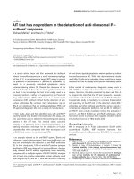
Histology revealed mild eosinophilic esophagitis and
moderate eosinophilic enterocolitis (Fig. 2). In addition,
the patient developed peripheral eosinophilia of 10.5%
compared to 2.9% on admission. Her symptoms
improved, and her albumin and eosinophilia normalized
gradually over the next 2 weeks with conservative support
and no steroids.
At 6 months follow-up, the patient has remained well,
with normal albumin levels and no symptoms of protein-
losing enteropathy. She developed an upper esophageal
stricture that required recurrent dilatation and steroid
injections. Her most recent endoscopic biopsies showed
moderate eosinophils in the esophageal mucosa. We pos-
tulate that this patient's symptoms were due to a manga-
nese leak from the 'retained' corroded disk battery before
or during the process of removal from the esophagus; this
caused an allergic response in her gut resulting in a pro-
tein-losing enteropathy.
Discussion
The best known manifestations of chronic Mn exposure
are neurological symptoms such as hypokinesia, rigidity
and tremor that resemble Parkinson's disease [3]. Rarely,
allergic responses have been described as well. Metal
allergy to stainless steel wire containing Mn has been
reported after coronary artery bypass grafting. A refractory
pruritic erythematous wheal over the body with positive
Mn patch testing and peripheral eosinophilia proved this
to be a systemic allergic reaction to Mn [4]. Mn used in the
manufacture of dental prosthesis has also been reported
to cause contact dermatitis, manifested by diffuse oral
edema, erythema and ulcerations; this was confirmed by
positive patch testing [5,6].
Epidemiological studies have reported an acute impact of
particulate Mn on the pulmonary system, including
reversible decrement of pulmonary functions and increase
Lithium disk batteryFigure 1
Lithium disk battery.
Colonoscopic biopsy showing eosinophilic infiltration of crypts in transverse colonFigure 2
Colonoscopic biopsy showing eosinophilic infiltration
of crypts in transverse colon.
Journal of Medical Case Reports 2008, 2:286 />Page 3 of 3
(page number not for citation purposes)
in bronchial hyperreactivity [7,8]. In children, peak expir-
atory flow was decreased with a high concentration of Mn
in the air, suggesting an obstructive allergic response
rather than restrictive airway disease [9]. Exposure to high
inhaled Mn concentrations has demonstrated an
increased incidence of cough, rhinitis, bronchitis, and
pneumonitis [10]. A study in rhesus monkeys docu-
mented subacute bronchiolitis and alveolar duct inflam-
mation with lymphocytes, neutrophils, and a few
eosinophils following inhalational exposure to Mn [11].
In general, serum or blood Mn does not serve as a reliable
indicator of the total body burden of Mn because of its
intracellular distribution and relatively short half-life
[12].
We postulate that our patient developed an allergic ente-
rocolitis and protein losing enteropathy in response to the
Mn exposure in the gastrointestinal (GI) tract. The
ingested battery was composed of lithium perchlorate and
manganese dioxide. The possibility that some other com-
ponent of the battery could have contributed to the patho-
genesis cannot be ruled out, but in the literature, Mn is the
only constituent that has been attributed to the allergic
responses. This is supported by the previously suggested
evidence that Mn can cause rhinitis, pneumonitis, and
bronchial hyperreactivity. Manganese exposure from a
cardiac stenting wire and dental prosthesis has also caused
allergic symptoms with peripheral eosinophilia. Our
patient's most recent esophageal biopsies suggest that she
either had a baseline mild asymptomatic eosinophilic
esophagitis that acutely worsened with exposure to Mn or
the Mn was a trigger to her eosinophilic esophagitis.
Conclusion
This case shows strong circumstantial evidence that the
eosinophilic enterocolitis and protein-losing enteropathy
were caused by the Mn leak from a retained disk battery;
she was completely asymptomatic before battery inges-
tion with normal albumin levels and eosinophil counts
before battery removal. Additionally, there was complete
resolution without any treatment aside from removal of
the Mn-containing disk battery. Clinicians should be vig-
ilant about this rare complication while managing chil-
dren with ingested disk batteries as symptoms might not
appear immediately after battery removal.
Abbreviations
CT: Computed Tomography; GI: Gastrointestinal; Mn:
Manganese; NG: Nasogastric.
Competing interests
The authors declare that they have no competing interests.
Authors' contributions
All authors (MAA, PSG, GT) contributed in the manage-
ment of the patient, writing of the manuscript and review-
ing of the literature. All authors read and approved the
final manuscript.
Consent
Written informed consent was obtained from the parent
for publication of this case report, as the child was a
minor. A copy of the written consent is available for
review by the Editor-in-Chief of this journal.
References
1. Temple DM, McNeese MC: Hazards of battery ingestion. Pediat-
rics 1983, 71(1):100-103.
2. Reilly DT: Mercury battery ingestion. Br Med J 1979,
1(6167):859.
3. Pal PK, Samii A, Calne DB: Manganese neurotoxicity: a review of
clinical features, imaging and pathology. Neurotoxicology 1999,
20(2–3):227-238.
4. Takazawa K, Ishikawa N, Miyagawa H, Yamamoto T, Hariya A, Dohi
S: Metal allergy to stainless steel wire after coronary artery
bypass grafting. J Artif Organs 2003, 6(1):71-72.
5. Pardo J, Rodriguez-Serna M, De La Cuadra J, Fortea JM: Allergic
contact stomatitis due to manganese in a dental prosthesis.
Contact Dermatitis 2004, 50(1):41.
6. Menezes LM, Campos LC, Quintao CC, Bolognese AM: Hypersen-
sitivity to metals in orthodontics. Am J Orthod Dentofacial Orthop
2004, 126(1):58-64.
7. Boezen M, Schouten J, Rijcken B, Vonk J, Gerritsen J, Zee S van der,
Hoek G, Brunekreef B, Postma D: Peak expiratory flow variabil-
ity, bronchial responsiveness, and susceptibility to ambient
air pollution in adults. Am J Respir Crit Care Med 1998,
158(6):1848-1854.
8. Sharma M, Kumar VN, Katiyar SK, Sharma R, Shukla BP, Sengupta B:
Effects of particulate air pollution on the respiratory health
of subjects who live in three areas in Kanpur, India. Arch Envi-
ron Health 2004, 59(7):348-358.
9. Hong YC, Hwang SS, Kim JH, Lee KH, Lee HJ, Lee KH, Yu SD, Kim
DS: Metals in particulate pollutants affect peak expiratory
flow of school children. Environ Health Perspect 2007,
115(3):430-434.
10. Roels H, Lauwerys R, Buchet JP, Genet P, Sarhan MJ, Hanotiau I, de
Fays M, Bernard A, Stanescu D: Epidemiological survey among
workers exposed to manganese: effects on lung, central
nervous system, and some biological indices. Am J Ind Med
1987, 11(3):307-327.
11. Dorman DC, Struve MF, Gross EA, Wong BA, Howroyd PC: Sub-
chronic inhalation of high concentrations of manganese sul-
fate induces lower airway pathology in rhesus monkeys.
Respir Res 2005, 6:121.
12. Lu L, Zhang LL, Li GJ, Guo W, Liang W, Zheng W: Alteration of
serum concentrations of manganese, iron, ferritin, and
transferrin receptor following exposure to welding fumes
among career welders. Neurotoxicology 2005, 26(2):257-265.