Báo cáo y học: " Broad ligament cystic lymphangioma: A case report" doc
Bạn đang xem bản rút gọn của tài liệu. Xem và tải ngay bản đầy đủ của tài liệu tại đây (1.25 MB, 3 trang )
BioMed Central
Page 1 of 3
(page number not for citation purposes)
Journal of Medical Case Reports
Open Access
Case report
Broad ligament cystic lymphangioma: A case report
K Harish*
1,2
, SR Karthik
3
and CS Manjunath
4
Address:
1
Department of Surgical Oncology, MS Ramaiah Medical College and Hospital, Bangalore 560054, India,
2
Bangalore Institute of
Oncology, Raja Ram Mohan Roy Extension, Bangalore 560027, India,
3
Department of Oncology, MS Ramaiah Medical College and Hospital,
Bangalore 560054, India and
4
Gokula Metropolis Lab, MS Ramaiah Memorial Hospital, Bangalore 560054, India
Email: K Harish* - ; SR Karthik - ; CS Manjunath -
* Corresponding author
Abstract
Introduction: Cystic lymphangiomas are uncommon tumors that can arise from any part of the
body. They can pose a diagnostic and therapeutic challenge. They are more common in infants and
children than adults. Broad ligament cystic lymphangioma is extremely rare.
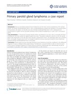
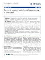
Case presentation: A 70-year-old multiparous woman presented with an abdominal mass of 20-
year duration. A large cystic swelling was detected on computed tomography scan that was found
to arise from the left adnexal region. This 19 kg lesion was found arising from the broad ligament.
It was successfully removed. A detailed pathological study, including immunohistochemistry, was
required to diagnose the lesion as a cystic lymphangioma.
Conclusion: Lymphangiomas should be treated with total surgical excision. Broad ligament
lymphangiomas are extremely rare but must be considered as a differential diagnosis of cystic
lesions in that region.
Introduction
Cystic lymphangiomas are common in infants and chil-
dren, but adult cystic lymphangiomas are rare. Although
they can occur at any site in the body, cystic lymphangi-
omas of the broad ligament are extremely rare [1].
Case presentation
A 70-year-old multiparous woman presented with a huge
abdominal swelling of 20-year duration. The patient had
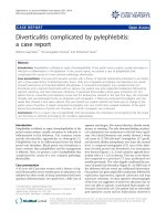
been unable to walk for 3 months. A computed tomogra-
phy (CT) scan revealed a huge cystic swelling in the abdo-
men, possibly arising from the left adnexal region (Figure
1). After presurgical workup, the patient underwent an
exploratory laparotomy. A large cystic mass was found
occupying the entire abdomen. The lower limit of the
mass was in close relation and adherent to the uterus on
its left side. The left ovary and fallopian tube were not sep-
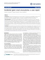
arately visualized. The surgery performed included pan-
hysterectomy and right salpingo-oophorectomy along
with the excision of the cystic mass (Figure 2). The tumor
weighed 19 kg. The postoperative period was uneventful.
Pathological gross findings were those of a very large mul-
tiseptate cystic lesion covered with serosa. Microscopy
revealed that the cyst wall had bundles of smooth muscle
with connective tissue. In addition, the cyst wall was lined
internally with epithelium (Figure 3). This tumor, arising
from the left broad ligament, was found to be benign.
Although the uterus showed multiple leiomyomata, in
view of the epithelial lining, cystic degeneration of leio-
myoma was considered unlikely. On immunohistochem-
ical study, the tumor cyst wall stained positively with the
Published: 23 September 2008
Journal of Medical Case Reports 2008, 2:310 doi:10.1186/1752-1947-2-310
Received: 16 January 2008
Accepted: 23 September 2008
This article is available from: />© 2008 Harish et al; licensee BioMed Central Ltd.
This is an Open Access article distributed under the terms of the Creative Commons Attribution License ( />),
which permits unrestricted use, distribution, and reproduction in any medium, provided the original work is properly cited.
Journal of Medical Case Reports 2008, 2:310 />Page 2 of 3
(page number not for citation purposes)
lymphatic marker D2-40 (Figure 3, right panel); hence,
the tumor was diagnosed as cystic lymphangioma. The
patient has had regular follow-up for 2 years, and there
have been no signs of recurrence.
Discussion
Lymphangiomas are benign tumors of the lymphatic sys-
tem. They are classified as cavernous, lymphangioma sim-
plex, or cystic lymphangioma [2]. Cystic lymphangioma,
first described in 1828 by Redenbacker, is a malformation
of the lymphatic system. It can affect any site in the body
but is seen more commonly in the head and neck region
and the axilla. It is also reported to occur in the mediasti-
num, retroperitoneum, and other regions [2,3]. Cystic
lymphangiomas most commonly affect children. About
90% of these lymphangiomas manifest before 2 years of
age and are very rarely encountered in adults [3]. The
reported patient was aged 70 years at presentation.
Lymphangiomas in children are considered to arise from
sequestered lymphatic sacs that fail to communicate with
the draining lymphatic channels. This is a widely accepted
theory. However, the etiology in the adult population is
controversial. Some authors believe that the adult mani-
festations are a result of delayed proliferation of congeni-
tal or acquired lymphoid nests after stimuli such as
respiratory infection or local trauma [4]. Others dispute
the congenital origin and propose that adult cystic lym-
phangiomas arise as a result of trauma alone [5]. There
was no history of trauma in this patient.
Radiographic evaluation with magnetic resonance imag-
ing or CT is invaluable for the diagnosis and determina-
tion of the extent of the lesion. In addition, it is essential
in defining normal anatomical structures that need to be
preserved when surgical excision is performed [4]. Accu-
rate pre-operative diagnosis of cystic lymphangioma is
uncommon [6] and was a problem faced in the reported
case. The CT scan revealed a huge cystic lesion, but identi-
fication of the site of origin and diagnosis were not possi-
ble.
Lymphangiomas are treated by surgical excision. Com-
plete excision of the mass with negative surgical margins
is the optimal treatment, and the results are excellent [7].
Intra-abdominal lymphangiomas have a 10% postopera-
tive recurrence rate for incompletely excised lesions. We
were able to achieve total excision of the cyst.
Effective immunohistochemical markers specific for lym-
phatic endothelial cells have been reported including lym-
phatic vessel endothelial receptor 1, vascular endothelial
growth factor receptor 3, and Prox-1. However, the anti-
bodies against these markers are available only for frozen
section specimens. More recently, a new monoclonal anti-
body, D2-40, has become available; this is a specific
marker of lymphatic endothelium, since it does not stain
vascular endothelium [8]. In this case, diagnosis was
made only after immunostaining with the lymphatic
marker D2-40.
Computed tomography scan of the abdomen and pelvis showing a large cystic mass occupying the entire abdomen and pelvisFigure 1
Computed tomography scan of the abdomen and
pelvis showing a large cystic mass occupying the
entire abdomen and pelvis.
Excision of the tumorFigure 2
Excision of the tumor. Note that the origin is from the
left broad ligament. The uterus is indicated with a bold white
arrow. The inset shows the entire tumor, measuring 47 cm
across.
Journal of Medical Case Reports 2008, 2:310 />Page 3 of 3
(page number not for citation purposes)
We could only find one case of broad ligament cystic lym-
phangioma reported in the literature [1]. The case pre-
sented here is probably only the second case of broad
ligament lymphangioma to be reported. The lesion
weighed 19 kg and is probably one of the largest to be
reported. Malignant transformations of cysts are rare and
have been reported only once [9]. Such a transformation
is an exception rather than a rule.
Conclusion
Adult cystic lymphangiomas of the broad ligament are
very rare benign tumors. Total surgical removal is the
treatment of choice. This is a report of one such case diag-
nosed with the help of the lymphatic marker D2-40,
treated successfully with surgery, and recurrence-free 2
years later. Although rare, cystic lymphangiomas must be
considered in the differential diagnosis of cystic lesions in
the abdomen and pelvis.
Abbreviations
CT: computed tomography.
Competing interests
The authors declare that they have no competing interests.
Authors' contributions
KH contributed to the conception, design, gathering data,
and revision of the manuscript draft. SRK contributed to
obtaining the data and drafting the manuscript. CSM con-
tributed to obtaining the data, the pathological review,
and drafted part of the manuscript. All authors read and
approved the final manuscript.
Consent
Written informed consent was obtained from the patient
for publication of this case report and any accompanying
images. A copy of the written consent is available for
review by the Editor-in-Chief of this journal.
Acknowledgements
The authors would like to thank Dr Dhanpat Jain, MD, Associate Professor
of Pathology, Yale New Haven Hospital Department of Pathology, for
reviewing the case and pathology.
References
1. Koo J, Joo H, Lee D, Jeon J, Lee J, Lee I: A case of cystic lymphangi-
oma of the broad ligaments. Korean J Obstet Gynecol 2002,
45:2312-2314.
2. Yildirim E, Dural K, Kaplan T, Sakinci U: Cystic lymphangioma:
report of two atypical cases. Interact Cardiovasc Thorac Surg 2004,
3:63-65.
3. Gelal F, Yucel K, Tugsel E, Guney S: Axillary cystic lymphangioma
presenting in pregnancy. Turk J Med Sci 1998, 28:571-572.
4. Lev S, Lev MH: Imaging of cystic lesions. Radiol Clin North Am
2000, 38:1013-1027.
5. Antoniades K, Kiziridou A, Psimopoulou M: Traumatic cervical
cystic hygroma. Int J Oral Maxillofac Surg 2000, 29:47-48.
6. Hornick JL, Fletcher CD: Intraabdominal cystic lymphangiomas
obscured by marked superimposed reactive changes: clinico-
pathological analysis of a series. Hum Pathol 2005, 36:426-432.
7. Guinier D, Denue PO, Mantion GA: Intra-abdominal cystic lym-
phangioma. Am J Surg 2006, 191:706-707.
8. Yonemura Y, Endou Y, Tabachi K, Kawamura T, Yun HY, Kameya T,
Hayashi I, Bandou E, Sasaki T, Miura M: Evaluation of lymphatic
invasion in primary gastric cancer by a new monoclonal anti-
body, D2-40. Hum Pathol 2006, 37:1193-1199.
9. Bury TF, Pricolo VE: Malignant transformation of benign
mesenteric cyst. Am J Gastroenterol 1994, 89:2085-2087.
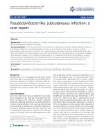
Histology and immunohistochemistryFigure 3
Histology and immunohistochemistry. The left panel shows a microphotograph (magnification ×400, hematoxylin and
eosin stain) showing smooth muscle in the wall of the cyst, indicated by a white bold arrow, and flattened endothelial-like cells
lining the cyst wall, indicated by a bold black arrow. The right panel shows immunohistochemistry by marker D2-40 identifying
the lymphatic endothelium, indicated by bold black arrows.