Báo cáo y học: " Oral cavity metastasis of renal cell carcinoma: A case report" pdf
Bạn đang xem bản rút gọn của tài liệu. Xem và tải ngay bản đầy đủ của tài liệu tại đây (444.23 KB, 4 trang )
BioMed Central
Page 1 of 4
(page number not for citation purposes)
Journal of Medical Case Reports
Open Access
Case report
Oral cavity metastasis of renal cell carcinoma: A case report
Thomas Anthony Will*
1
, Neena Agarwal
2
and Guy Joseph Petruzzelli
3
Address:
1
Department of Urology, Loyola University Medical Center, 2160 S. First Ave., Bldg 54 – 2nd Floor, Maywood, IL 60153, USA,
2
Department of Otolaryngology, Loyola University Medical Center, 2160 S. First Ave., Maguire Bldg, Maywood, IL 60153, USA and
3
Rush
University Medical Center, 1725 West Harrison Street, Professional Building 1, Suite 218, Chicago, IL 60612, USA
Email: Thomas Anthony Will* - ; Neena Agarwal - ; Guy Joseph Petruzzelli -
* Corresponding author
Abstract
Introduction: Despite being reported rarely, renal cell carcinoma is the third most frequent
neoplasm to metastasize to the head and neck region preceded only by breast and lung cancer.
Little information exists regarding the presentation and work-up of metastatic renal cell carcinoma
in the oral cavity.
Case presentation: We report the case of a 63-year-old Caucasian man presenting with an oral
cavity lesion that was painful and that had grown substantially over several months. Biopsy resulted
in persistent bleeding requiring cautery and manual pressure. Immunoperoxidase testing was
necessary to make the diagnosis of metastatic renal cell carcinoma and rule out other clear cell
carcinomas of salivary gland origin.
Conclusion: Metastatic renal cell carcinoma is part of the differential diagnosis for patients
presenting with a new head or neck lesion in the setting of a history of kidney cancer. The physician
needs to be prepared for the increased risk of bleeding and understand the importance of
immunohistochemical staining to differentiate between metastatic renal cell carcinoma and
malignancies of salivary origin. Unfortunately, the prognosis is invariably poor in these patients.
Introduction
Metastatic lesions of the oral cavity are extremely rare,
accounting for approximately 1% of all malignant oral
tumors. Renal cell carcinoma (RCC) is the third most fre-
quent neoplasm to metastasize to the head and neck
region preceded only by breast and lung cancer. It
accounts for nearly 3% of all adult malignancies and is the
most lethal urologic cancer. Approximately one-third of
patients present with metastatic disease and 40% to 50%
will develop distant metastases (asynchronous metastatic
disease) after the initial diagnosis. The expected 5- and 10-
year survival rates for these patients are 5–30% and 0–5%,
respectively. The most common sites of metastasis include
the lungs, regional lymph nodes, bone, liver, adrenal
glands, contralateral kidney and brain [1].
Despite being reported infrequently, head and neck
region metastases may be linked to RCC in up to 8–15%
of cases [2]. The nose and paranasal sinuses are most com-
monly affected followed by the oral cavity. Within the oral
cavity, the tongue is a frequent target for RCC metastasis
while isolated spread to the floor of mouth is rarely
reported. Lesions in the tongue or floor of mouth area can
cause severe pain, bleeding, difficulty with eating and
even complete oral obstruction. Unfortunately, oral cavity
metastasis from RCC is usually a manifestation of wide-
Published: 29 September 2008
Journal of Medical Case Reports 2008, 2:313 doi:10.1186/1752-1947-2-313
Received: 24 December 2007
Accepted: 29 September 2008
This article is available from: />© 2008 Will et al; licensee BioMed Central Ltd.
This is an Open Access article distributed under the terms of the Creative Commons Attribution License ( />),
which permits unrestricted use, distribution, and reproduction in any medium, provided the original work is properly cited.
Journal of Medical Case Reports 2008, 2:313 />Page 2 of 4
(page number not for citation purposes)
spread disease. The following is a case study of a patient
with oral cavity metastasis of renal adenocarcinoma.
Case presentation
A 63-year-old Caucasian man presented to his primary
care physician with a 6-month history of intermittent
right anterior neck and intraoral pain. The patient noted a
tongue mass, which had grown substantially over the last
several months. The mass made eating difficult at times
and resulted in one episode of mild oral bleeding that
resolved spontaneously. He was referred to our institu-
tion's department of otolaryngology/head and neck sur-
gery for further evaluation.
The patient's past medical history is significant for RCC of
the right kidney diagnosed 4 years prior and treated with
right radical nephrectomy. An appropriate work-up at that
time included a CT scan of the chest, abdomen, and pelvis
and liver functions tests, all of which were negative for
metastatic disease. He did not follow-up with his urolo-
gist as recommended after the surgery.
The physical exam revealed an erythematous, indurated 3
cm mass in the right anterior floor of mouth region that
was tender to palpation. It was not fixed to the mandible
and appeared vascular. The neck exam was positive for a 3
cm firm mass in the right thyroid lobe with no pathologic
lymphadenopathy otherwise.
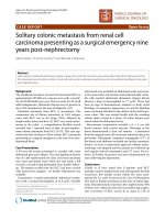
Biopsy of his anterior floor of mouth lesion was notable
for persistent bleeding and revealed clear cell carcinoma,
consistent with the patient's previous history of renal cell
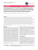
cancer (Figure 1). Histologic evaluation revealed the pres-
ence of a solid nest of epithelial cells with clear cytoplasm
and small, round hyperchromatic nuclei (Figure 2). A rich
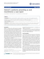
vascular network was also noted. Immunoperoxidase test-
ing was positive for CD10 and vimentin and negative for
gross cystic disease fluid protein (GCDFP), S-100, HMB-
45, muscle-specific antigen, and desmin, supporting the
diagnosis of metastatic RCC (Figure 3).
Original surgical, pathology and postoperative records
were eventually obtained revealing the discovery of suspi-
cious lymph nodes near the renal hilum during his origi-
nal nephrectomy. The resected lymph nodes were found
to harbor metastatic carcinoma and the patient was
referred to a medical oncologist at that time to discuss
additional therapeutic options. Unfortunately, he did not
Renal cell carcinoma; ulceration of mucosal epithelium noted secondary to tumor cell infiltrationFigure 1
Renal cell carcinoma; ulceration of mucosal epithe-
lium noted secondary to tumor cell infiltration.
Histologic features of renal cell carcinoma; epithelial cellular network shown with clear cytoplasm and hyperchromatic nuclei surrounded in a rich vascular networkFigure 2
Histologic features of renal cell carcinoma; epithelial
cellular network shown with clear cytoplasm and
hyperchromatic nuclei surrounded in a rich vascular
network.
Staining for clear cell carcinoma; carcinomatous cells are pos-itive for vimentin by immunohistochemical stainingFigure 3
Staining for clear cell carcinoma; carcinomatous cells
are positive for vimentin by immunohistochemical
staining.
Journal of Medical Case Reports 2008, 2:313 />Page 3 of 4
(page number not for citation purposes)
seek further care for his metastatic RCC. After receiving the
news of his biopsy results in our clinic, the patient decided
to return to his original institution for further care and
died of metastatic disease within several months.
Discussion
Metastatic tumors to the oral cavity are extremely rare.
Therefore, existing literature is based largely on sporadic
case reports. Possible routes of metastasis to the oral cavity
include arterial, venous and lymphatic circulations. In the
case of head and neck metastasis without lung involve-
ment, several theories exist to address a route of dissemi-
nation that avoids pulmonary vascular filtration. These
include spread via Batson's venous plexus or through the
thoracic lymphatic duct. Batson's paraspinal plexus is a
valveless, venous system extending from the skull to the
sacrum allowing tumor emboli to bypass the pulmonary
venous system with minimal resistance resulting in metas-
tasis to the head and neck region in the absence of obvi-
ous lung lesions [3].
Reports of RCC to the head and neck region involve the
nose, tongue, paranasal sinuses, larynx, mandible, tempo-
ral bone, thyroid gland, and parotid glands [4]. The loca-
tion of metastasis usually dictates the presenting
symptoms.
Metastatic RCC will often behave similarly to that of the
primary renal lesion in terms of morphology and histol-
ogy. The risk of bleeding after fine needle aspiration
biopsy of RCC in the kidney is well documented, with up
to 90% of patients bearing evidence of perinephric bleed-
ing on CT imaging and 5–7% developing clinically signif-
icant hemorrhage [5]. Therefore, clinical suspicion of RCC
metastasis or other vascular lesions should prompt a
biopsy in a controlled setting should hemorrhage ensue.
Histologically, differentiating among clear cell tumors
with conventional light microscopy can be challenging. It
can be especially difficult to distinguish between RCC
metastasis and clear cell malignancies of salivary glands.
Clear cell carcinomas of salivary gland origin are usually
nests of clear cells divided by thin, fibrous connective
septa and irregular vascular tissue. Immunohistochemical
staining helps in this distinction, with RCC metastasis
exhibiting focal cytokeratin positivity (versus minor sali-
vary gland cancers showing diffuse positivity) and a
strong reaction for vimentin [6].
Treatment of renal adenocarcinoma metastasis to the
head and neck is directed mainly toward palliation [7].
Excision has been performed primarily to control pain
and prevent bleeding and infection. Azam et al describe
surgically debulking a rapidly growing metastatic tongue
lesion in order to relieve pain and allow the patient to
swallow. They then administered radiotherapy to the oral
cavity at a dose of 60 Grays to treat any remaining micro-
scopic disease [8]. Although RCC is traditionally known
as a radioresistant tumor, radiotherapy can aid in local
symptom control for perhaps a few months [2]. Little data
exist regarding the use of systemic therapy in the setting of
RCC metastasis to the oral cavity. Kyan and Kato report
the surgical resection of a lingual mass followed by the
administration of interferon-alpha and interleukin-II
without recurrence of disease at 2 years [9]. Still, most
patients die within 1 year after detection of head and neck
metastasis; therefore, therapeutic decisions should maxi-
mize comfort and minimize morbidity considering the
poor long-term prognosis at this stage of the disease.
Of interest in this patient is whether other measures taken
at the time of the initial diagnosis and treatment period
may have slowed disease progression or prolonged sur-
vival. Certainly, the finding of lymph node involvement
portends a poor prognosis with 5- and 10-year survival
rates similar to those with systemic metastasis (5–30%
and 0–5%, respectively). An extended lymph node dissec-
tion has been suggested to benefit patients with early, iso-
lated or microscopic involvement of the nodes. However,
this remains controversial with a lack of randomized data
to support any survival advantage [10].
Immunologic therapy following radical nephrectomy in
the setting of metastatic disease may improve time to pro-
gression in properly selected patients. Prospective trials
have demonstrated that, in patients with synchronous
metastatic disease, cytoreductive nephrectomy and a sys-
temic cytokine give a distinct survival advantage over
those treated with immunotherapy alone (17.4 versus
11.7 months in patients with an Eastern Cooperative
Oncology Group performance status of 0–1) [11]. Unfor-
tunately, complete durable responses with systemic ther-
apy are infrequent and the significant toxicity of
immunologic agents may limit their use.
Finally, newer agents targeting the VEGF pathway such as
bevacizumab and sorafenib may provide hope for
patients with metastatic RCC. Early trials have shown a
prolongation of progression-free survival with the use of
these targeted molecular therapies in cytokine-refractory
patients [12].
Conclusion
RCC has been shown to metastasize to the head and neck
region in rare instances. Therefore, the work-up of a new
oral or neck lesion in light of a history of RCC should
include metastatic RCC as part of the differential diagno-
sis. The physician needs to be prepared for the increased
risk of bleeding involved in the biopsy of RCC metastasis.
Should the biopsy specimen reveal clear cell carcinoma of
Publish with BioMed Central and every
scientist can read your work free of charge
"BioMed Central will be the most significant development for
disseminating the results of biomedical research in our lifetime."
Sir Paul Nurse, Cancer Research UK
Your research papers will be:
available free of charge to the entire biomedical community
peer reviewed and published immediately upon acceptance
cited in PubMed and archived on PubMed Central
yours — you keep the copyright
Submit your manuscript here:
/>BioMedcentral
Journal of Medical Case Reports 2008, 2:313 />Page 4 of 4
(page number not for citation purposes)
the mouth, it is vital to perform immunohistochemical
staining to differentiate between metastatic RCC and
malignancies of salivary origin. If a diagnosis of metastatic
RCC is established, additional therapeutic options,
including immunotherapy, tyrosine kinase inhibitors,
and participation in a clinical trial, should be discussed
with the patient despite the poor overall prognosis.
Abbreviations
CT: computed tomography; RCC: renal cell carcinoma;
VEGF: vascular endothelial growth factor.
Competing interests
The authors declare that they have no competing interests.
Authors' contributions
TW collected and interpreted all of the urologic data per-
tinent to the case report and was the major contributing
author of the final written report. NA collected and inter-
preted all of the otolaryngologic data pertinent to the case
including the pathology specimens deemed Figures 1, 2,
3. GP performed the initial biopsy on the patient and crit-
ically revised the content and structure of the manuscript.
All authors read and approved the final manuscript.
Consent
Written informed consent was obtained from the patient
for publication of this case report and accompanying
images. A copy of the written consent is available for
review by the Editor-in-Chief of this journal.
References
1. Campbell SC, Novick AC, Bukowski RM: Renal tumors. In Camp-
bell-Walsh Urology Volume 2. 9th edition. Edited by: Wein AJ. Philadel-
phia, PA: Saunders Elsevier; 2007:1582-1632.
2. Pritchyk KM, Schiff BA, Newkirk KA, Krowiak E, Deeb ZE: Meta-
static renal cell carcinoma to the head and neck. Laryngoscope
2002, 112:1598-1601.
3. Boles R, Cerny J: Head and neck metastases from renal carci-
nomas. Mich Med 1971, 90:616-618.
4. Torres-Carranza E, Garcia-Perla A, Infante-Cossio P, Belmonte-Caro
R, Loizaga-Iriondo JM, Gutierrez-Perez JL: Airway obstruction due
to metastatic renal cell carcinoma to the tongue. Oral Surg
Oral Med Oral Pathol Oral Radiol Endod 2006, 101:E76-78.
5. Vassiliades VG, Bernardino ME: Percutaneous renal and adrenal
biopsies. Cardiovasc Intervent Radiol 1991, 14:50-54.
6. Marioni G, Gaio E, Poletti A, Derosas F, Staffieri A: Uncommon
metastatic site of renal adenocarcinoma: the oral tongue.
Acta Otolaryngol 2004, 124:197-201.
7. Fukuda M, Miyata M, Okabe K, Sakashita H: A case series of 9
tumors metastatic to the oral and maxillofacial region. J Oral
Maxillofac Surg 2002, 60:942-944.
8. Azam F, Abubakerr M, Gollins S: Tongue metastasis as an initial
presentation of renal cell carcinoma: a case report and liter-
ature review. J Med Case Reports 2008, 2:249.
9. Kyan A, Kato SN: Renal cell carcinoma to the base of tongue:
a case report. Hinyokika Kiyo 2004, 50(11):791-793.
10. Phillips CK, Taneja SS: The role of lymphadenectomy in the sur-
gical management of renal cell carcinoma. Urol Oncol 2004,
22(3):214-223.
11. Flanigan RC, Salmon SE, Blumenstein BA, Bearman SI, Roy V, McGrath
PC, Caton JR Jr, Munshi N, Crawford ED: Nephrectomy followed
by interferon alfa-2b compared with interferon alfa-2b alone
for metastatic renal-cell carcinoma.
N Engl J Med 2001,
345(23):1655-1659.
12. Escudier B, Eisen T, Stadler WM, Szczylik C, Oudard S, Siebels M,
Negrier S, Chevreau C, Solska E, Desai AA, Rolland F, Demkow T,
Hutson TE, Gore M, Freeman S, Schwartz B, Shan M, Simantov R,
Bukowski RM, TARGET Study Group: Sorafenib in advanced
clear-cell renal-cell carcinoma. N Engl J Med 2007,
356(2):125-134.