Báo cáo y học: "Congenital intrarenal arteriovenous malformation presenting with gross hematuria after endoscopic intervention: a case report" ppt
Bạn đang xem bản rút gọn của tài liệu. Xem và tải ngay bản đầy đủ của tài liệu tại đây (450.91 KB, 4 trang )
BioMed Central
Page 1 of 4
(page number not for citation purposes)
Journal of Medical Case Reports
Open Access
Case report
Congenital intrarenal arteriovenous malformation presenting with
gross hematuria after endoscopic intervention: a case report
Michael Seitz*
1
, Tobias Waggershauser
2
and Wael Khoder
1
Address:
1
Department of Urology, University Hospital Grosshadern, Marchioninistrasse, 81377 Munich, Germany and
2
Department of Radiology,
University Hospital Grosshadern, Marchioninistrasse, 81377 Munich, Germany
Email: Michael Seitz* - ; Tobias Waggershauser - ;
Wael Khoder -
* Corresponding author
Abstract
Introduction: Although diagnostic ureterorenoscopy is a minimally invasive and effective
diagnostic procedure, it has the potential for significant postoperative complications. We report
the first case in the literature of intrarenal arteriovenous fistulas causing hemodynamic effective
anemia 4 days after ureterorenoscopic biopsy.
Case presentation: A 63-year-old Caucasian woman presented with hemodynamic effective
macrohematuria (hemoglobin 70 g/liter) 4 days after ureterorenoscopy and biopsy of the upper
pole collecting system due to recurrent microhematuria. Duplex-sonography and computed
tomography angiography revealed multiple arteriovenous fistulas and erosions into the calyceal
system. Intra-arterial digital subtraction angiography confirmed this condition. After superselective
embolization of the arteriovenous fistulas, the patient had no further episodes of bleeding or
microhematuria.
Conclusion: If malignancies, urolithiasis or urinary tract infections are ruled out by common
diagnostic procedures as the cause of recurrent minor or gross hematuria, the possibility of
arteriovenous fistulas should be included in the differential diagnosis and Duplex-Sonography or the
more invasive selective renal arteriography should be performed as this is the most definitive
method for diagnosing arteriovenous fistula.
Introduction
Although arteriovenous fistulas are rare conditions, they
have a considerable clinical impact. In fact they may cause
hypertension, local thrombosis, peripheral embolization,
high output cardiac failure and hematuria. Although ure-
terorenoscopy is a minimally invasive and effective diag-
nostic and therapeutic procedure, it has the potential for
significant postoperative complications. We report a case
of intrarenal arteriovenous fistulas causing hemodynamic
effective anemia 4 days after ureterorenoscopic biopsy.
Case presentation
A 63-year-old woman presented with recurrent microhe-
maturia. She had no history of flank pain, macrohematu-
ria, hypertension, renal trauma or percutaneous
instrumentation. Physical examination was normal and
specifically, there was no abdominal bruit on ausculta-
tion. She had a blood pressure of RR 130/80 mmHg. Rou-
tine laboratory tests were within normal limits. Urinalysis
showed no evidence of infection but was positive for
erythrocytes. An initial renal ultrasound revealed a dis-
Published: 12 October 2008
Journal of Medical Case Reports 2008, 2:326 doi:10.1186/1752-1947-2-326
Received: 6 October 2007
Accepted: 12 October 2008
This article is available from: />© 2008 Seitz et al; licensee BioMed Central Ltd.
This is an Open Access article distributed under the terms of the Creative Commons Attribution License ( />),
which permits unrestricted use, distribution, and reproduction in any medium, provided the original work is properly cited.
Journal of Medical Case Reports 2008, 2:326 />Page 2 of 4
(page number not for citation purposes)
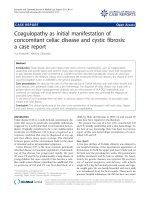
crete hypoechogeneity of the left upper renal pole. An
intravenous pyelogram was performed demonstrating an
irregular configuration of the upper pole collecting sys-
tem, which was also seen in a retrograde ureteropyelogra-
phy (Figure 1). Cystoscopy as well as ureterorenoscopy
(URS) revealed no suspicious formation within the blad-
der or along the left ureter or in the renal pelvis. Tissue
around blood clots in the upper calyceal group was biop-
sied. Cytology and histology did not identify malignant
cells. The patient was discharged with a ureteral stent.
Four days after the intervention, emergency admission
was necessary due to a hemodynamic effective macrohe-
maturia (hemoglobin 70 g/liter) causing a bladder coagu-
lum, which made transurethral evacuation necessary.
Duplex-sonography and computed tomography angiogra-
phy (CTA) were then carried out and revealed multiple
arteriovenous fistulas (AVFs) and erosions into the calyc-
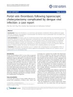
eal system. Intra-arterial digital subtraction angiography
(i.a. DSA, Figure 2) in the early arterial phase showed arte-
riovenous fistulas between a subsegmental branch of the
renal artery and the renal vein and these were superselec-
tively embolized by 8 Platin-coils with cotton filaments.
Angiographically, no significant differences in parenchy-
mal perfusion were noted before and after intervention.
Pathologic neoplastic vessels were ruled out radiomor-
phologically. Five months after intervention, a control
computed tomogram showed no recurrent AVF or malig-
nancy. The patient had no further episodes of bleeding or
microhematuria.
Discussion
Arteriovenous fistulas, first described by Varela in 1928,
are rare conditions, which, however, have a considerable
clinical impact [1]. In fact, they may cause hypertension,
local thrombosis, peripheral embolization, high output
cardiac failure and hematuria [2]. There are two types of
AVF, classified as congenital and acquired [3]. In total, 70
to 80% of all AVFs are of the acquired type and may be
secondary to trauma, renal surgery, inflammation, neo-
plasia or percutaneous needle biopsy, the latter contribut-
ing to recent increased incidence. Acquired renal AVFs
may be located throughout the whole kidney. Angio-
graphically, they appear as solitary communications
between arteries and veins. More than 70% of these fistu-
las close spontaneously within a few weeks or months
without active intervention. Therefore the common strat-
egy in asymptomatic patients with incidental detection of
AVFs is to 'wait and watch' [4].
In 20 to 30% of all cases, an arteriovenous fistula is a con-
genital condition usually located in the upper pole (45%
of cases) but may also appear in the midportion or the
lower pole of the kidney in equal ratio topographically
beneath the calyceal or pelvic mucosa. Congenital AVFs
are characterized angiographically, as in our patient, by
their cirsoid configuration with multiple communications
between arteries (main or segmental renal arteries) and
veins [2,4].
Based on the angiographic criteria, a second form of con-
genital AVF exists which is classified as the aneurysmal
type and has been mentioned in the literature as a sponta-
neous or idiopathic fistula [4]. While the latter predomi-
nantly present with cardiovascular symptoms, the cirsoid
forms show a high incidence of gross hematuria [2].
In the pericalyceal renal parenchyma, the small interlobu-
lar arteries and their corresponding veins as well as exist-
ing AVFs are in close proximity to the collecting system.
This explains recurrent hematuria in more than 75% of
individuals and possible filling defects or reduced func-
tion of the affected kidney in the excretory urography, but
Retrograde ureteropyelographyFigure 1
Retrograde ureteropyelography. Retrograde uretero-
pyelography demonstrates an irregular configuration of the
upper pole collecting system.
Journal of Medical Case Reports 2008, 2:326 />Page 3 of 4
(page number not for citation purposes)
these are absent in 50% of cases [2]. In our patient, we
postulate that, due to the biopsy during endoscopic inter-
vention, a perforation had occurred and venous dilata-
tions of the AVFs eroded into the collecting system
causing gross hematuria. Active management was neces-
sary due to hemodynamic effective gross hematuria. Selec-
tive renal arteriography, as the most definitive method for
diagnosing the lesion, was performed with simultaneous
superselective coil embolization. This treatment method
is well accepted in such conditions since it avoids surgery.
Parenchymal infarction secondary to embolization can be
limited to the region which is supplied by the artery con-
taining the lesion. This is especially important in patients
with only one functioning kidney or renal insufficiency.
The technique is also indicated in patients who are con-
sidered poor surgical candidates since the procedure is
performed under local anesthesia with low morbidity and
low risk of complications [5-7].
In contrast to patients presenting with hematuria, we sug-
gest nephrectomy or partial nephrectomy as the treatment
of choice in individuals with symptoms of alterations in
the cardiovascular system such as renin-mediated hyper-
tension due to fistula-related relative ischemia or high-
output cardiac failure caused by increased venous return.
Conclusion
Congenital AVFs are rare conditions which may cause car-
diovascular complications (in 50% of cases) and recurrent
hematuria in more than 75% of individuals.
If malignancies, urolithiasis or urinary tract infections are
ruled out by common diagnostic procedures as the cause
of recurrent minor or gross hematuria, the possibility of
AVFs should be included in the differential diagnosis and
Duplex-Sonography, or the more invasive selective renal
arteriography, as the most definitive method for diagnos-
ing AVF, should be performed. Depending on the general
condition of the patient and their symptoms, the treat-
ments of choice include nephrectomy and partial
nephrectomy but most urologists aim for superselective
embolization.
Abbreviations
AVF: arteriovenous fistula; CTA: computed tomography
angiography; DSA: digital subtraction angiography; i.a.
DSA: intra-arterial digital subtraction angiography; RR:
blood pressure (measured by the technique of Riva
Rocci); URS: ureterorenoscopy
Competing interests
The authors declare that they have no competing interests.
Authors' contributions
MS made substantial contributions to acquisition and
interpretation of the data and drafted the manuscript. TW
carried out the imaging studies and performed the embol-
ization. WK managed the critically ill patient clinically
and also contributed substantially to the interpretation of
the literature.
Consent
Written informed consent was obtained from the patient
for publication of this case report and any accompanying
images. A copy of the written consent is available for
review by the Editor-in-Chief of this journal.
AngiographyFigure 2
Angiography. In the early arterial phase, intra-arterial digital subtraction angiography demonstrates arteriovenous fistulas
between a subsegmental branch of the renal artery and the renal vein (left, central) and these were superselectively embolized
by 8 Platin-coils with cotton filaments (right). Angiographically, no significant differences are noted in parenchymal perfusion
before and after intervention. Pathologic neoplastic vessels are ruled out radiomorphologically.
Publish with BioMed Central and every
scientist can read your work free of charge
"BioMed Central will be the most significant development for
disseminating the results of biomedical research in our lifetime."
Sir Paul Nurse, Cancer Research UK
Your research papers will be:
available free of charge to the entire biomedical community
peer reviewed and published immediately upon acceptance
cited in PubMed and archived on PubMed Central
yours — you keep the copyright
Submit your manuscript here:
/>BioMedcentral
Journal of Medical Case Reports 2008, 2:326 />Page 4 of 4
(page number not for citation purposes)
Acknowledgements
We thank Christian Stief for help in critically revising the manuscript and in
giving final approval of the version to be published.
References
1. Varela ME: Aneurisma arteriovenoso de los vasos renales y
asistolia consecutiva. Rev Med Latino-Am 1928, 14:3244.
2. Walsh PC, Retik AB, Vaughan ED, Wein AJ, Kavoussi LR, Novick AC,
Partin AW, Peters CA: Campbell's Urology 8th edition. Elsevier Sci-
ence; 2002:3422-3423.
3. Maldonado JE, Sheps SG, Bernatz PE, Deweerd JH, Harrison EG Jr:
Renal arteriovenous fistula. A reversible cause of hyperten-
sion and heart failure. Am J Med 1964, 37:499-513.
4. Takaha M, Matsumoto A, Ochi K, Takeuchi M, Takemoto M, Sonoda
T: Intrarenal arteriovenous malformation. J Urol 1980,
124:315-318.
5. Savastano S, Feltrin GP, Miotto D, Chiesura-Corona M: Renal aneu-
rysm and arteriovenous fistula: Management with transcath-
eter embolization. Acta Radiol 1990, 31:73-76.
6. Husstedt H, Chavan A, Ghadban F, Leppert A, Galanski M: Percuta-
neous superselective coil-embolization of intrarenal arterio-
venous fistulas. Acta Radiol 1996, 37:539-541.
7. Crotty KL, Orihuela E, Warren MM: Recent advances in the diag-
nosis and treatment of renal arteriovenous malformations
and fistulas. J Urol 1993, 150:1355.