Báo cáo y học: " Double rupture of interventricular septum and free wall of the left ventricle, as a mechanical complication of acute myocardial infarction: a case report" pot
Bạn đang xem bản rút gọn của tài liệu. Xem và tải ngay bản đầy đủ của tài liệu tại đây (390.18 KB, 5 trang )
BioMed Central
Page 1 of 5
(page number not for citation purposes)
Journal of Medical Case Reports
Open Access
Case report
Double rupture of interventricular septum and free wall of the left
ventricle, as a mechanical complication of acute myocardial
infarction: a case report
Elias I Rentoukas, George A Lazaros*, Andreas P Kaoukis and
Evangellos P Matsakas
Address: Cardiology Department, Athens General Hospital, Athens, Greece
Email: Elias I Rentoukas - ; George A Lazaros* - ; Andreas P Kaoukis - ;
Evangellos P Matsakas -
* Corresponding author
Abstract
Introduction: Cardiac ruptures following acute myocardial infarction include rupture of the left
ventricle free-wall, ventricular septal defects, and papillary muscle rupture. Double myocardial
rupture is a rare complication of acute myocardial infarction (0.3 %) and the report of such cases
is exclusively limited to a small series of autopsy studies.
Case presentation: In this report we present the unusual case of a 70-year-old woman with
acute anteroseptal myocardial infarction, which was complicated by a combined rupture of the
interventricular septum near the apex, and the free wall of the left ventricle with concomitant
formation of a pseudoaneurysm. The double myocardial rupture was accidentally discovered 10
days later with echocardiography, when the patient, complaining only of mild exertional dyspnea,
was hospitalized for a scheduled coronary angiography. The patient underwent successful surgical
correction of the double myocardial rupture along with by-pass grafting.
Conclusion: This report highlights the importance of comprehensive noninvasive predischarge
diagnostic evaluation of all postinfarct patients, since serious and potentially life-threatening
complications might have not been suspected on clinical grounds.
Introduction
Cardiac ruptures are serious and life-threatening mechan-
ical complications of acute myocardial infarction (AMI).
Types of rupture include left ventricle (LV) free-wall rup-
ture (FWR), ventricular septal defect (VSD), and papillary
muscle rupture (PMR). Double myocardial rupture
(DMR) is defined as the coexistence of two of the above-
mentioned forms of rupture. It complicates approxi-
mately 0.3% of AMI with the most frequent combination
being FWR and VSD [1]. Small autopsy series report that
DMR is seen in 13% of patients with FWR and in approx-
imately 16% of patients with VSD [1]. The contribution of
2-D echocardiography and color Doppler in the early
diagnosis of these lesions is well established [2]. Since
DMR carries a high mortality, surgical correction, even in
advanced age, constitutes the treatment of choice [3].
We present the case of a female patient whose recent AMI
was complicated by a combination of VSD and FWR of the
LV with formation of a pseudoaneurysm, which were suc-
Published: 17 March 2008
Journal of Medical Case Reports 2008, 2:85 doi:10.1186/1752-1947-2-85
Received: 27 June 2007
Accepted: 17 March 2008
This article is available from: />© 2008 Rentoukas et al; licensee BioMed Central Ltd.
This is an Open Access article distributed under the terms of the Creative Commons Attribution License ( />),
which permits unrestricted use, distribution, and reproduction in any medium, provided the original work is properly cited.
Journal of Medical Case Reports 2008, 2:85 />Page 2 of 5
(page number not for citation purposes)
cessfully surgically corrected. This case is interesting due
to the scarcity of such reports and the authors wish to
emphasize both the contribution of echocardiography in
identifying the above complications and the favorable
outcome of our surgically treated patient, despite the seri-
ousness of this complication and its relatively late diagno-
sis.
Case presentation
A 70-year-old-female, with a history of diabetes, arterial
hypertension and mild chronic renal failure, fifteen days
before her admission to our Department, had been admit-
ted to another hospital, because of substernal squeezing
pain of ten hours duration and an electrocardiogram com-
patible with acute anteroseptal myocardial infarction (ST-
segment elevation in leads V
1
to V
4
). Moreover, an
echocardiographic study on admission was reported to
show regional wall motion abnormalities in the territory
of distribution of the left anterior descending coronary
artery (LAD). In the absence of contraindications, she was
administered fibrinolysis with Tenekteplase, which was
considered successful using the current clinical and elec-
trocardiographic criteria. On hospital day 5, the patient
had an episode of hypotension, which was treated with
infusion of normal saline but no further investigation due
to its short duration and her relatively prompt recovery.
On the 9
th
post-infarct day, the patient was discharged
with the recommendation for follow-up coronary angiog-
raphy.
Six days later, the patient was admitted to our Department
for the scheduled coronary arteriography. She reported
mild exertional dyspnea and fatigue until about 3 days
ago. On examination the patient was an obese woman
who appeared well. Her blood pressure was 115/80 and
her pulse 80. The only remarkable finding on chest exam-
ination was a grade 1-2/6 parasternal holosystolic mur-
mur without gallop or rub. The electrocardiogram was
compatible with her recent anteroseptal infarction. An
echocardiographic study was performed and disclosed a
DMR, consisting of VSD [due to the rupture of the inter-
ventricular septum (IVS), with a maximal pressure gradi-
ent of approximately 90 mmHg], and rupture of the apical
part of the LV free wall with pseudoaneurysm formation
(Fig. 1 and 2). The global LV contractility was affected
with the ejection fraction being approximately 40%,
whereas the anterior wall appeared hypokinetic and the
apex akinetic. A coronary arteriography performed on the
same day showed a total occlusion of the LAD branch in
its proximal part along with an 80% stenosis of the first
obtuse marginal branch of the left circumflex coronary
artery. The right coronary angiogram disclosed a 50% ste-
nosis in the midportion of the right coronary artery. In
addition left ventriculography confirmed the abnormal
communication between left and right ventricle (Fig. 3).
On the next day, the patient was transferred to a Cardiac
Surgery Center, and underwent surgical closure of the
DMR along with double bypass-grafting (left internal
mammary artery grafting applied to the LAD and saphen-
ous vein bypass grafting to the obtuse marginal branch).
Pulsed wave Doppler showing a systolic flow (SF) from the LV cavity to the pseudoaneurysm and a diastolic regurgitant flow (DF) in the opposite directionFigure 2
Pulsed wave Doppler showing a systolic flow (SF)
from the LV cavity to the pseudoaneurysm and a
diastolic regurgitant flow (DF) in the opposite direc-
tion. In the right part of the picture, colour Doppler depicts
a flow between right and left ventricle (white arrow).
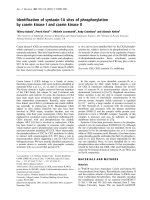
Modified left parasternal short axis view that shows a discon-tinuity of the apical part of the interventricular septum and the LV apex, a communication between the left and the right ventricle, and a small cavity contained by epicardium (pseu-doaneurysm) through a narrow neckFigure 1
Modified left parasternal short axis view that shows a
discontinuity of the apical part of the interventricular
septum and the LV apex, a communication between
the left and the right ventricle, and a small cavity
contained by epicardium (pseudoaneurysm) through
a narrow neck. (LV: left ventricle, RV: right ventricle, PA:
pseudoaneurysm, PE: pericardial effusion).
Journal of Medical Case Reports 2008, 2:85 />Page 3 of 5
(page number not for citation purposes)
Discussion
VSD complicates 1–2% of all AMIs and approximately 0.2
% of fibrinolysed AMIs. In the later case it is seen earlier
in the post infarct period (within the first 24 hours or so)
in contrast with the non-fibrinolysed AMIs, where it is
commonly seen after two to five days [4]. VSD is more
common in females, and those with advanced age, ante-
rior AMI and single-vessel disease with poorly developed
collaterals to the IVS [4]. In cases of anterior transmural
AMI, the rupture is usually located in the anteroapical part
of IVS, whilst in inferior infarctions the defect occurs in its
basal part [5]. The complication of VSD carries a high
mortality and early surgical closure is the treatment of
choice, even if the patient's condition is stable. Surgical
mortality is high among patients with inferior AMIs
(58%), as compared to that of anterior AMIs (25%) [6].
Sporadic reports have also shown that, in selected severely
sick and haemodynamically unstable patients with large
defects (and consequently shunts), either percutaneous
transcatheter closure of the defect or insertion of a left
ventricular assist device may improve clinical condition
and allow a subsequent surgical repair under better hemo-
dynamics and more favourable local conditions [7,8].
FWR occurs 10 times more frequently than VSD or PMR.
Its incidence is higher among patients subjected to late
fibrinolysis (i.e. several hours after the onset of symp-
toms), in comparison to those with early administration
of fibrinolysis (within 6–8 hours of the onset of symp-
toms) [9]. Higher rates of FWR have been also observed in
patients taking anti-inflammatory agents (steroids or non-
steroidal) [9]. Most patients present with electromechani-
cal dissociation and sudden death or, less frequently, with
cardiac tamponade. Some patients may have a subacute
course as a result of a contained rupture with pseudoaneu-
rysm formation. In this case there are symptoms of pul-
monary congestion, recurrent tachyarrhythmias or
systemic embolism. Occasionally, patients may be com-
pletely asymptomatic (10–13%) [10]. Spontaneous rup-
ture of a pseudoaneurysm occurs in approximately one
third of patients with FWR (as opposed to true LV aneu-
rysms where rupture is quite uncommon), and as a result,
surgical resection is recommended regardless of the symp-
toms or the size of the pseudoaneurysm [11].
DMR is defined as the combination of any two of the three
forms of cardiac rupture, with VSD and FWR being the
most common (in 17% of patients with VSD there is con-
comitant FWR) [12]. Tanaka et al. studied a series of ten
patients with DMR and concluded that advanced age
(mean age 69 years), absence of history of coronary artery
disease (90%), anterior AMI (60%), arterial hypertension
(60%), and male sex (male/female ratio:8/2) were risk
factors for the development of this complication [1].
There are two forms of DMR: a) true, with rupture of both
IVS and LV free wall, and b) junctional, located at the
junction between IVS and free wall [13]. The analysis of
similar cases has shown that the coexistence of FWR is fre-
quently established only at the time of operation for the
correction of VSD [3]. Tanaka et al. reported that the
majority of patients had an apical AMI and VSD near the
junction between IVS and LV apical free wall and con-
cluded that this combination might be a precursor of
DMR [1].
Two-dimensional echocardiography, in combination
with Doppler study, being an accessible and non-invasive
method, contributes significantly both to the diagnosis of
every form of cardiac rupture, and the determination of
the size of the defect and the magnitude of the left-to-right
shunt (as far as VSD is concerned) [2]. Magnetic resonance
imaging (MRI) is also a useful tool for the confirmation of
diagnosis, particularly when there is a pseudoaneurysm.
Before the 1980s, there was a vogue for managing patients
with cardiac rupture non-surgically in the first instant.
After a period of perhaps six weeks, often with intraaortic
balloon counterpulsation support, the patient underwent
surgery. The main advantage of this strategy for the sur-
geon was that the remaining septum was no longer mushy
necrotic muscle, but it had begun to fibrose and thus, was
more receptive to sutures. However, the literature in the
late 1970s and early 1980s established that there was no
Right anterior oblique left venticulography during systole showing simultaneous opacification of the aorta (red arrows) and the pulmonary artery (yellow arrows)Figure 3
Right anterior oblique left venticulography during systole
showing simultaneous opacification of the aorta (red arrows)
and the pulmonary artery (yellow arrows).
Journal of Medical Case Reports 2008, 2:85 />Page 4 of 5
(page number not for citation purposes)
place for procrastination, as the great majority of patients
died while waiting for the surgical procedure and the
mood shifted to early surgical correction of every form of
cardiac rupture (including DMR), even in hemodynami-
cally stable patients [14]. Conservative measures such as
diuretics, inotropes, nitroprusside and intraaortic balloon
counterpulsation are used for the initial stabilization of
these patients, as a bridge to surgery. Inferior AMI, right
ventricular dysfunction, cardiogenic shock, advanced age,
and delay of surgery, are all considered as intraoperative
risk factors [6]. In the GUSTO-I study, the 30 day-mortal-
ity of surgically managed patients with VSD was 47% as
opposed to 94% of conservatively managed patients [4].
Surgical correction of DMR is also accompanied by a high
mortality (Tanaka et al. report a 4 month-survival of
37.5%), which nonetheless is less than the mortality of
conservative treatment [1]. In the international medical
literature, there are mostly case reports of successful surgi-
cal correction of DMR, while studies comparing surgical
with conservative management have not been performed
[3].
The most possible scenario concerning our patient is that
the anteroseptal AMI was complicated by VSD near the LV
apex. The episode of hypotension at the fifth post-infarct
day was probably the manifestation of the second cardiac
rupture (FWR), which was easily managed as it resulted in
pseudoaneurysm formation without extensive hemoperi-
cardium and tamponade. It was quite impressive that the
pseudoaneurysm did not rupture during the following ten
day period and that the patient, despite the seriousness of
this complication, had only mild symptoms. In addition,
the detection of DMR was virtually accidental.
Conclusion
This report emphasizes both the significance and the
necessity of the detailed non-invasive evaluation (such as
echocardiographic study), in all post-infarct patients, as it
may sometimes reveal serious complications that have
not been suspected on clinical grounds. Routine pre-dis-
charge echocardiographic evaluation seems also to be a
cost-effective approach, as it provides unique information
that can significantly impact on patient management deci-
sions.
Abbreviations
AMI: acute myocardial infarction; LV: left ventricle; FWR:
free-wall rupture; VSD: ventricular septal defect; PMR:
papillary muscle rupture; DMR: double myocardial rup-
ture; LAD: left anterior descending coronary artery; RV:
right ventricle; PA: pseudoaneurysm; PE: pericardial effu-
sion; SF: systolic flow; DF diastolic flow.
Competing interests
The author(s) declare that they have no competing inter-
ests.
Authors' contributions
EIR was involved in the conception and final reviewing of
this report. GAL was involved in the manuscript prepara-
tion, editing, and submission. APK was involved in the lit-
erature review and manuscript preparation. EPM was
involved in the patient's evaluation. All authors read and
approved the final manuscript.
Consent
Written informed consent was obtained from the patient
for publication of this case report and accompanying
images. A copy of the written consent is available for
review by the Editor-in-Chief of this journal.
References
1. Tanaka K, Sato N, Yasutake M, Takeda S, Takano T, Ochi M, Tanaka
S, Tamura K: Clinicopathological characteristics of 10 patients
with rupture of both ventricular free wall and septum (dou-
ble rupture) after acute myocardial infarction. J Nippon Med
Sch 2003, 70:21-27.
2. Helmcke F, Mahan EF III, Nanda NC, Jain SP, Soto B, Kirklin JK,
Pacifico AD: Two-dimensional echocardiography and Doppler
color flow mapping in the diagnosis and prognosis of ven-
tricular septal rupture. Circulation 1990, 81:1775-1783.
3. Ide H, Ino T, Mizuhara A, Yamaguchi A: Successful repair of com-
bined ventricular septal rupture and free wall rupture. Ann
Thorac Surg 1993, 55:762-763.
4. Crenshaw B, Granger C, Birnbaum Y, Pieper K, Morris D, Kleiman N,
Vahanian A, Califf RM, Topol EJ: Risk factors, angiographic pat-
terns, and outcomes in patients with ventricular septal
defect complicating acute myocardial infarction. GUSTO-I
(Global Utilization of Streptokinase and TPA for Occluded
Coronary Arteries) trial investigators. Circulation 2000,
101:27-32.
5. Cummings RG, Reimer KA, Califf R, Hackel D, Boswick J, Lowe JE:
Quantitative analysis of right and left ventricular infarction
in the presence of postinfarction ventricular septal defect.
Circulation 1988, 77:33-42.
6. Jones MT, Schofield PM, Dark JF, Moussalli H, Deiraniya AK, Lawson
RA, Ward C, Bray CL: Surgical repair of acquired ventricular
septal defect. Determinants of early and late outcome. J Tho-
rac Cardiovasc Surg 1987, 93:680-686.
7. Costache V, Chavanon O, Bouvaist H, Blin D: Early Amplatzer
occluder closure of a postinfarct ventricular septal defect as
a bridge to surgical procedure Interact. CardioVasc Thorac Surg
2007, 6:503-504.
8. Patane F, Zingarelli F, Sansone F, Rinaldi M: Acute ventricular sep-
tal defect treated with an Impella recovery as a 'bridge ther-
apy' to heart transplantation. Interact CardioVasc Thorac Surg
2007, 6:818-819.
9. Massel DR: How sound is the evidence that thrombolysis
increases the risk of cardiac rupture? Br Heart J 1993,
69:284-287.
10. Castellanos S, Zezas S, Kalfoglou Th, Papadimitriou K: Left ventricu-
lar pseudoaneurism after a silent myocardial infarction. Hell
J Cardiol 2003, 44:97-101.
11. Martin RH, Almond CH, Saab S, Watson LE: True and false aneu-
rysms of the left ventricle following myocardial infarction.
Am J Med 1977, 62:418-424.
12. Edwards BS, Edwards WD, Edwards JE: Ventricular septal rup-
ture complicating acute myocardial infarction: identification
of complex and simple types in 53 autopsied hearts. Am J Car-
diol 1984, 54:1201-1205.
13. Mann JM, Roberts WC: Fatal rupture of both left ventricular
free wall and ventricular septum (double rupture) during
Publish with BioMed Central and every
scientist can read your work free of charge
"BioMed Central will be the most significant development for
disseminating the results of biomedical research in our lifetime."
Sir Paul Nurse, Cancer Research UK
Your research papers will be:
available free of charge to the entire biomedical community
peer reviewed and published immediately upon acceptance
cited in PubMed and archived on PubMed Central
yours — you keep the copyright
Submit your manuscript here:
/>BioMedcentral
Journal of Medical Case Reports 2008, 2:85 />Page 5 of 5
(page number not for citation purposes)
acute myocardial infarction: analysis of seven patients stud-
ied at necropsy. Am J Cardiol 1987, 60:722-724.
14. Norell MS, Gershlick AH, Pillai R, Walesby R, Magee PG, Wright J,
Layton C, Balcon R: Ventricular septal rupture complicating
myocardial infarction: is earlier surgery justified? Eur Heart J
1987, 8:1281-1286.