Báo cáo y học: " Valsalva retinopathy in pregnancy: a case report" ppsx
Bạn đang xem bản rút gọn của tài liệu. Xem và tải ngay bản đầy đủ của tài liệu tại đây (209.47 KB, 3 trang )
BioMed Central
Page 1 of 3
(page number not for citation purposes)
Journal of Medical Case Reports
Open Access
Case report
Valsalva retinopathy in pregnancy: a case report
Abdullah S Al-Mujaini* and Carolina C Montana
†
Address: Department of Ophthalmology, Sultan Qaboos University Hospital, Alkhod, Muscat, Sultanate of Oman
Email: Abdullah S Al-Mujaini* - ; Carolina C Montana -
* Corresponding author †Equal contributors
Abstract
Introduction: Valsalva retinopathy is a unilateral or bilateral condition that occurs when increased
intra-thoracic or intra-abdominal pressure transmitted to the eye causes a sharp rise in the intra-
ocular venous pressure, and rupture of superficial retinal capillaries. The patient often gives a
history of a recent strenuous physical act, which could have increased the intra-thoracic pressure.
Pregnancy is known to be a risk factor for Valsalva retinopathy.
Case presentation: A 23-year-old woman in her seventh month of pregnancy presented with a
history of decreased vision in her left eye of one-week duration. Examination of the affected eye
showed best corrected visual acuity of 20/50, and fundus examination revealed a pre-retinal
hemorrhage located in the macula. Based on clinical findings, the diagnosis of Valsalva retinopathy
was made.
Conclusion: Retinal hemorrhages can be generated by Valsalva maneuvers. Pregnancy is a known
risk factor for Valsalva retinopathy; however, the diagnosis should be made only after excluding
other causes of retinal hemorrhages. It is a self-limited event. We report a case of Valsalva
retinopathy complicating normal pregnancy and confirm that, to date, there is no evidence to
indicate that there is a risk of recurrence following spontaneous vaginal delivery.
Introduction
Valsalva retinopathy is a unilateral or bilateral condition
that occurs when increased intra-thoracic or intra-abdom-
inal pressure transmitted to the eye causes a sharp rise in
the intra-ocular venous pressure, and rupture of superfi-
cial retinal capillaries [1]. The patient often gives a history
of a recent strenuous physical act, which could have
increased the intra-thoracic pressure. Pregnancy is known
to be a risk factor for Valsalva retinopathy [2]. We hereby
report a case of Valsalva retinopathy in a woman in her
seventh month of pregnancy.
Case presentation
A 23-year-old woman in her seventh month of pregnancy
presented with a history of decreased vision in her left eye
of one week duration. Her previous medical history was
unremarkable but for constipation. She was not on medi-
cations. Ophthalmic examination showed best corrected
visual acuity of 20/20 in the right eye (OD) and 20/50 in
the left eye (OS). The anterior segment was unremarkable
and intra-ocular pressure was 10 mm Hg in both eyes.
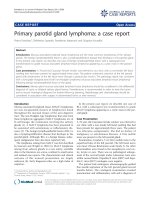
Fundus examination was normal OD, and revealed a pre-
retinal hemorrhage located in the macula OS (Figure 1).
Since the patient was pregnant, fluorescein angiography
was not performed. Blood pressure, full blood count,
Published: 7 April 2008
Journal of Medical Case Reports 2008, 2:101 doi:10.1186/1752-1947-2-101
Received: 4 November 2007
Accepted: 7 April 2008
This article is available from: />© 2008 Al-Mujaini and Montana; licensee BioMed Central Ltd.
This is an Open Access article distributed under the terms of the Creative Commons Attribution License ( />),
which permits unrestricted use, distribution, and reproduction in any medium, provided the original work is properly cited.
Journal of Medical Case Reports 2008, 2:101 />Page 2 of 3
(page number not for citation purposes)
coagulation profile, fasting blood sugar and sickle cell
tests were within normal limits. Additional tests for a
hypercoagulable state and autoimmune diseases were
negative. A clinical diagnosis of Valsalva retinopathy was
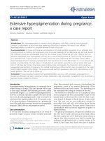
made and it was decided to observe the patient. Follow-up
one month later revealed improvement in the best cor-
rected visual acuity to 20/20 OS, with complete resolution
of the macular hemorrhage (Figure 2). The patient had a
spontaneous vaginal delivery, was seen immediately after
the delivery, with no recurrence of the retinal hemorrhage.
Discussion
Pregnancy exerts multiple hormonal, metabolic, hemato-
logical and immunological alterations in the mother that
represent risk factors for Valsalva retinopathy. Elevation of
intra-abdominal pressure during pregnancy, with a fur-
ther increase during labor, leads to a considerable eleva-
tion in intravenous pressure, which increases the potential
for retinal hemorrhages following a Valsalva maneuver.
Hematological changes during pregnancy such as throm-
bocytopenia add to the risk of Valsalva retinopathy in
pregnancy.
It is important to rule out all systemic diseases that may
result in retinal hemorrhages, such as diabetes, hyperten-
sion, sickle cell disease, anemia, coagulopathy, blood dys-
crasias and previous ocular vein occlusions.
Whether vaginal delivery poses a risk of recurrence or
exacerbation of the hemorrhage is unclear [2]. A review of
the literature showed no recurrence of retinopathy follow-
ing spontaneous vaginal delivery [2,3].
Valsalva maneuvers typically result in sub-internal limit-
ing membrane hemorrhages with a predilection for the
macula, but sub-retinal, retinal or intravitreal hemor-
rhages can occur. The prognosis in general is good and the
condition in most patients resolves spontaneously over
several months. Some patients may have a poor visual
outcome, which has been attributed to retinal pigmentary
changes at the macula [3]. In most cases, conservative
management is indicated with periodic observation. A
YAG laser has been employed in selective cases to disperse
the pre-retinal hemorrhages and speed up resolution [4].
Conclusion
Retinal hemorrhages can be generated by Valsalva maneu-
vers. Pregnancy is a known risk factor for Valsalva retinop-
athy; however, the diagnosis should be made only after
excluding other causes of retinal hemorrhages. It is a self-
limited event. We have reported a case of Valsalva retinop-
athy complicating normal pregnancy and confirm that, to
date, there is no evidence to indicate that there is a risk of
recurrence following spontaneous vaginal delivery.
Competing interests
The author(s) declare that they have no competing inter-
ests.
Authors' contributions
All authors participated in the design of the manuscript.
AM was involved in drafting the manuscript for important
intellectual content. All authors read and approved the
final manuscript.
Consent
Written informed consent was obtained from the patient
for publication of this case report and accompanying
images. A copy of the written consent is available for
review by the Editor-in-Chief of this journal.
Fundus photograph of the left eye a month laterFigure 2
Fundus photograph of the left eye a month later.
Fundus photograph of the left eye at presentationFigure 1
Fundus photograph of the left eye at presentation.
Publish with BioMed Central and every
scientist can read your work free of charge
"BioMed Central will be the most significant development for
disseminating the results of biomedical research in our lifetime."
Sir Paul Nurse, Cancer Research UK
Your research papers will be:
available free of charge to the entire biomedical community
peer reviewed and published immediately upon acceptance
cited in PubMed and archived on PubMed Central
yours — you keep the copyright
Submit your manuscript here:
/>BioMedcentral
Journal of Medical Case Reports 2008, 2:101 />Page 3 of 3
(page number not for citation purposes)
References
1. Chapman-Davies A, Lazarevic A: Valsalva maculopathy. Clin Exp
Optom 2002, 85:42-45.
2. Deane JS, Ziakas N: Valsalva retinopathy in pregnancy. Eye
1997, 11:137.
3. Wickremasinghe SS, Tranos PG, Davey C: Valsalva hemorrhagic
retinopathy in a pregnant woman: implications for delivery.
Acta Ophthalmol Scand 2003, 81:420-422.
4. Bourne RA, Talks SJ, Richards AB: Treatment of preretinal Val-
salva hemorrhages with neodymium: YAG laser. Eye 1999,
13:791-793.