Báo cáo y học: " Esophageal squamous cell carcinoma presenting with extensive skin lesions: a case report" docx
Bạn đang xem bản rút gọn của tài liệu. Xem và tải ngay bản đầy đủ của tài liệu tại đây (282.86 KB, 3 trang )
BioMed Central
Page 1 of 3
(page number not for citation purposes)
Journal of Medical Case Reports
Open Access
Case report
Esophageal squamous cell carcinoma presenting with extensive skin
lesions: a case report
GB Iwanski*
1
, A Block
1
, G Keller
1
, J Muench
1
, S Claus
2
, W Fiedler
1
and
C Bokemeyer
1
Address:
1
Department of Internal Medicine, Oncology and Hematology, University Hospital Hamburg, Eppendorf, Martinistrasse, 20246
Hamburg, Germany and
2
Department of Internal Medicine, Bethesda Hospital, Hamburg, Germany
Email: GB Iwanski* - ; A Block - ; G Keller - ;
J Muench - ; S Claus - ; W Fiedler - ;
C Bokemeyer -
* Corresponding author
Abstract
Introduction: Esophageal squamous cell carcinoma (ESCC) is the most common histological
subtype of cancer in the upper and middle esophagus and is characterized by a high rate of
mortality. The incidence of esophageal cancer varies greatly among regions of the world and occurs
at a high frequency in Asia and South America.
Case presentation: In our department, a 51-year-old man was diagnosed with ESCC after
presenting with extensive disseminated skin nodules. Biopsy of the nodules showed metastatic
ESCC. Cutaneous manifestations of esophageal neoplasia are very rare and are mainly described
for esophageal adenocarcinoma (EADC). Here we report a very uncommon case of extensive skin
metastases of ESCC.
Conclusion: Early biopsies of suspicious skin lesions are important and should be performed in
patients with unclear symptoms such as weight loss or dysphagia and especially in patients with a
history of cancer, since they can reveal the existence of a distant malignant disease leading to
diagnosis and prompt therapy.
Introduction
Cancer of the esophagus is the ninth most common
malignancy and ranks as the sixth most frequent cause of
cancer death in the world, constituting 7% of all gastroin-
testinal cancers [1]. Patients with esophageal cancer usu-
ally present with disease that is locally advanced and
which has already metastasized stage at the time of initial
diagnosis. Cancer of the esophagus exists in two main
forms with different etiological and pathological charac-
teristics: esophageal squamous cell carcinoma (ESCC)
and esophageal adenocarcinoma (EADC). ESCC is the
predominant histological subtype, comprising about 70%
of cases [2].
In general, skin metastases from malignant tumors of the
internal organs are rarely seen, with a frequency of
between 0.7 and 9% [3-5]. The overall survival rate varies
from 4.3 to 4.7 months [6]. The cancer types most com-
monly associated with cutaneous metastases are breast,
lung and melanoma [4,7,8]. Metastatic spread to the skin
occurs either hematogenously or via the lymphatic system
and presents in the form of rapidly growing papules or
Published: 21 April 2008
Journal of Medical Case Reports 2008, 2:115 doi:10.1186/1752-1947-2-115
Received: 26 September 2007
Accepted: 21 April 2008
This article is available from: />© 2008 Iwanski et al; licensee BioMed Central Ltd.
This is an Open Access article distributed under the terms of the Creative Commons Attribution License ( />),
which permits unrestricted use, distribution, and reproduction in any medium, provided the original work is properly cited.
Journal of Medical Case Reports 2008, 2:115 />Page 2 of 3
(page number not for citation purposes)
nodules [9,10]. On histopathology, clusters of atypical
cells infiltrating the dermis without connection to the
adjacent epidermis can be seen [6]. Here we report an
uncommon case of massive cutaneous metastases of
ESCC in a 51-year-old man.
Case presentation
A 51-year-old man was admitted to our department with
a four-week history of dysphagia, weight loss and nausea.
He had a medical history of multiple sclerosis since April
2004 and a smoking history of 30 pack-years. The patient
underwent esophagogastroduodenoscopy resulting in the
diagnosis of esophageal carcinoma located in the mid-
thoracic part of the esophagus. Histology of an
endosonography-guided biopsy showed an intermediate
grade ESCC according to the criteria of the American Joint
Committee of Cancer (AJCC). Moreover, the patient pre-
sented with approximately 20 diffuse, painless and solid
skin nodules that were about 1–3 cm in diameter, found
all over his body surface including the scalp, upper
extremities, axillae, back, chest and abdominal wall.
According to the patient they had been growing rapidly
over the previous four weeks, and he had noticed the first
skin lesion more than two months earlier. Excisional
biopsy of one representative prominent cutaneous forma-
tion on the abdominal wall was performed. On macro-
scopic inspection, the lesion was superficially ulcerated
and measured 2 cm × 3 cm (Figure 1). Histopathology
revealed nodulous skin infiltration of intermediate grade
ESCC (Figure 2). Interestingly, staging by thoracoabdom-
inal computed tomography (CT) scan showed some of
these skin lesions (Figure 3). Extensive mediastinal lymph
nodes and multiple osteolytic lesions of the spine were
also detected without signs of any other tumor manifesta-
tion (T1-2, N1, M1, G2; ESCC state IV). The patient sub-
sequently received palliative chemotherapy with cisplatin
(80 mg/sqm) and 5-fluoruracil (1,000 mg/sqm) given
over four days every three weeks. After three cycles of
chemotherapy, the cutaneous metastases became smaller,
but some appeared in new areas.
Discussion
Due to the extreme rarity of cutaneous metastases from
ESCC, there are only limited data in the literature regard-
ing their incidence. Fereidooni and colleagues reported a
solid facial skin metastasis of EACC [11]. Two additional
cases have been published discussing solitary metastases
on a digit from an unusual variant of ESCC, the basaloid
squamous cell carcinoma [12,13]. Schoenlaub and col-
leagues reviewed the clinical findings and overall survival
of 200 patients with cutaneous metastases of various can-
cers. The incidence of cutaneous metastases from EACC
was 2 out of the 200 cases studied [6]. The cancers most
frequently causing cutaneous metastases were breast can-
cers (n = 64), pulmonary cancers (n = 36) and melanomas
(n = 31) [6]. Reingold reported clinical and necropsy find-
ings of 32 cases out of 2,300 internal carcinomas. The
most common primary site was the lungs (50%). The
esophagus was the primary tumor site in just one case and
this was an adenocarcinoma. The most common sites of
skin metastases were on the chest and abdomen [14].
Lookingbill et al reviewed 420 patients with cutaneous
metastases from melanoma and carcinoma [9]. In this
study, tumor registry data from 7,608 patients was evalu-
ated; 4,020 of these patients had metastatic disease and
420 (10.4%) had cutaneous metastases. The most com-
mon primary tumors causing cutaneous metastases were
melanoma (n = 77) and breast cancer (n = 212). The
esophagus was the primary site in only three cases, spread-
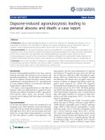
HistopathologyFigure 2
Histopathology. Infiltration of cutis and subcutaneous fat
by intermediate grade atypical squamous cell clusters (HE).
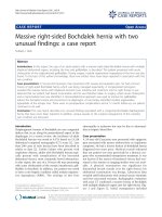
Representative ESCC skin metastasis on the abdominal wall Diameter 2–3 cmFigure 1
Representative ESCC skin metastasis on the abdomi-
nal wall Diameter 2–3 cm.
Publish with BioMed Central and every
scientist can read your work free of charge
"BioMed Central will be the most significant development for
disseminating the results of biomedical research in our lifetime."
Sir Paul Nurse, Cancer Research UK
Your research papers will be:
available free of charge to the entire biomedical community
peer reviewed and published immediately upon acceptance
cited in PubMed and archived on PubMed Central
yours — you keep the copyright
Submit your manuscript here:
/>BioMedcentral
Journal of Medical Case Reports 2008, 2:115 />Page 3 of 3
(page number not for citation purposes)
ing mainly to the chest and abdomen [9]. Tharakaram
described five cases of skin metastases from ESCC in male
patients [15].
Conclusion
Skin manifestations of ESCC are extremely rare and only
a small number of cases with solid skin metastases have
been reported. A case of ESCC with such diffuse and mas-
sive skin metastases, most likely indicating highly aggres-
sive disease, has not been described previously. Our
patient complained about these unusual cutaneous
metastases before any of the more usual symptoms such
as dysphagia or weight loss were manifested.
Competing interests
The authors declare that they have no competing interests.
Authors' contributions
GBI initiated the report and undertook the majority of the
writing of the manuscript. AB made substantial contribu-
tions to the conception and design of the report and was
involved in drafting the manuscript. GK, JM and SC made
contributions to the conception and design of the report
and was involved in drafting the manuscript. WF and CB
made substantial contributions to the conception and
design of the manuscript and revised it critically for
important intellectual content. All authors read and
approved the final manuscript.
Consent
Written informed consent was obtained from the patient
for publication of this case report and any accompanying
images. A copy of the written consent is available for
review by the Editor-in-Chief of this journal.
Acknowledgements
We thank A Niendorf MD and K Hamper MD for providing histopatholog-
ical figures, T Göttsche MD for radiological diagnosis and HANSERAD for
providing the CT scan pictures.
References
1. Levine MS, Halvorsen RA: Carcinoma of the esophagus. In Text-
book of Gastrointestinal Radiology Edited by: Gore RM, Levine MS. Phil-
adelphia, PA: Saunders; 2000:403-433.
2. Parkin DM, Bray F, Ferlay J, Pisani P: Estimating the world cancer
burden: Globocan 2000. Int J Cancer 2001, 94:153-156.
3. Spencer PS, Helm TN: Skin metastases in cancer patients. Cutis
1987, 39:119-121.
4. Lookingbill DP, Spangler N, Sexton FM: Skin involvement as the
presenting sign of internal carcinoma. J Am Acad Dermatol 1990,
22:19-26.
5. Rosen T: Cutaneous metastases. Med Clin North Am 1980,
64:885-900.
6. Schoenlaub P, Sarraux A, Grosshans E, Heid E, Cribier B: Survival
after the occurrence of cutaneous metastasis: a study of 200
cases. Ann Dermatol Venereol 2001, 128:1310-1315.
7. Brownstein MH, Helwig EB: Patterns of cutaneous metastasis.
Arch Dermatol 1972, 105:862-868.
8. Schoenlaub P, Meyer P, Heid E, Grosshans E, Cribier B: Métastases
cutanées révélatrices d'un cancer méconnu. Étude anatomo-
clinique de 40 cas. Ann Dermatol Venereol 1998, 3S:90-91.
9. Lookingbill DP, Spangler N, Helm KF: Cutaneous metastases in
patients with metastatic carcinoma: A retrospective study of
4020 patients. J Am Acad Dermatol 1993, 29:228-236.
10. Schwartz RA: Cutaneous metastatic disease. J Am Acad Dermatol
1995, 31:161-182.
11. Fereidooni F, Kovacs K, Azizi MR, Nikoo M: Skin metastasis from
an occult esophageal adenocarcinoma. Can J Gastroenterol 2005,
19:673-676.
12. Houston JD, Telepak RJ: An isolated digital metastasis of
esophageal basaloid squamous cell carcinoma. Clin Nucl Med
2000, 25:557-558.
13. Silfen R, Amir A, Tobar A, Hauben DJ: The digital pulp as a pre-
senting site of metastatic esophageal carcinoma. Ann Plast
Surg 2001, 46:183-184.
14. Reingold IM: Cutaneous metastases from internal carcinoma.
Cancer 1966, 19:162-168.
15. Tharakaram S: Metastases to the skin. Int J Dermatol 1988,
27:240-242.
Thoraco-abdominal CT scan showing two representative cutaneous metastases (arrows)Figure 3
Thoraco-abdominal CT scan showing two represent-
ative cutaneous metastases (arrows).