Báo cáo y học: " Gigantic retroperitoneal hematoma as a complication of anticoagulation therapy with heparin in therapeutic doses: a case report" docx
Bạn đang xem bản rút gọn của tài liệu. Xem và tải ngay bản đầy đủ của tài liệu tại đây (1.03 MB, 5 trang )
BioMed Central
Page 1 of 5
(page number not for citation purposes)
Journal of Medical Case Reports
Open Access
Case report
Gigantic retroperitoneal hematoma as a complication of
anticoagulation therapy with heparin in therapeutic doses: a case
report
Stavros I Daliakopoulos*
1
, Andreas Bairaktaris
1
, Dimitrios Papadimitriou
2
and Perikles Pappas
2
Address:
1
Herz- und Diabeteszentrum Nordrhein, Westfalen, Georgstrasse, Bad Oeynhausen, Universitätsklinikum der Ruhr-Universität Bochum,
Germany and
2
Department of Vascular and Endovascular Surgery, 424 Military Hospital, Thessaloniki, Greece
Email: Stavros I Daliakopoulos* - ; Andreas Bairaktaris - ;
Dimitrios Papadimitriou - ; Perikles Pappas -
* Corresponding author
Abstract
Introduction: Spontaneous retroperitoneal hemorrhage is a distinct clinical entity that can
present as a rare life-threatening event characterized by sudden onset of bleeding into the
retroperitoneal space, occurring in association with bleeding disorders, intratumoral bleeding, or
ruptures of any retroperitoneal organ or aneurysm. The spontaneous form is the most infrequent
retroperitoneal hemorrhage, causing significant morbidity and representing a diagnostic challenge.
Case presentation: We report the case of a patient with coronary artery disease who presented
with transient ischemic attack, in whom anticoagulant therapy with heparin precipitated a massive
spontaneous atraumatic retroperitoneal hemorrhage (with international normalized ratio 2.4),
which was treated conservatively.
Conclusion: Delay in diagnosis is potentially fatal and high clinical suspicion remains crucial. Finally,
it is a matter of controversy whether retroperitoneal hematomas should be surgically evacuated
or conservatively treated and the final decision should be made after taking into consideration
patient's general condition and the possibility of permanent femoral or sciatic neuropathy due to
compression syndrome.
Introduction
Hemorrhage is the most important complication of
unfractionated heparin in patients with atrial fibrillation
(AF) treated with oral vitamin K antagonist (VKA) during
hospitalization or among those receiving anticoagulants
in terms of emergency or elective cardiac surgery [1,2] or
in the initial treatment of deep venous thrombosis [3,4].
Analysis of the data presented by the European AF Trial
Study Group [5] shows that as the international normal-
ized ratio (INR) increased, there was an increase in the risk
of major bleeding, such that at INR ≥ 5.0, the risk of bleed-
ing increased 3.6-fold relative to INR ≤ 2. The optimal
intensity of anticoagulation that achieved maximum ther-
apeutic effect with minimum risk was determined to be at
Published: 17 May 2008
Journal of Medical Case Reports 2008, 2:162 doi:10.1186/1752-1947-2-162
Received: 6 November 2007
Accepted: 17 May 2008
This article is available from: />© 2008 Daliakopoulos et al; licensee BioMed Central Ltd.
This is an Open Access article distributed under the terms of the Creative Commons Attribution License ( />),
which permits unrestricted use, distribution, and reproduction in any medium, provided the original work is properly cited.
Journal of Medical Case Reports 2008, 2:162 />Page 2 of 5
(page number not for citation purposes)
INR = 3.0. These data, along with recommendations from
the recent American College of Chest Physicians (ACCP)
guidelines, indicate that the optimal intensity of anticoag-
ulation for balancing efficacy in presenting stoke, while
minimizing the risk of bleeding, is within the range INR =
2.0–3.0 (see [6]). Among outpatients receiving oral anti-
coagulants those with INR ≥ 6.0 face a significant risk of
major hemorrhage [7].
Retroperitoneal hemorrhage is most frequently seen after
femoral artery catheterization or pelvic and lumbar
trauma [8-10]. In the absence of trauma, retroperitoneal
hemorrhage most frequently results from a ruptured
abdominal aortic aneurysm or bleeding from an underly-
ing condition in the kidneys or adrenal glands. Spontane-
ous retroperitoneal hemorrhage (SRH) denotes bleeding
without any known inciting trauma or underlying retro-
peritoneal pathology. SRH is uncommon and is almost
exclusively seen in association with anticoagulation states,
coagulopathies and hemodialysis [11,12].
A plethora of conditions have been used as a possible
hypothesis of the pathophysiology of SRH. Unrecognized
minor trauma in the microcirculation in the presence of
coagulopathy has been suggested [13,14].
The surgeons' quiver contains various types of approach
to the treatment of this relatively uncommon complica-
tion such as conservative management, angiographic eval-
uation, percutaneous embolization or surgical
intervention.
Case presentation
A 57-year-old Caucasian male was admitted to our hospi-
tal presenting with focal ischemic cerebral neurological
deficit of acute onset. The patient had had an acute non-
Q-wave myocardial infarction episode 11 years ago and
post-infarct had undergone percutaneous transluminal
coronary angioplasty: ramus circumflexus in 1995 and
ramus diagonalis I in 1997. Eleven months before admis-
sion, an evaluation elsewhere had revealed persistent
atrial fibrillation and since this evaluation the patient had
been receiving Warfarin and had maintained INR = 2.0–
2.5.
On the day of admission, examination of the patient
revealed intense dizziness with diplopia, instability and
complete left-sided homonymous hemianopsia. The find-
ings suggested a transient ischemic attack involving the
anterior circulation: carotid artery territory.
There was no personal or family history of coagulopathy
or stroke, valvular heart disease trauma, chest pain or
illicit intravenous drug usage. He smoked 20 cigarettes
daily and consumed alcohol in moderation in the past.
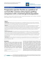
The prothrombin was normal, INR = 2.4, the partial
thromboplastin time was 45 seconds, the values for urea,
nitrogen, creatinine, glucose, uric acid, bilirubin, phos-
phorus, electrolytes, creatinine kinase, lactate dehydroge-
nase, amylase and alkaline phosphatase were normal. An
electrocardiogram (ECG) revealed atrial fibrillation at a
rate of 110, with nonspecific ST-segment and T-wave
abnormalities. A radiograph of the chest showed clear
lungs and slight cardiac enlargement. A cardiac ultrasono-
graphic examination showed no vegetations, intracardiac
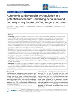
MRI – axial plan showing a large, mixed density mass in the right side of the abdomen suggestive of a large retroperito-neal hematoma, with areas of hyperdensity (arrows) indicat-ing ongoing hemorrhageFigure 2
MRI – axial plan showing a large, mixed density mass
in the right side of the abdomen suggestive of a large
retroperitoneal hematoma, with areas of hyperden-
sity (arrows) indicating ongoing hemorrhage.
MRI – transverse plan (L4) with IV contrast gadolinium-BOPTA, revealing a well-defined mass, a huge retroperito-neal hematomaFigure 1
MRI – transverse plan (L4) with IV contrast gadolin-
ium-BOPTA, revealing a well-defined mass, a huge
retroperitoneal hematoma.
Journal of Medical Case Reports 2008, 2:162 />Page 3 of 5
(page number not for citation purposes)
thrombus, segmental wall-motion abnormalities or int-
racardiac shunts. A test for the erythrocyte sedimentation
rate was normal, as were tests for antinuclear antibodies,
lupus anticoagulant and antiphospholipid antibody.
Computed tomography (CT) brain imaging was per-
formed without the use of contrast material, but failed to
indicate hemorrhage, infarct, abscess, tumor or cerebral
metastasis. Heparin 20,000IE/24 hours intravenously and
Metoprolol 100 mg by mouth were administered.
Repeated physical examinations and ECGs showed no
changes.
On the second hospital day the patient awoke with a
slight neurologic deficit that gradually progressed in a
stepwise fashion. Hemiplegia (upper left extremity and
face were involved), hemianesthesia and Babinski sign
contralateral to the hemiparesis were established. He had
mild dysarthria, but his speech was fluent and his compre-
hension, repetition and naming abilities were intact. CT
brain imaging was performed and no hemorrhagic trans-
formation was found. Dipyridamole 200 mg/day, Aspirin
25 mg/day, Heparin 20,000IE/24 hours and Mannitol
20% solution (1 g/kg) were administered. Daily monitor-
ing of ECG, vital signs, electrolytes, blood urea nitrogen,
creatinine, urine output showed no changes.
On the fifth hospital day the patient noted the acute onset
of pain in the lower right abdominal quadrant and lum-
bar region accompanied by mild nausea. The patient held
the right hip in flexion and external rotation. Any attempt
to straighten the leg aggravated the pain with radiation to
the medial and anterior portions of the lower extremity
Weakness of the right quadriceps femoris muscle, par-
esthesia over the anterior thigh and right flank were evi-
dent. The partial thromboplastin time was 43 seconds and
INR = 2.4.
Hematocrit level fell as did hemoglobin (Table 1). CT and
magnetic resonance imaging (MRI) scan of the abdomen
and the pelvis was obtained (Figures 1 and 2) and
revealed extensive enlargement and heterogeneity of the
right iliopsoas muscle as well as displacement of the right
kidney. The high-attenuation component in the absence
of intravenous contrast enhancement (Figure 3) is a find-
ing that is usually consistent with the presence of a large
retroperitoneal hematoma.
Transfusion of six units of packed red cells and the admin-
istration of two units of fresh frozen plasma was followed
by fluid overload. The patient was treated conservatively
and his condition promptly stabilized after the restoration
of normal blood coagulation; however, he remained in
the hospital for 38 days. Three months later he had signs
of partial lateral paresis of the right quadriceps muscle and
thigh adductors and, at 1-year follow-up, the only find-
ings were suggestive of the previous transient ischemic
Table 1: Hematologic laboratory values
On admission On fifth hospital day
Variable Value Variable Value
Hematocrit (%) 43 Hematocrit (%) 24.3
Hemoglobin (g/dl) 13.7 Hemoglobin (g/dl) 8.7
Mean corpuscular volume (μm
3
) 92 Platelet count (per mm
3
)85,000
Erythrocyte sedimentation rate (mm/hour) 130 White-cell amount (per mm
3
)14,900
White-cell amount (per mm
3
) 10,200 Prothrombin time (s) 16.1
Differential count (%) Partial thromboplastin time (s) 63
Neutrophilis 64 D-dimer test (μg/l) Negative
a
Lymphocytes 27 Fibrinogen (mg/dl) 446
b
Monocytes 7 Antithrombin III (mg/dl) 28
c
Eosinophilis 1 Factor II (mg/dl) 14
d
Basophilis 1 Factor V (mg/dl) 0.8
e
Platelet count (per mm
3
) 265,000 Factor VII (mg/dl) 0.3
f
Factor X (mg/dl) 0.8
g
Prekallikrein (mg/dl) 5
h
a
Normal values less than 250 μg/l.
b
Normal values in the range 200–400 mg/dl.
c
Normal immunologic assay range 17–30 mg/dl.
d
Normal values in the range 10–15 mg/dl.
e
Normal values in the range 0.5–1 mg/dl.
f
Normal value 0.2 mg/dl.
g
Normal values in the range 0.6–0.8 mg/dl.
h
Normal value 5 mg/dl.
Journal of Medical Case Reports 2008, 2:162 />Page 4 of 5
(page number not for citation purposes)
attack involving the carotid artery territory; he had recov-
ered completely from the femoral neuropathy.
Discussion
The large study of Sasson et al. [15] showed that patients
who are receiving Heparin anticoagulation therapy, even
in therapeutic doses, should be carefully monitored for
the development of groin pain or leg weakness.
The most common symptoms are the acute onset, the
severity and the persistence of the patient's pain in the
lower abdominal quadrant, inguinal or lumbar region,
and its radiation to the scrotum. Pain and paresthesia
extend over the anterior, medial or lateral aspects of the
lower extremities depending on the branches of the lum-
bar plexus that are involved. The most frequently involved
nerve is the femoral nerve, the largest branch of the lum-
bar plexus which arises from the dorsal branches of L2, L3
and L4 ventral rami. It descends through the psoas major,
emerging low on its lateral border and then passes
between the psoas and iliacus, which makes the nerve vul-
nerable to traction injury from an underlying iliacus mus-
cle hematoma [16,17], deep to the iliac fascia, passing
behind the inguinal ligament into the thigh.
The diagnosis of atraumatic retroperitoneal hemorrhage
remains challenging even when high-resolution MRI and
CT imaging are used, because a large number of benign or
malignant lesions can mimic this condition [18,19].
However, despite these limitations, MRI and CT imaging
are superior to ultrasound and should be the preferred pri-
mary investigation [20-22].
The mainstay management currently consists of modifica-
tion or cessation of anticoagulation therapy according to
its clinical requirement, correction of the anticoagulation
state, volume resuscitation and hemodynamic stabiliza-
tion with adequate hematology and transfusion therapy
and supportive measures [23]. Small hematomas with
mild symptoms of neuropathy, without resultant obscura-
tion, displacement or compression of normal retroperito-
neal structures, without the need for multiple transfusions
and without signs of infection may be treated conserva-
tively.
On the other hand the effectiveness and safety of surgical
intervention and evacuation of the hematoma should be
considered as a potential strategy in uncontrollable hemo-
dynamic collapse or when the nerve involved in the
decompression might be effective in that the direct pres-
sure and pressure-induced ischemic effects are reversible
[24,25]. The latter is limited by the inability to localize or
control the bleeding vessel and the risk of worsening the
bleeding by releasing the tamponade [26].
Conclusion
The rarity of this possible complication of the intravenous
use of Heparin in patients with INR < 4.5 means that it
remains a challenge for surgeons. We strongly suggest
that, according to our experience, daily measurement of
INR and activated partial thromboplastin time (aPTT) in
patient's receiving Heparin intravenously as an anticoagu-
lation agent is of great importance. In deep vein thrombo-
sis or acute myocardial infarction, the usual protocol
requires injection of Heparin monitored by the pro-
thrombin time, aPTT or both followed by long-term ther-
apy with oral anticoagulants. As the half-life of Heparin is
3 hours, we suggest that aPTT to be measured 3 hours after
Heparin administration or 1 hour before the next dose.
Some of the most important factors for the diagnosis are
acute onset of pain, a dramatic change in the patient's
clinical status and high clinical suspicion. CT and MRI
remain the most powerful diagnostic tools. The complex
challenge for the surgeon is the choice of clinical pathway
in the management of this rare entity and this choices
should only be made after taking two key points into con-
sideration: (i) the patient's general condition; (ii) in the
presence of permanent femoral or sciatic neuropathy due
to a compression syndrome, hemodynamically unstable
patients should be managed with an emergency laparot-
omy.
Competing interests
The authors declare that they have no competing interests.
Authors' contributions
SID participated in the sequence alignment, in the design
of the case report and drafted the manuscript. AB partici-
MRI – coronar planFigure 3
MRI – coronar plan.
Publish with BioMed Central and every
scientist can read your work free of charge
"BioMed Central will be the most significant development for
disseminating the results of biomedical research in our lifetime."
Sir Paul Nurse, Cancer Research UK
Your research papers will be:
available free of charge to the entire biomedical community
peer reviewed and published immediately upon acceptance
cited in PubMed and archived on PubMed Central
yours — you keep the copyright
Submit your manuscript here:
/>BioMedcentral
Journal of Medical Case Reports 2008, 2:162 />Page 5 of 5
(page number not for citation purposes)
pated in the design of the case report. DP participated in
the design of the case report and coordination. PP partic-
ipated in the design of the study. All authors read and
approved the final manuscript.
Consent
Written informed consent was obtained from the patient
for publication of this case report and accompanying
images. A copy of the written consent is available for
review by the Editor-in-Chief of this journal.
References
1. Juergens CP, Semsarian C, Keech AC, Beller EM, Harris PJ: Hemor-
rhagic complications of intravenous heparin use. Am J Cardiol
1997, 80:150-154.
2. Mant MJ, O'Brien BD, Thong KL, Hammond GW, Birtwhistle RV,
Grace MG: Haemorrhagic complications of heparin therapy.
Lancet 1977, 1:1133-1135.
3. Leizorovicz A: Comparison of the efficacy and safety of low
molecular weight heparins and unfractionated heparin in the
initial treatment of deep venous thrombosis: an updated
meta-analysis. Drugs 1996, 52(Suppl 7):30-37.
4. Montoya JP, Pokala N, Melde SL: Retroperitoneal hematoma and
enoxaparin. Ann Intern Med 1999, 131:796-797.
5. The European Atrial Fibrillation Trial Study Group: Optimal oral
anticoagulant therapy in patients with nonrheumatic atrial
fibrillation and recent cerebral ischemia. N Engl J Med 1995,
333:5-10.
6. Singer Daniel E MD, Chair, Albers Gregory W MD, Dalen James E
MD, MPH, Master FCCP, Go Alan S MD, Halperin Jonathan L MD,
Manning Warren J MD: Antithrombotic therapy in atrial fibril-
lation: the Seventh ACCP Conference on Antithrombotic
and Thrombotic Therapy. Chest 2004, 126:429S-456S.
7. Hylek EM, Chang YC, Skates SJ, Hughes RA, Singer DE: Prospective
study of outcomes of ambulatory patients with excessive
Warfarin anticoagulation. Arch Inter Med 2000, 160:1612-1617.
8. Panetta T, Sclafani SJ, Goldstein AS, Phillips TF, Shaftan GW: Percu-
taneous Transcatheter embolization for massive bleeding
from pelvic fractures. J Trauma 1985, 25:1021-1029.
9. Illescas FF, Baker ME, McCann R, Cohan RH, Silverman PM, Dunnick
NR: CT evaluation of retroperitoneal hemorrhage associ-
ated with femoral arteriography. AJR Am J Roentgenol 1986,
146:1289-1292.
10. Sclafani Salvatore JA, Florence Lauren O, Phillips Thomas F, Scalea
Thomas M, Glanz Sidney, Goldstein Alan S, Duncan Albert O, Shaftan
Gerald W: Lumbar arterial injury: radiologic diagnosis and
management. Radiology 1987,
165:709-714.
11. Bhasin HK, Dana CL: Spontaneous retroperitoneal hemor-
rhage in chronically hemodialyzed patients. Nephron 1978,
22:322-327.
12. Fernadez-Palazzi F, Hernandez SR, De Bosch NB, De Saez AR:
Hematomas within the iliopsoas muscles in hemophilic
patients: the Latin American experience. Clin Orthop Relat Res
1996, 328:19-24.
13. McCort JJ: Intraperitoneal and retroperitoneal hemorrhage.
Radiol Clin North Am 1976, 14:391-405.
14. Heim M, Horoszowski H, Seligsohn U, Martinowitz U, Strauss S: Ili-
opsoas hematoma – its detection, and treatment with spe-
cial reference to hemophilia. Arch Orthop Trauma Surg 1982,
99(3):195-197.
15. Sasson Z, Mangat I, Peckham KA: Spontaneous iliopsoas
hematoma in patients with unstable coronary syndromes
receiving intravenous heparin in therapeutic doses. Can J Car-
diol 1996, 12:490-494.
16. Nobel W, Marks SC Jr, Kubik S: The anatomical basis for femo-
ral nerve palsy following iliacus hematoma. J Neurosurg 1980,
52:533-540.
17. Susens GP, Hendrickson CG, Mulder MJ, Sams B: Femoral nerve
entrapment secondary to a heparin hematoma. Ann Intern
Med 1968, 69:575.
18. Nishimura H, Zhang Y, Ohkuma K, Uchida M, Hayabuchi N, Sun S:
MR imaging of soft-tissue masses of the extraperitoneal
spaces. Radiographics 2001, 21:1141-1154.
19. Lenchik L, Dovgan DJ, Kier R: CT of the iliopsoas compartment:
value in differentiating tumor, abscess and hematoma. AJR
Am J Roentgenol 1994, 162(1):83-86.
20. Takebayashi S, Matsui K, Hidai H: Nontraumatic hemorrhage in
abdomen and retroperitoneum-CT, sonographic and clinical
findings. Nippon Igaku Hoshasen Gakkai Zasshi 1990, 50:1206-1214.
21. Pless T, Loertzer H, Brandt S, Radke J, Fornara P, Soukup J: Atrau-
matic retroperitoneal hemorrhage: interdisciplinary and dif-
ferential diagnostic considerations based on a case report.
Anaesthesiol Reanim
2003, 28:50-53.
22. Nazarian LN, Lev-Toaff AS, Spettell CM, Wechsler RJ: CT assess-
ment of abdominal hemorrhage in coagulopathic patients:
impact on clinical management. Abdom Imaging 1999,
24:246-249.
23. Sherer DM, Dayal AK, Schwartz BM, Oren R, Abufalia O: Extensive
spontaneous retroperitoneal hemorrhage: an unusual com-
plication of heparin anticoagulation during pregnancy. J
Matern Fetal Med 1999, 8:196-199.
24. Baker BH, Baker MS: Indications for exploring the retroperito-
neal space. South Med J 1980, 73:969-970.
25. Topgul K, Uzun O, Anadol AZ, Gok A: Surgical management of
enoxiparin- and/or warfarin- induced massive retroperito-
neal bleeding: report of a case and review of the literature.
South Med J 2005, 98:104-106.
26. Grimm MR, Vrahas MS, Thomas KA: Pressure volume character-
istics of the intact and disrupted pelvic retroperitoneum. J
Trauma 1998, 44:454-459.