báo cáo khoa học:" Histological analysis of the effects of a static magnetic field on bone healing process in rat femurs" pptx
Bạn đang xem bản rút gọn của tài liệu. Xem và tải ngay bản đầy đủ của tài liệu tại đây (1.91 MB, 9 trang )
BioMed Central
Page 1 of 9
(page number not for citation purposes)
Head & Face Medicine
Open Access
Research
Histological analysis of the effects of a static magnetic field on bone
healing process in rat femurs
Edela Puricelli*
†1
, Lucienne M Ulbrich
†2
, Deise Ponzoni
†2
and João Julio da
Cunha Filho
†2
Address:
1
Oral and Maxillofacial Surgery Unit, Hospital de Clinicas de P.A., School of Dentistry, UFRGS, Porto Alegre, RS, Brazil and
2
Dept. of Oral
Maxillofacial Surgery, School of Dentistry, UFRGS, Porto Alegre, RS, Brazil
Email: Edela Puricelli* - ; Lucienne M Ulbrich - ; Deise Ponzoni - ;
João Julio da Cunha Filho -
* Corresponding author †Equal contributors
Abstract
Background: The aim of this study was to investigate, in vivo, the quality of bone healing under
the effect of a static magnetic field, arranged inside the body.
Methods: A metallic device was developed, consisting of two stainless steel washers attached to
the bone structure with titanium screws. Twenty-one Wistar rats (Rattus novergicus albinus) were
used in this randomized experimental study. Each experimental group had five rats, and two animals
were included as control for each of the groups. A pair of metal device was attached to the left
femur of each animal, lightly touching a surgically created bone cavity. In the experimental groups,
washers were placed in that way that they allowed mutual attraction forces. In the control group,
surgery was performed but washers, screws or instruments were not magnetized. The animals
were sacrificed 15, 45 and 60 days later, and the samples were submitted to histological analysis.
Results: On days 15 and 45 after the surgical procedure, bone healing was more effective in the
experimental group as compared to control animals. Sixty days after the surgical procedure,
marked bone neoformation was observed in the test group, suggesting the existence of continued
magnetic stimulation during the experiment.
Conclusion: The magnetic stainless steel device, buried in the bone, in vivo, resulted in increased
efficiency of the experimental bone healing process.
Background
Bone neoformation is of primary importance for the suc-
cess of dental clinical-surgical treatments. Much attention
has been given to the research of new strategies to improve
oral maxillofacial surgical techniques, as well as on the
knowledge and application of biomaterials [1] an their
possible chemical and physical consequences on the
patients.
Electromagnetic fields have been used for the stimulation
of bone neoformation processes. Their effects are
observed in the treatment of osteoporosis, osteonecrosis,
osteotomized areas, integration of bone grafts and post-
traumatic pseudarthrosis [2]. Several cell functions were
also shown to be influenced by electromagnetic fields
[3,4]. Electromagnetism affects osteogenesis through
mechanisms such as neovascularization, collagen produc-
Published: 24 November 2006
Head & Face Medicine 2006, 2:43 doi:10.1186/1746-160X-2-43
Received: 15 February 2006
Accepted: 24 November 2006
This article is available from: />© 2006 Puricelli et al; licensee BioMed Central Ltd.
This is an Open Access article distributed under the terms of the Creative Commons Attribution License ( />),
which permits unrestricted use, distribution, and reproduction in any medium, provided the original work is properly cited.
Head & Face Medicine 2006, 2:43 />Page 2 of 9
(page number not for citation purposes)
tion, proliferation and differentiation of osteogenic cells,
and the maintenance of the molecular structure of the
extracellular matrix [5-7].
The objective of the present study is to contribute to the
understanding of processes involved in the response of
bone to electromagnetic fields, by evaluation of cortical
and trabecular bone neoformation. Cell stimulation was
induced by static, in vivo buried magnetic fields.
Methods
Twenty-one male Wistar rats (Rattus novergicus albinus)
were used in this randomized experimental study, aiming
at the use of permanent magnetic fields buried in vivo. The
animals were six-months old and weighed in average 450
grams. They were divided into three experimental and
control groups, which were analyzed on days 15, 45 and
60 after beginning of the experiment.
The metal devices consisted of commercially pure marten-
sitic stainless steel washers and titanium screws. The
screws measured 1.0 mm in diameter, 0.5 mm in thread
pitch and 2.0 mm in length. The pre-made magnetized
washers were 3.0 mm in outer diameter, 1.5 mm in core
diameter and 0.5 mm in thick. They were held over a 60
mm × 12 mm × 5 mm magnet during the sterilization
process and surgery. The magnetic field was 41 Gauss (G).
Calculations were performed at the Electromagnetism
Laboratory, Physics Institute from Universidade Federal
do Rio Grande do Sul.
The animals were anesthetized by intraperiotoneal injec-
tion of sodium tiopenthal in a dose of 25 mg/kg body
weight and local infiltration of 3% prilocaine with fely-
pressin.
After reaching the medial portion of the left femur dia-
phisis, a surgical bone cavity was produced with a tre-
phine (PROMM
®
, Comércio de Implantes Cirúrgicos Ltda.
Porto Alegre, RS, Brazil) measuring 2.0 mm diameter
active region, with low rotation and constant irrigation.
Two holes were drilled with a drill guide (PROMM
®
) 1.0
mm away from the osteotomized border, one of them
proximal and the other one distal to the surgical bone cav-
ity. The washers were attached to the bone structure with
titanium screws (Figure 1). A magnetic field was created in
animals of the test groups, by placing up the north and
south poles of the distal and proximal washers. In control
animals, surgery included non-magnetized instruments,
washers and screws.
The placement and stability of implants were confirmed
by radiographic examination at the end of the experi-
ments. Samples were submitted to longitudinal section-
ing of the femur, which allowed simultaneous
examination of the surgical cavity between the screw
holes. The samples were prepared in hematoxylin and
eosin stain (HE) for histological analysis.
Results
On day 15, extensive trabecular formation with marked
osteoblastic activity was seen on the cortical marginal
zone of samples from animals of the control group, begin-
ning in the endosteum close to the osteotomized cortical
surface. On the external surface, its predominantly hori-
zontal and flat direction maintained continuity and shape
of the remaining cortical levels. Trabecular proliferation
was also apparent in a centripetal direction relative to the
surgical cavity. Medullary spaces showed connective tissue
which was richly populated with cells and intense osteob-
lastic activity (Figure 2). In animals from the test group,
trabecular formations presented a marginal centripetal
direction relative to the magnetic field. The cortical wall
on the osteotomized area presented a tendency to convex-
ity, escaping from the horizontal outer border where the
remaining cortical walls were located (Figure 3). Regular
bone formation was observed following the limits of the
magnetic washer. Medullary spaces were filled with
numerous trabecular bone formations, showing a ten-
dency for more abundant vertical growth. Rich hemat-
opoietic tissue, with marked cell activity, could also be
observed.
On day 45, little activity was observed in the cortical zone
of samples derived from the control group, which main-
tained convexity and showed predominant lamellar dep-
osition. Areas limited by the washers showed fibrous
tissue associated to osteoclastic activity. Medullary space
was extensively invaded by bone trabecules and vascular-
ized hematopoietic tissue (Figure 4). In the test group, the
bone structure showed well organized areas of trabecular
bone interspersed with medullary tissue. Blood vessels,
adipose degeneration and osteoclastic activity were
observed in medullary spaces, suggesting bone remodel-
ling (Figure 5).
On day 60, active remodelling of the surgical cavity was
apparent in samples from the control group, with normal
cortical bone structures, trabecular spaces and hematopoi-
etic tissue. Important thickening of the fibrous connective
tissue was observed. This fibrous capsule is possibly due
to an inflammatory reaction to the foreign body repre-
sented by the buried metallic device (Figure 6). In speci-
mens from the test group, centrifugal growth,
approximately symmetrical and bilateral in relation to the
wound borders and reproducing the washers layout, was
seen (Figure 7). Bone formation, surpassing the cortical
level, showed recovery with normal characteristics. Dis-
tinct alterations were no longer present when the original
Head & Face Medicine 2006, 2:43 />Page 3 of 9
(page number not for citation purposes)
and healed bone were compared, at the level of the med-
ullary channel.
Discussion
As in many other studies reported, rat was also used as a
model in this study [1,6,8-10]. The advantages include
easy manipulation, maintenance and adaptation to the
objectives of the study. Other animals have been used,
such as rabbits [7,11,12] or dogs [13].
This experimental study was based on investigations
reported by Brighton (apud Christian) [14]; Burkitt,
Young and Heath [15]; Hunter (apud Christian) [14]; and
Lane and Davis (apud Christian) [14]. The surgically pre-
pared bone cavity presented only one ruptured cortical,
maintaining thus the reproducibility of a fixed fracture
[16].
The metallic washers were attached to the bone structure
with titanium screws. Biocompatibility of titanium with
the spongeous medullary area has already been shown by
Veeck, Puricelli and Souza [1]. Due to technical difficul-
ties, the stainless steel washers were not protected against
corrosion, differing thus from those used by Lemons and
Natiella [17]. Martensitic stainless steel relates to the clas-
sification described by Chiaverini [18]. The need for exter-
nally adapted electric currents was avoided by the
generation of a magnetic field through buried magnets,
which resulted in a constant field with no need for reacti-
vation during the experimental period.
A 41 G magnetic field was used, significantly higher than
that of previously reported studies such as those of Grace,
Revell and Brookes [5]; Matsumoto et al. [7]; Fini et al.
[11]; Aaron, Wang and Ciombor [9]; and Ciombor et al.
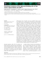
Distribution of screws and washers, outlining the borders of the surgical bone cavityFigure 1
Distribution of screws and washers, outlining the borders of the surgical bone cavity. A distance of 1.3 mm separates the wash-
ers over the surgical cavity, corresponding to the area where the magnetic field operates. P and D mark, respectively, the
proximal and distal regions of the left femur.
Head & Face Medicine 2006, 2:43 />Page 4 of 9
(page number not for citation purposes)
[10], in which intensities of 12 G, 2 G, 16 G, 16 G and 16
G were employed respectively. The expressive difference
in charge was due to lack of calibration information in lit-
erature reports, and to the novelty represented by devices
which keep an active, isolated field with no possible reac-
tivation.
Different in vitro and in vivo experimental systems have
been used for the investigation of electric fields effects in
vital tissues. Bodamyali et al. [19] and Ishisaka et al. [3]
described the use of weak magnets for in vitro cell stimula-
tion, but observed little activity in this system. In vivo stud-
ies were performed by Grace, Revell and Brookes [6];
Matsumoto et al. [7]; Fini et al. [11]; Aaron, Wang and
Ciombor [9]; Ciombor et al. [10] and Inoue et al. [13],
with daily application of electromagnetic fields during 2,
8, 6, 1, 8 and 8 hours respectively. Experiments were con-
ducted during periods between 2 days and 8 weeks, and
the studies were characterized by the use of an electromag-
netic field with continuous stimulation.
According to Halliday et al. [20], the electric neutrality of
a body is modified when it is submitted to a magnetic
field. Reports by Oishi and Onesti [2] and Teló [4] suggest
that cell electronegativity at bone fractures and after can-
cer treatment should be regarded as a possible indication
of electric modifications on the local wound.
The extensive trabecular formation beginning in the
endosteum, histologically observed in the surgical bone
cavity in samples from the test groups as early as 15 days
later, suggests that the magnetic field stimulates bone
healing.
Control group, day 15Figure 2
Control group, day 15. Surgical cavity (SC) limited on the upper side by cortical neoformation linearly continuous to borders
(CB). Beginning of bone trabeculae in centripetal direction (BT). Medullary spaces showing connective tissue of great cellularity
(MS). (HE 40×).
Head & Face Medicine 2006, 2:43 />Page 5 of 9
(page number not for citation purposes)
On day 45, neoformed bone was rather similar to the sur-
rounding bone tissue in test and control groups, showing
the presence of a first intention healing process as stated
by Lane and Danis (apud Christian) [14]. In the test
group, however, stronger neovascularization as well as
osteoclastic and bone remodelling activities were
observed.
On day 60, besides marked external configuration of the
magnetic washers with cortical bone, the establishment of
bone projections beyond the external border of the previ-
ously osteotomized cortical was observed. These results
suggest that the magnetic field was active during all the
experimental period. Even though they cannot be strictly
compared to the studies of Grace, Revell and Brookes [6];
Matsumoto et al. [7]; Fini et al. [11]; and Fredericks et al.
[12], since these authors used intermittent electromag-
netic fields, the results of the present work agree with the
accelerated bone neoformation reported.
The histological observation of hematopoietic activity in
the bone marrow is an important result. Urist, Delange
and Finermann [21] and Grace, Revell and Brookes [6]
suggested that cartilage formation is due to a shortage of
blood supply. The results of the present study, with in vivo
observations during a period of 60 days, show that blood
supply to the region was not impaired, but on the contrary
was stimulated, which may explain the absence of carti-
lage formation during the healing process.
The results of the present experimental work indicate that
further studies are needed for the detailed analysis of the
in vivo activity and best intensity of magnetic stimulation
on healing bone tissue.
Test group, day 15Figure 3
Test group, day 15. Surgical cavity (SC) with extensive centrifugal trabecular formation. Beginning of bone trabeculae in centrif-
ugal direction (BT, CD). In (OC), osteotomized cortical bone marks the border of the cavity, supporting the magnetized
washer (MW) (HE 40×).
Head & Face Medicine 2006, 2:43 />Page 6 of 9
(page number not for citation purposes)
Conclusion
The experimental approach used in this study allows the
following conclusions:
1. The magnetized stainless steel material used in these
studies is able to affect the bone healing process;
2. The comparison of test and control groups indicates
that bone healing was accelerated by the effect of mag-
netic fields in all the conditions analyzed;
3. The marked configuration of a bone outline involving
the metallic devices in the test group, observed until the
end of the experimental period, suggests that the magnetic
field exerted a constant local activity on the surgical
wound.
Competing interests
The author(s) declare that they have no competing inter-
ests.
Authors' contributions
EP suggested the original idea for the study; initiated the
investigations leading to these results; wrote the protocols
for the study and for the Research and Ethics in Health
Committee; participated in discussions on the undertak-
ing of the study; conceived, designed, and supervised the
study; interpreted the data; reviewed all iterations of the
paper. LMU developed the dissertation on which this
work is based; participated in discussions on the under-
taking of the study, supervised and participated in obtain-
ing the results, interpreted the data, reviewed the paper for
content, and reviewed and contributed to the writing of
Control group, day 45Figure 4
Control group, day 45. Surgical cavity (SC) with mature bone tissue, blood vessels and areas of internal remodelling. Fibrous
capsules (FC) can be observed on the upper and lateral regions of the slide (HE 40×).
Head & Face Medicine 2006, 2:43 />Page 7 of 9
(page number not for citation purposes)
all iterations of the paper. DP collaborated with labora-
tory experimental procedures and observation of animal
bioethics guidelines; reviewed and contributed to the
writing of all iterations of the paper, including the final
version of the manuscript. JJCF participated in the analysis
of results and implementation of material and other con-
ditions for development of the project. All authors
approved the final report.
Acknowledgements
We would like to thank Prof. Dr. Paulo Pureur Neto (Physics Institute,
UFRGS), Marcel Fasolo de Paris (Oral and Maxillofacial Surgeon, Hospital
Moinhos de Vento) and Isabel Regina Pucci (Manager, Instituto Puricelli &
Associados).
This study is in accordance with the guidelines for animal research estab-
lished by the State Code for Animal Protection and Normative Rule 04/97
from the Research and Ethics in Health Committee/GPPG/HCPA.
References
1. Veeck EB, Puricelli E, Souza MAL: Análise do comportamento do
osso e da medula hemopoética em relação a implantes de
titânio e hidroxiapatita: estudo experimental em fêmur de
rato. Odonto Ciência 1995, 10:235-291.
2. Oishi M, Onesti ST: Electrical Bone Graft Stimulation for Spi-
nal Fusion: A Review. Neurosurgery 2000, 47:1041-1056.
3. Ishisaka R, Kanno T, Inai Y, Nakahara H, Akiyama J, Yoshioka T,
Utsumi K: Effects of a magnetic field on the various functions
of subcellular organelles and cells. Pathophysiology 2000,
7:149-152.
4. Teló M, et al.: O uso da corrente elétrica no tratamento do
câncer. Edited by: Teló M et al. Porto Alegre: Edipucrs; 2004.
5. Aaron RK, Ciombor DM: Therapeutic Effects of Electromag-
netic Fields in the Stimulation of Connective Tissue Repair.
J Cell Biochem 1993, 52:42-46.
6. Grace KL, Revell WJ, Brookes M: The Effects of Pulsed Electro-
magnetism on Fresh Fracture Healing: Osteochondral
Repair in the Rat Femoral Groove. Orthopedics 1998,
21:297-302.
7. Matsumoto H, Ochi M, Abiko Y, Hirose Y, Kaku T, Sakaguchi K:
Pulsed Electromagnetic Fields Promote Bone Formation
Around Dental Implants Inserted into the Femur of Rabbits.
Clin Oral Implants Res 2000, 11:354-360.
Test group, day 45Figure 5
Test group, day 45. Surgical cavity (SC) with mature cortical and trabecular bone tissue characterized by lamellar structure.
Blood vessels (BV) and areas of internal remodelling can be seen. Bone trabeculae remodeling (BT). Screw Space (SS). The neo-
formed area and the surrounding bone tissue (MW) show similar patterns (HE 40×).
Head & Face Medicine 2006, 2:43 />Page 8 of 9
(page number not for citation purposes)
Control group, day 60Figure 6
Control group, day 60. Surgical cavity (SC) covered by neoformed cortical bone in remodelling with similar process in the
femur residual cortical. Inflammatory response to a foreign body (IR) is apparent. Medullary spaces (MS). Cortical Bone (CB).
Space corresponding to the washer (MW) (HE 40×).
Test group, day 60Figure 7
Test group, day 60. Photograph showing the surgical cavity sequence (SC). Centrifugal growth (↑), limiting the space corre-
sponding to the magnetized washers are observed (MW). Bone remodelling with normal histological patterns, going beyond
the cortical external border, is observed (HE 40×).
Publish with Bio Med Central and every
scientist can read your work free of charge
"BioMed Central will be the most significant development for
disseminating the results of biomedical research in our lifetime."
Sir Paul Nurse, Cancer Research UK
Your research papers will be:
available free of charge to the entire biomedical community
peer reviewed and published immediately upon acceptance
cited in PubMed and archived on PubMed Central
yours — you keep the copyright
Submit your manuscript here:
/>BioMedcentral
Head & Face Medicine 2006, 2:43 />Page 9 of 9
(page number not for citation purposes)
8. Nagai N, Inoue M, Ishiwari Y, Nagatsuka H, Tsujigiwa H, Nakano K,
Nagaoka N: Age and Magnetic Effects on Ectopic Bone For-
mation Induced by Purified Bone Morphogenetic Protein.
Pathophysiology 2000, 7:107-114.
9. Aaron RK, Wang S, Ciombor DM: Upregulation of Basal
TGFbeta1 Levels by EMF Coincident with Chondrogenesis:
Implications for Skeletal Repair and Tissue Engineering. J
Orthop Res 2002, 20:233-240.
10. Ciombor DM, Lester G, Aaron RK, Neame P, Caterson B: Low Fre-
quency EMF Regulates Chondrocyte Differentiation and
Expression of Matrix Proteins. J Orthop Res 2002, 20:40-50.
11. Fini M, Cadossi R, Cane V, Cavani F, Giavaresi G, Krajewski A, Martini
L, Aldini NN, Ravaglioli A, Rimondini L, Torricelli P, Giardino R: The
Effect of Pulsed Electromagnetic Fields on the Osteointegra-
tion of Hydroxyapatite Implants in Cancellous Bone: A Mor-
phologic and Microstructural In Vivo Study. J Orthop Res 2002,
20:756-763.
12. Fredericks DC, Nepola JV, Baker JT, Abbott J, Simon B: Effect of
Pulsed Electromagnetic Field Stimulation on Distraction
Osteogenesis in the Rabbit Tibial Leg Lengthening Model. J
Pediatr Orthop 2003, 23:478-483.
13. Inoue N, Ohnishi I, Chen D, Deitz LW, Schwardt JD, Chao EY: Effect
of Pulsed Electromagnetic Fields (PEMF) on Late-phase
Osteotomy Gap Healing in a Canine Tibial Model. J Orthop Res
2002, 20:106-114.
14. Christian CA: General Principles of Fracture Treatment. In
Campbell's Operative Orthopaedics Volume 3. 9th edition. Edited by:
Canale ST, Daugherty K, Jones L. St. Louis: Mosby; 1996:1993-2041.
15. Burkitt HG, Young B, Heath JW: Wheater Histologia Funcional.
3rd edition. Rio de Janeiro: Guanabara Koogan; 1994.
16. Feinberg SE, Steinberg B, Helman JI: Healing of Traumatic Inju-
ries. In Oral and Maxillofacial Trauma Volume 1. Edited by: Fonseca RJ,
Walker RV. Philadelphia: Saunders; 1997:13-57.
17. Lemons J, Natiella J: Biomaterials, Biocompatibility, and Peri-
Implant Considerations. Dent Clin North Am 1986, 30:3-23.
18. Chiaverini V: Aços Resistentes à Corrosão. In Aços e Ferros Fun-
didos
5th edition. São Paulo: Associação Brasileira de Metais;
1982:322-357.
19. Bodamyali T, Bhatt B, Hughes FJ, Winrow VR, Kanczler JM, Simon B,
Abbott J, Blake DR, Stevens CR: Pulsed Electromagnetic Fields
Simultaneously Induce Osteogenesis and Upregulate Tran-
scription. Biochem Biophys Res Commun 1998, 250:458-461.
20. Halliday , et al.: Eletromagnetismo. In Fundamentos de Física Volume
3. 3rd edition. Edited by: Halliday et al. Rio de Janeiro: Livros Técnicos
e Científicos; 1994.
21. Urist MR, Delande RJ, Finerman GAM: Bone Cell Differentiation
and Growth Factors. Science 1983, 220:680-686.