báo cáo khoa học:" Combination of surgical excision and custom designed silicon pressure splint therapy for keloids on the helical rim" ppt
Bạn đang xem bản rút gọn của tài liệu. Xem và tải ngay bản đầy đủ của tài liệu tại đây (1.3 MB, 4 trang )
BioMed Central
Page 1 of 4
(page number not for citation purposes)
Head & Face Medicine
Open Access
Case report
Combination of surgical excision and custom designed silicon
pressure splint therapy for keloids on the helical rim
Michael Sand*
1
, Daniel Sand
2
, Pejman Boorboor
3
, Benno Mann
1
,
Peter Altmeyer
4
, Klaus Hoffmann
4
and Falk G Bechara
4
Address:
1
Department of General and Visceral Surgery, Augusta Kranken Anstalt, Academic Teaching Hospital of the Ruhr-University Bochum,
Germany,
2
Department of Physiological Science, University of California Los Angeles (UCLA), Los Angeles, California, USA,
3
Department of
Plastic and Reconstructive Surgery, Hannover Medical School, Hannover, Germany and
4
Department of Dermatology and Allergology, Ruhr-
University Bochum, Germany
Email: Michael Sand* - ; Daniel Sand - ; Pejman Boorboor - ;
Benno Mann - ; Peter Altmeyer - ; Klaus Hoffmann - ;
Falk G Bechara -
* Corresponding author
Abstract
Keloids are defined as dermal fibrotic lesions which are considered an aberration of the wound
healing process. Their etiology and pathogenesis are poorly understood. Different treatment
modalities are described in the literature depending on the morphology and size of the keloid. We
report a case of a large ear keloid on the helical rim which was successfully treated with surgery
and a custom designed silicon pressure clip.
Background
Keloids are defined as dermal fibrotic lesions which are
considered an aberration of the wound healing process.
They are included in the spectrum of fibroproliferative
disorders and can potentially occur anywhere on the
body. Areas more commonly affected are the anterior
chest, shoulders, flexor surfaces of the extremities, and the
ears.
Keloids on the ears present several therapeutic challenges.
They are common after small skin excisions and other
procedures, including drainage of auricular hematomas,
repair of other auricular traumas, or as secondary keloid
formation after prior keloid excision.
Several procedures have been described for effective treat-
ment of keloid scars. They include silicon occlusive dress-
ings, mechanical compression, radiation, cryosurgery,
topical Imiquimod application, bleomycin tattooing, int-
ralesional injections of steroids, 5-floururacil, as well as
interferon-alpha, -beta or -gamma in combination with
excisional surgery [1-7]. Although optimal conditions for
the prevention of keloid formation are still unknown the
combination of exicisional surgery and the placement of
a silicone gel sheet over the wound surface with the appli-
cation of light pressure are known to be advantageous [8-
10].
In the following case report we describe a custom
designed silicon pressure splint which was successfully
used for preventive, postoperative treatment of a large kel-
oid formation on the helical rim.
Case
A 25-year-old Caucasian female with skin type 2 (Fitz-
patrick classification) presented because of a plum-sized
Published: 12 March 2007
Head & Face Medicine 2007, 3:14 doi:10.1186/1746-160X-3-14
Received: 28 December 2006
Accepted: 12 March 2007
This article is available from: />© 2007 Sand et al; licensee BioMed Central Ltd.
This is an Open Access article distributed under the terms of the Creative Commons Attribution License ( />),
which permits unrestricted use, distribution, and reproduction in any medium, provided the original work is properly cited.
Head & Face Medicine 2007, 3:14 />Page 2 of 4
(page number not for citation purposes)
pedunculated keloid on the upper part of her left helical
rim. She reported that 10 years ago she had already expe-
rienced formation of a nodule in this area which became
evident 6 months after an ear piercing. This keloid-like
nodule was excised twice and injected with steroids. At the
time of presentation the plum-sized keloid on her helical
rim had been increasing in size and was accompanied by
severe pruritus (Fig 1). We introduced our patient to an
audiology technician in order to design and build a spe-
cially silicon pressure splint for her left ear (Fig 2).
The keloid was then excised with cold steel. Immediately
after the operation a combination of 0.5 ml triamci-
nolonacetonid and scandicain 2 % was intralesionally
injected. The custom made silicon splint was applied
directly after surgery and steroid injection (Fig 3). The
injections were repeated at intervals of 8 weeks for 12
months. The patient was instructed to wear the splint for
24 h a day, 7 days a week. A clinical check-up one year and
24 months after the last injection showed no tendency to
relapse (Fig 4 and Fig 5).
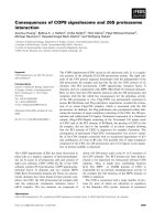
Patients' left ear after keloid excision with silicon pressure splint on the left helical rimFigure 3
Patients' left ear after keloid excision with silicon pressure
splint on the left helical rim.
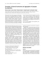
Plum-sized keloid on the left helical rimFigure 1
Plum-sized keloid on the left helical rim.
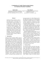
Custom-build silicon pressure splint for the left helical rimFigure 2
Custom-build silicon pressure splint for the left helical rim.
Head & Face Medicine 2007, 3:14 />Page 3 of 4
(page number not for citation purposes)
Discussion
Although the incidence of keloid formation is predomi-
nantly in darkly pigmented individuals, who form keloids
up to 19 times more than Caucasians, those Caucasians
who do are among skin types I and II and, as in our
patient, are the most difficult ones to treat [11]. After an
earring piercing, our patient experienced the third relapse
of a keloid on the helical rim which had been unsuccess-
fully treated with excision and intralesional steroid-injec-
tion before.
After surgery, the combination of several preventive steps
is essential for a successful treatment plan. It is known that
surgical monotherapy results in a high incidence of recur-
rence (50–100%) [12,13]. Additionally surgical excision
and primary closure should be performed with as little
wound tension as possible which is not always an easy
task where the amount of skin is limited, as on the ante-
rior side of the ear. Hence, we utilized multi-modal stand-
ard therapy forms in this patient.
Surgical excision and postoperative intralesional injection
of steroid was combined with silicon gel sheeting and
compression therapy with an individually designed sili-
con pressure splint for the helical rim. The procedure
combines the advantageous effects of pressure and silicon
gel sheeting. Silicon has been described as effective in pre-
venting the development of keloids. It reduces keloid scar
formation by 70% when used consistently [14]. There are
several theories of the action mechanism. Although some
authors propose that silicon diffuses from the surface of
the silicon gel sheets and reduces keloid ground substance
it is more likely that retardation of epidermal water loss
and a subsequent increase of wound hydration is respon-
sible for the keloid-inhibiting [15,16].
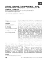
Posterior view on the patients left ear 24 months after the last injectionFigure 5
Posterior view on the patients left ear 24 months after the
last injection.
Lateral view on the patients left ear 24 months after the last injectionFigure 4
Lateral view on the patients left ear 24 months after the last
injection.
Publish with Bio Med Central and every
scientist can read your work free of charge
"BioMed Central will be the most significant development for
disseminating the results of biomedical research in our lifetime."
Sir Paul Nurse, Cancer Research UK
Your research papers will be:
available free of charge to the entire biomedical community
peer reviewed and published immediately upon acceptance
cited in PubMed and archived on PubMed Central
yours — you keep the copyright
Submit your manuscript here:
/>BioMedcentral
Head & Face Medicine 2007, 3:14 />Page 4 of 4
(page number not for citation purposes)
Compression therapy with dressings or devices that apply
more than 24 mmHg, the capillary pressure, create a
hypoxic microenvironment which results in fibroblast,
and, subsequently, collagen degradation. Pressure ear-
rings with compression plates which are available in dif-
ferent sizes are successfully used for ear lobe keloids. It is
obvious that the helical rim with its concave anterior and
convex posterior surface is not easily amenable for com-
pression. The silicon pressure splint introduced here not
only enjoys all the advantages of silicon dressings but also
successfully delivers pressure on the helical rim.
We suggest that in cases of keloids on the helical rim the
above described custom designed silicon pressure splint
combined with subsequent steroid injections respects the
delicate anatomy of the helical rim and can be a therapeu-
tic approach with strong benefit for the patient.
Authors' contributions
MS: Surgeon who performed the operation, documented
and prepared the draft
DS: Literature search, revision of bibliography and helped
with editing of the manuscript
PB: Helped in preparing the draft
BM: Edited most of the manuscript
PA: Revised and edited the manuscript and helped in pre-
paring the draft
KH: Literature search and edited part of the manuscript
FGB: Surgeon who performed the operation and edited
part of the manuscript and helped in preparing the draft
Acknowledgements
The written consent was obtained from the patient.
References
1. Kauh YC, Rouda S, Mondragon G, Tokarek R, diLeonardo M, Tuan
RS, Tan EM: Major suppression of pro-alpha1(I) type I collagen
gene expression in the dermis after keloid excision and
immediate intrawound injection of triamcinolone acetonide.
J Am Acad Dermatol 1997, 37(4):586-9.
2. Ogawa R, Mitsuhashi K, Hyakusoku H, Miyashita T: Postoperative
electron-beam irradiation therapy for keloids and hyper-
trophic scars: retrospective study of 147 cases followed for
more than 18 months. Plast Reconstr Surg 2003, 111(2):547-53.
3. Naeini FF, Najafian J, Ahmadpour K: Bleomycin tattooing as a
promising therapeutic modality in large keloids and hyper-
trophic scars. Dermatol Surg 2006, 32(8):1023-9.
4. Maarouf M, Schleicher U, Schmachtenberg A, Ammon J: Radiother-
apy in the management of keloids. Clinical experience with
electron beam irradiation and comparison with X-ray ther-
apy. Strahlenther Onkol 2002, 178(6):330-5.
5. Rusciani L, Rossi G, Bono R: Use of cryotherapy in the treat-
ment of keloids. J Dermatol Surg Oncol 1993, 19(6):529-34.
6. Berman B, Kaufman J: Pilot study of the effect of postoperative
imiquimod 5% cream on the recurrence rate of excised kel-
oids. J Am Acad Dermatol 2002, 47(4):209-11.
7. Berman B, Villa A: Imiquimod 5% cream for keloid manage-
ment. Dermatol Surg 2003, 29(10):1050-1.
8. Ahn ST, Monafo WW, Mustoe TA: Topical silicone gel: A new
treatment for hypertrophic scars. Surgery 1989, 106:781-6.
9. Sproat JE, Dalcin A, Weitauer N, Roberts RS: Hypertrophic ster-
nal scars: Silicone gel sheet versus Kenalog injection treat-
ment. Plast Reconstr Surg 1992, 90:988-92.
10. Fulton JE Jr: Silicone gel sheeting for the prevention and man-
agement of evolving hypertrophic and keloid scars. Dermatol
Surg 1995, 21:947-51.
11. Alhady SM, Sivanantharajah K:
Keloids in various races. A review
of 175 cases. Plast Reconstr Surg 1969, 44(6):564-6.
12. Darzi MA, Chowdri NA, Kaul SK, Khan M: Evaluation of various
methods of treating keloids and hypertrophic scars: a 10-
year follow-up study. Br J Plast Surg 1992, 45(5):374-9.
13. Lawrence WT: In search of the optimal treatment of keloids:
report of a series and a review of the literature. Ann Plast Surg
1991, 27(2):164-78.
14. Fulton JE: Silicone gel sheeting for the prevention and man-
agement of evolving hypertrophic and keloid scars. Dermatol
Surg 1995, 21(11):947-51.
15. Ahn ST, Monafo WW, Mustoe TA: Topical silicone gel for the
prevention and treatment of hypertrophic scar. Arch Surg
1991, 126(4):499-504.
16. de Oliveira GV, Nunes TA, Magna LA, Cintra ML, Kitten GT, Zarpel-
lon S, Raposo Do Amaral CM: Silicone versus nonsilicone gel
dressings: a controlled trial. Dermatol Surg 2001, 27(8):721-6.