báo cáo khoa học:" Standardization of surface electromyography utilized to evaluate patients with dysphagia" pot
Bạn đang xem bản rút gọn của tài liệu. Xem và tải ngay bản đầy đủ của tài liệu tại đây (737.29 KB, 7 trang )
BioMed Central
Page 1 of 7
(page number not for citation purposes)
Head & Face Medicine
Open Access
Methodology
Standardization of surface electromyography utilized to evaluate
patients with dysphagia
Michael Vaiman*
1,2
Address:
1
Department of Otolaryngology, Assaf Harofe Medical Center, Affiliated to the Sackler Faculty of Medicine, Tel Aviv University, Tel Aviv,
Israel and
2
33 Shapiro Street, Bat Yam, 59561, Israel
Email: Michael Vaiman* -
* Corresponding author
Abstract
Backgorund: Patients suspected of having swallowing disorders, could highly benefit from simple
diagnostic screening before being referred to specialist evaluations. We introduce surface
electromyography (sEMG) to carry out rapid assessment of such patients and propose suggestions
for standardizing sEMGs in order to identify abnormal deglutition.
Methods: Specifics steps for establishing standards for applying the technique for screening
purposes (e.g., evaluation of specific muscles), the requirements for diagnostic sEMG equipment,
the sEMG technique itself, and defining the tests suitable for assessing deglutition (e.g., saliva,
normal, and excessive swallows and uninterrupted drinking of water) are presented in detail. A
previously described normative database for single swallowing and drinking and standard approach
to analysis was compared to data on the duration and electric activity of muscles involved in
deglutition and with sEMG recordings in order to estimate stages of a swallow.
Conclusion: SEMG of swallowing is a simple and reliable method for screening and preliminary
differentiation among dysphagia and odynophagia of various origins. This noninvasive radiation-free
examination has a low level of discomfort, and is simple, timesaving and inexpensive to perform.
With standardization of the technique and an established normative database, sEMG can serve as
a reliable screening method for optimal patient management.
Background
Swallowing disorders (dysphagia) occurs in approxi-
mately 14% of patients in acute care setting an up to 50%
of patients in nursing homes [1]. Its prevalence is related
to the fact that dysphagia often is present in patients who
have sudden-onset neurologic disorders, chronic neuro-
degenerative diseases, and patients with general medical
problems, but in general it is an interdisciplinary phe-
nomenon. It is frequently associated with painful swal-
lowing, or odynophagia. It is estimated that 15 million
patients suffered from a swallowing disorder during each
year in the United States alone [2,3]. Patients with sus-
pected dysphagia and/or odynophagia could highly bene-
fit from preliminary screening for confirmation of
diagnosis before being referred for more extensive clinical
and instrumental evaluations by a specialist. Currently
videofluorographic swallow study (VFSS) is the most
commonly used tool in the assessment of oropharyngeal
dysphagia, and it is considered the gold standard in the
dysphagia workup. Unfortunately, VFSS has several draw-
backs, as the patient must be transported to the radiology
suite, must be able to cooperate with the examination,
Published: 6 June 2007
Head & Face Medicine 2007, 3:26 doi:10.1186/1746-160X-3-26
Received: 13 March 2007
Accepted: 6 June 2007
This article is available from: />© 2007 Vaiman; licensee BioMed Central Ltd.
This is an Open Access article distributed under the terms of the Creative Commons Attribution License ( />),
which permits unrestricted use, distribution, and reproduction in any medium, provided the original work is properly cited.
Head & Face Medicine 2007, 3:26 />Page 2 of 7
(page number not for citation purposes)
and will be exposed to radiation. Also, VFSS does not
always identify neuromuscular abnormalities in pharyn-
geal or laryngeal physiology. A reliable, noninvasive,
time-saving and inexpensive procedure that might be eas-
ily learned and applied by primary health clinicians as
well as by nurses would be a valuable addition to our
diagnostic armamentarium.
Given that the swallowing mechanism by which food is
transmitted to the stomach is a complex action involving
26 muscles and five cranial nerves, electromyography
(EMG) would appear especially suitable for screening and
early diagnosis of dysphagia and odynophagia. Indeed,
surface EMG (sEMG) provides information on the timing
of selected muscle contraction patterns during swallowing
[4-6], on the amplitude of electric activity of the muscles
[7], and was shown to be easily learned by medical per-
sonnel [8,9]. EMG had already been proposed for screen-
ing purposes in neurogenic dysphagia [10].
For the past five years, we have been investigating degluti-
tion by means of sEMG. Numerous studies on EMG activ-
ity of face and neck muscles during swallowing had
appeared in the 1990s [3-11] and revealed a lack of agree-
ment among experts regarding some of the basic aspects
of the act of swallowing common to all subjects as well as
in differentiating between the values that represent nor-
mal and abnormal function. Thus, we first established a
normative database for deglutition for adults [12,13] and
children [14] and now embarked on devising standards
for sEMG in diagnosing it. It emerged that there is a large
variation in examination techniques, strategies, interpre-
tations and diagnostic criteria among electromyographers
[15], further reinforcing the need for international stand-
ardization.
In the current article, we introduce sEMG as a rapid
screening method for patients with complaints suggestive
of dysphagia or odynophagia that need to be differenti-
ated and localized in oral, laryngeal and esophageal
causes. We also suggest steps for standardization of sEMG
assessment of normal and abnormal deglutition, as had
been done for electrocardiograms one hundred years ago.
Indeed, as patients with chest pains are more likely to
approach their primary health provider before consulting
a cardiologist, we expect patients with swallowing disor-
ders to be seen first by family physicians before consulting
an otolaryngologist.
Standardization of the diagnostic procedure
Any diagnostic method designed for use in different areas
of medicine requires standards, and we propose the fol-
lowing for EMG evaluation of deglutition
Standards for test application
Ever since Magendie's publications in 1813, physicians
have adapted the concept of three stages in swallowing,
oral, pharyngeal, and esophageal [16,17]. This was altered
to four stages in the 1980s, with the oral stage having been
divided into oral initial (for solids, the "oral preparation"
stage) and oral final stages [18]. This latter staging can be
helpful in diagnosing disorders that lead to dysphagia and
odynophagia. In liquid swallowing, the water intake takes
place during the oral initial stage starting with sealing of
the labia. The oral final stage occurs when the tongue
squeezes the liquid volume against the hard palate so that
it is propelled past the anterior faucial arches, whereupon
the automatic reflexive gesture of swallowing is triggered.
During the pharyngeal stage, the liquid volume is trans-
ferred from the level of the faucial arches through the
pharynx to the cricopharyngeal sphincter at the rostral
aspect of the esophagus. In the esophageal stage of the
swallow, the water volume is transferred in a continuation
of the peristaltic movement from the cricopharyngeal to
the gastroesophageal sphincter at the entrance to the
stomach.
Each of these stages can be impaired and the screening
evaluation should be capable of indicating which is the
impaired stage. (Surface EMG recordings cannot trace
esophageal activity, only the initial esophageal stage.)
Standards for the equipment
To carry out rapid and accurate assessments, the diagnos-
tic tool should be reliable, preferably noninvasive, prefer-
ably radiation-free, inexpensive, time saving and simple
and easy to operate. Surface EMG devices meet all these
criteria. We propose a fourchannel computerbased EMG
unit equipped with surface electrodes. We use standard
surface electrodes AE-131 and AE-178 which are silver-
coated discs with 11 mm diameters and placed 10 mm
from each other. Other surface electrodes with similar
characteristics can be used as well. In our earlier studies,
sEMG recordings were performed by a NeuroDyne Neu-
romuscular Sys/3 fourchannel computerbased EMG unit
with NeuroDyne Medical software (NeuroDyne, Cam-
bridge, MA, USA) and AE-204 active sensors attached to
AE-131 or AE-178 electrodes [12].
Any other EMG device with similar characteristics can be
used as long as the EMG recording is full-wave rectified
and low-passed filtered in such a way that it resembles a
single EKG line. EMG records with numerous closely
packed spikes are almost impossible to interpret rapidly.
A 2-channel EMG is not enough for rapid testing, and 8-
channel EMG records are difficult to perform and take a
considerable amount of time to interpret: testing with the
4 channel device can take only 5–7 minutes when the
patient is fully cooperative.
Head & Face Medicine 2007, 3:26 />Page 3 of 7
(page number not for citation purposes)
Standards for the electromyographic technique
The four examined muscle groups are the superior and
inferior orbicularis oris (OO), the masseter (MS), the sub-
mental muscle (SUB) group, which includes the anterior
belly of the digastric, mylohyoid, and geniohyoid, and the
infrahyoid group (INF), which includes also the laryngeal
strap muscles and the thyrohyoid, all covered by the
platysma. These muscles are superficial and are thought to
be involved in the oral and pharyngeal phases of a swal-
low.
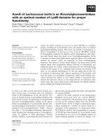
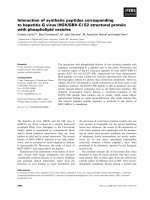
The suggested standard electrode positions are as follows
(Fig. 1):
1. Two bipolar stick-on surface electrodes applied at the
right or left angle of the mouth, one electrode above the
upper lip, and another electrode below the lower lip (OO-
location);
2. Two electrodes parallel to the MS fibers on the left or
right side of the face, preferably on the opposite side from
the OO-location (MS-location);
3. Two surface electrodes on the skin beneath the chin on
the right or left side of midline to record SUB myoelectri-
cal activity over the platysma (SUB-location);
4. Two electrodes on the left or right side of the thyroid
cartilage to record from the laryngeal strap and infrahyoid
muscles (INF-location).
The exact electrode positions for each muscle group have
been known since the 19
th
century [20,21], and can be
adjusted to accommodate anatomical exceptions [22].
Each pair of electrodes has a third electrode as ground.
Standards for testing procedures (Fig. 2)
The proposed set of four tests includes voluntary single
swallows of saliva ("dry" swallow), voluntary single water
swallows from an open cup ("normal"), voluntary single
swallows of an excessive amount of water (20 ml, "stress
test"), and continuous drinking of 100 cc of tap water
from an open cup. Subjects are permitted to move their
chins slightly upwards while swallowing if needed when
it emerges that there is no changes of the graphic and
numerical baseline associated with this movement. (This
movement involves the mm. rectus capitis posterior
minor and minor, as well as some other posterior neck
muscles, and does not affect signals from the abovemen-
tioned electrode locations.) The tasks to be performed are:
1. Three trials of "dry" swallowing. Instruction: "Swallow
your saliva".
2. Three trials of swallowing normal volume of tap water
with a mean volume of 16.5 cc. Instruction: "Swallow
once in your usual way".
3. Three trials of swallowing 20 cc of tap water to check
adaptation abilities of the patients ("stress test", larger
bolus volume accommodation). Instructions: "Swallow
in one gulp".
4. One trial of continuous drinking of 100 ml of tap water.
Instruction: "Drink this in your usual way".
Normative database and standards for analysis
Standards for analysis include assessment of duration (in
sec) of the swallowing act, amplitude of electric activity
(mean, in μV), graphic patterns and number of swallows
(in the continuous drinking task) (Tables 1 and 2). A nor-
mative database for these variables in adults [12] and chil-
dren [14] was introduced earlier. (Figs. 3 and 4).
There was no visible difference between the shapes of
EMG recordings of swallows based on gender [12,13].
Elderly patients (aged 70+ years) showed age-related
peculiarities in the recorded swallows. For them, the mus-
cle activity is usually longer in duration, and suggests a
lack of coordination between activities of different mus-
cles involved in deglutition. For children, the duration of
muscle activity during swallows and drinking in all tests
decreased significantly with age [14]. There was, however,
no statistically significant difference in electric amplitude
measurements between children and adults (p = 0.05).
Discussion
Swallowing disorders comprise an interdisciplinary phe-
nomenon. Practitioners in various fields of medicine,
such as otorhinolaryngology, neurology, general medi-
cine, gastroenterology, head and neck surgery, dentistry
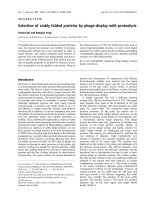
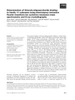
The electroneuromyograph NS/3 in operation.Figure 1
The electroneuromyograph NS/3 in operation.
Head & Face Medicine 2007, 3:26 />Page 4 of 7
(page number not for citation purposes)
and facial surgery, pediatrics and psychiatry deal with
these disorders regularly, but family doctors and emer-
gency department personnel might well be the first physi-
cians to evaluate these patients. The need for established
standards in various EMG investigations is well recog-
nized [23-25], and several attempts already have been
made to determine them [26,27]. To our knowledge, no
such standards were proposed for sEMG evaluations of
dysphagia and odynophagia. While an EMG evaluation of
deglutition is not a new diagnostic method, the lack of
standard requirements negatively impacts the value of this
investigative technique. The objectives of the current work
were to suggest potential solutions to this drawback by
determining optimal sEMG standards suitable for investi-
gation of face and neck muscles involved in deglutition, to
establish a normal database, and to set down investigative
standards to differentiate between specific pathological
EMG patterns of abnormal deglutition.
EMG records
To carry out the rapid assessment of patients, the EMG
record should be clear and easily understandable, and we
again stress a need for filtering EMG recordings. A recent
comprehensive study in which sEMG was used for moni-
toring functionally distinct muscle activation during swal-
lowing [27] supports our contention that raw sEMG
records should be rectified and filtered before evaluation.
EMG electrode locations
The proposed electrode locations were chosen in order to
cover all stages of a swallow. The staging of normal deglu-
tition can be clinically important as an additional tool for
establishing etiology and localization (oral, pharyngeal,
or esophageal) of the causes of dysphagia or
odynophagia. Each stage has its mean normal duration
and its specific graphic pattern. While additional research
is needed, our preliminary assumptions are that OO and
MS electrode locations represent the initial oral stage of
swallowing, that the MS and SUB locations represent the
final oral stage, that the MS, SUB and INF locations are
important for evaluating the pharyngeal stage, and that
the SUB and INF locations represent the initial esophageal
stage. Therefore, when the reflex part of a swallow is inves-
tigated in healthy volunteers, the OO location provides
less informative data and can be safely ignored. This loca-
tion, however, might be important for patients with dys-
phagia due to abnormal eating patterns, problems with
dentition, congenital abnormalities of the nasal and oral
cavity, and others. In these cases, even without reliable
normative data, the evaluation of OO sEMG activity
Locations of electromyogram electrodes for testing orbicula-ris oris (OO), masseter (MS), submental group (SUB) and laryngeal strap muscles (INF).Figure 2
Locations of electromyogram electrodes for testing orbicula-
ris oris (OO), masseter (MS), submental group (SUB) and
laryngeal strap muscles (INF).
Table 1: Simplified set of normative data for timing measures in reflex normal swallowing (oral final stage + pharyngeal stage + initial
esophageal stage) [age range: duration range, sec]
Age groups (y)
Saliva swallow 18–70: 1.0–5.44 70+: 1.44–6.24
Normal swallow 18–70: 1.0–5.74 70+: 2.3–6.7
20 cc swallow 18–70: 1.8–6.2 70+: 1.8–8.13
One swallow while drinking 18–70: 0.56–2.56 70+: 0.55–3.0
100 cc drinking 18–60: 6.2–15.4 61–70: 5.4–21.4 70+: 7.1–28.3
# swallows while drinking 18–40: 4–9 41–70: 4–12 70+: 4–16
Head & Face Medicine 2007, 3:26 />Page 5 of 7
(page number not for citation purposes)
might be important if different tests are compared within
the same patient.
Tests
The fundamental test is a single swallow of water in a nor-
mal manner. Saliva swallow test is especially relevant in
cases of salivary gland diseases, such as Sjögren syndrome
[28]. The stress test with an excessive amount of water
swallowed in one gulp might reveal a lack of larger bolus
volume accommodation abilities in cases of anatomical
changes of the pharynx or neurological problems. Testing
of continuous drinking is important not only in the eval-
uation of dysphagia but also of odynophagia and in the
differential diagnosis in cases of compulsive water drink-
ing, excessive water drinking, the malingering of dys-
phagia and psychogenic disorders expressed by symptoms
of dysphagia. This test is most suitable for cases of mild
dysphagia in easily tiring subjects for whom continuous
non-interrupted drinking is a stress test. The amount of
water for continuous drinking test was set at 100 cc, i.e.,
approximately onehalf of a standard glass, because a
smaller volume, e.g., 50 cc, can be swallowed in two gulps
and thus yield inadequate data while 200 cc of water
involves considerable swallowing/ventilation interactions
which can confound the validity of the obtained data.
Duration of a swallow
Older people (aged 70+ years) swallow and drink more
slowly as do patients with various neurological disorders
affecting deglutition. (Fig. 5) The times indicated by an
EMG device represent the duration of sEMG activity which
lasted longer than the actual time required to pass a bolus
from the oral cavity to the esophagus.
Electric amplitude
The range (amplitude, in μV) and mean of electric activity
are less important for stage-by-stage evaluation of a sEMG
recording. These data might be useful, however, when
abnormal swallows are investigated. For example, a per-
son usually presents low electric activity at the MS loca-
tion after undergoing a tooth extraction. We also observed
patients with a Zenker diverticulum [29] who presented
unusually high electric activity at the INF location, and
numerous patients with recurrent tonsillitis or during flu
with abnormally high electric activity of their infrahyoid
muscles. (Fig. 4). There are also numerous reports on
changes of the MS electric activity in patients with diseases
of the temporomandibular joint [30,31].
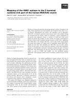
An example of normal drinking of 100 cc of waterFigure 4
An example of normal drinking of 100 cc of water. It took
this subject 15.04 sec to drink 100 cc of water in 7 swallows.
The last 8
th
peak is a dry swallow aftereffect. Upper line = the
submentalsubmandibular electrode location, lower line = the
masseter location. All these muscles are almost completely
relaxed before and after drinking.
Table 2: Quick reference simplified set of normative data for electric activity obtained by surface EMG for the masseter, submental
group (SUB) and laryngeal strap muscles (INF) during various tests, in μV. [age range: normal range of voltage values]
Saliva swallow
Masseter range 18–30: 4.5 – 15.9 31–70: 5.54 – 12.1 70+: 2.94–22.42
SUB range 18–30: 13.4–59.72 31–70: 9.52 – 49.5 70+: 10.2–42.32
INF range 18–40: 2.0–4.5 41–70: 2.5–4.3 70+: 4.3–7.25
Normal swallow
Masseter range 18–60: 2.2–31.0 61–70: 1.97 – 27.69 70+: 3.77–20.0
SUB range 18–30: 11.4–63.41 31–50: 12.58–51.6 51–70+: 7.4 – 44.8
INF range 18–40: 2.85–6.3 41–70: 3.8–5.6 70+: 4.33–8.0
Excessive swallow
Masseter range 18–40:1.5–37.0 41–70: 1.2 – 29.4 70+: 4.65–21.13
SUB range 18–30: 19.28–50.80 31–70+: 12.1 – 47.44
INF range 18–40: 3.8–6.55 41–70: 3.9–6.23 70+: 4.5–9.3
100 cc drinking
Masseter mean (real)* 18–70: 0.8 – 6.2 70+: 1.0 – 7.84
SUB mean (real) 18–60: 3.5 – 11.5 61 – 70+: 4.25 – 16.25
INF mean (real) 18–70: 1.4–2.8 70+: 1.0–3.85
*Raw mean = computer-calculated mean, while Real mean = raw mean minus the mean resting potential of an actual muscle covered by skin (2.808
μV = for the m. submental and m. infrahyoid, 2.495 μV = for the m. masseter, and 4.542 = for the m. orbicularis oris).
Head & Face Medicine 2007, 3:26 />Page 6 of 7
(page number not for citation purposes)
Published studies on normal subjects show a very wide
range of normal electric amplitudes for sEMG studies.
These variations are not only due to biologic causes but
are also greatly affected by such technical factors as skin/
electrode impedance, depth of the muscle from the skin
surface, location of the recording electrodes in relation to
anatomic structures, variation in muscle size among indi-
viduals, and temperature. It is because of the wide varia-
tion in the normal values that an absolute value of the
amplitude is considered less clinically useful. While we
feel that the amplitude data might be valuable for com-
parison across subjects, some additional information is
needed to clarify this issue. The EMG amplitude, however,
remains an important aspect in the relationship between
muscle force and the associated electric activity, although
there is no simple relationship between a sEMG signal
and muscle force. When all the different types of neu-
romuscular disorders are considered collectively, ampli-
tudes are by far the most informative features. Indeed,
some authors argue that amplitudes are the only compo-
nents that have a direct relationship to clinical symptoms
(muscle weakness) in neurogenic lesions [33].
During sEMG testing, there is a certain amount of imped-
ance noise that arises directly from the resistance of the
electrodes' connection to the skin. This feature makes skin
resistance a significant factor when working with the low-
level EMG signals typical of the small muscles involved in
swallowing. Wiping the skin with isopropyl alcohol in a
water solution has proven to be the best form of prepara-
tion for most situations. The alcohol removes the dead
skin and surface oils, and the water moistens the skin and
provides improved ion flow. The sEMG sensors we used
are designed so that the use of electrode gel is generally
not necessary.
Dysphagia is a very common finding in patients with neu-
rologic disturbances like amyotrophic lateral sclerosis,
Parkinson's disease, Huntington's disease, Multiple scle-
rosis, myasthenia gravis, stroke, laryngeal nerve injury and
many others. This study emphasizes that a sEMG analysis
of all the muscle groups involved in swallowing process,
following a proper placement of the electrodes and selec-
tion of tests, can give reliable indications of muscle activ-
ity and provides data for screening evaluation of
complaints a patient came with. Further investigation
might help to develop a proper combination of flexible
endoscopic evaluation of swallowing (FEES) with a
nasopharyngoscope (or flexible endoscopic evaluation of
swallowing with sensory testing, FEESST) [34] with SEMG
to achieve complete evaluation of swallowing without
exposure to radiation. Such screening might help a gen-
eral practitioner to direct a patient to a neurologist,
A prolonged drinking of a 75 year old subject suffering with influenza (MS = green line, SUB = blue line, and INF = red line locations)Figure 5
A prolonged drinking of a 75 year old subject suffering with
influenza (MS = green line, SUB = blue line, and INF = red
line locations). It took this subject 46 sec to drink 100 cc of
water in 11 swallows. The electric amplitude is characteristi-
cally low. INF muscles are more involved in swallowing than
usual, thus SUB and INF lines are almost identical.
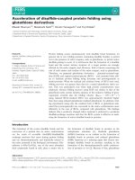
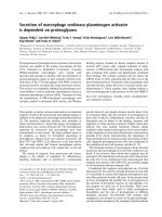
Stages of the normal swallow (reflex part)Figure 3
Stages of the normal swallow (reflex part). Horizontal mark 3
– water intake, 3.5–4 – final oral stage, 4–4.5 – pharyngeal
stage, 4.5–5.5 – initial esophageal stage. Upper peak – sub-
mental location, middle peak – masseter location, lower peak
– infrahyoid muscles location. Total electric activity duration
after water intake: 2.5 sec.
Publish with Bio Med Central and every
scientist can read your work free of charge
"BioMed Central will be the most significant development for
disseminating the results of biomedical research in our lifetime."
Sir Paul Nurse, Cancer Research UK
Your research papers will be:
available free of charge to the entire biomedical community
peer reviewed and published immediately upon acceptance
cited in PubMed and archived on PubMed Central
yours — you keep the copyright
Submit your manuscript here:
/>BioMedcentral
Head & Face Medicine 2007, 3:26 />Page 7 of 7
(page number not for citation purposes)
otolaryngologist or other proper specialist and to a spe-
cific radiographic or, perhaps, manofluorographic investi-
gation.
Conclusion
Surface EMG of swallowing is a simple and reliable
method for screening and initial evaluation of dysphagia
and odynophagia complaints of various origins. This non-
invasive radiation-free examination has low level of dis-
comfort, and is simple, time-saving and inexpensive. With
proper standard technique and established normative
database, sEMG can serve as a reliable screening method
for the assessment of dysphagia of unknown origin for
optimal patient management.
Acknowledgements
Esther Eshkol is thanked for editorial assistance.
References
1. Logemann J: Upper digestive tract anatomy and physiology. In
Head and Neck Surgery-Otolaryngology Edited by: Bailey B. Philadelphia,
JB Lippincott; 1993:485-491.
2. Byles J: The epidemiology of communication and swallowing
disorders. Advances in Speech Language Pathology 2005, 7,1:1-7.
3. Murry T, Carrau RL, Eibling DE: Epidemiology of swallowing dis-
orders (Chapter 1). In Comprehensive Management of Swallowing
Disorders Edited by: Carrau R, Murry T. San Diego, CA: Singular Pub-
lishing; 1998:3-7.
4. Perlman AL: Electromyography and the study of oropharyn-
geal swallowing. Dysphagia 1993, 8:351-5.
5. Palmer JB: Electromyography of the muscles of oropharyngeal
swallowing: basic concepts. Dysphagia 1989, 3:192-8.
6. Logemann JA: Non-imaging techniques for the study of swal-
lowing. Acta Otorhinolaryngol Belg 1994, 48:139-42.
7. Ertekin C, Palmer JB: Physiology and electromyography of swal-
lowing and its disorders. Suppl Clin Neurophysiol 2000, 53:148-54.
8. Gupta V, Reddy NP, Canilang EP: Surface EMG measurements at
the throat during dry and wet swallow. Dysphagia 1996,
11:173-9.
9. Crary MA, Baldwin BO: Surface electromyographic character-
istics of swallowing in dysphagia secondary to brainstem
stroke. Dysphagia 1997, 12:180-7.
10. Logemann JA: Screening, diagnosis and management of neuro-
genic dysphagia (review). Semin Neurol 1996, 16:319-327.
11. Schultz JL, Perlman AL, Van Daele DJ: Laryngeal movement,
oropharyngeal pressure, and submental muscle contraction
during swallowing. Arch Phys Med Rehabil 1994, 75:183-8.
12. Vaiman M, Eviatar E, Segal S: Evaluation of stages of normal
deglutition with the help of rectified surface electromyogra-
phy records. Dysphagia 2004, 19:125-32.
13. Vaiman M, Eviatar E, Gabriel Ch, Segal S: Rectified and filtered sur-
face electromyography of continuous drinking in healthy
adults. Laryngoscope 2005, 115(1):68-73.
14. Vaiman M, Segal S, Eviatar E: Surface electromyographic studies
of swallowing in normal children, age 4–12. J Pediatr Otorhi-
nolaryngol 2004, 68:65-73.
15. Fuglsang-Frederiksen A, Johnsen B, Vingtoft S, Carvalho M, Fawcett P,
Liguori R, Nix W, Schofield I, Veloso M, Vila A: Variation in per-
formance of the EMG examination at six European laborato-
ries. Electroencephalogr Clin Neurophysiol 1995, 97:444-50.
16. Magendie F: Vomissement. Procès-verb Acad d Sc 1813, 5:152-9.
174–83
17. Magendie F: L'epiglotte et ses usages dans le deglutition.
Procès-verb Acad d Sc 1813, 5:192-9. 205–16
18. Logemann JA: Evaluation and treatment of swallowing disor-
ders. San Diego, CA: College Hill Press; 1983.
19. Erb W: Handbuch der Electrotherapie. 2 Aufl., Leipzig: Vogel;
1886:34-45.
20. Remak E: Grundriss der Electrodiagnostik und Electrothera-
pie. 2 Aufl., Wien: Meyer; 1909:26-34.
21. Goodgold J: Anatomical correlates of clinical electromyogra-
phy. 1st edition. Baltimore: The Williams & Wilkins Company;
1975:2-3.
22. Stalberg E, Fuglsang-Frederiksen A, Bischoff C: Quantitation and
standardization in EMG and neurography. Suppl Clin Neurophys-
iol 2000, 53:101-11.
23. Bolton CF, Benstead TJ, Grand'Maison F, Tardif GS, Weston LE: Min-
imum standards for electromyography in Canada: a state-
ment of the Canadian Society of Clinical Neurophysiologists.
Can J Neurol Sci 2000, 27:288-91.
24. Stalberg E, Falck B, Gilai A, Jabre J, Sonoo M, Todnem K: Standards
for quantification of EMG and neurography. The Interna-
tional Federation of Clinical Neurophysiology. Electroencepha-
logr Clin Neurophysiol Suppl 1999, 52:213-20.
25. Farina D, Madeleine P, Graven-Nielsen T, Merletti R, Arendt-Nielsen
L: Standardising surface electromyogram recordings for
assessment of activity and fatigue in the human upper trape-
zius muscle. Eur J Appl Physiol 2002, 86:469-78. Epub 2002 Feb 19
26. Podnar S, Vodusek DB, Stalberg E: Standardization of anal
sphincter electromyography: normative data. Clin Neurophys-
iol 2000, 111:2200-7.
27. McKeown MJ, Torpey DC, Gehm WC: Non-invasive monitoring
of functionally distinct muscle activations during swallowing.
Clin Neurophysiol 2002, 113:354-66.
28. Vaiman M, Nahlieli O, Eviatar E, Segal S: Electromyography mon-
itoring of patients with salivary gland diseases. Otolaryngol
Head Neck Surg 2005, 133,6:869-73.
29. Vaiman M: Surface electromyography in Preoperative Evalua-
tion and Postoperative Monitoring of Zenker's diverticulum.
Dysphagia 2006, 21:1-7.
30. Wang K, Arendt-Nielsen L, Svensson P: Capsaicin-induced muscle
pain alters the excitability of the human jaw-stretch reflex. J
Dent Res 2002, 81(9):650-4.
31. Glaros AG, Burton E: Parafunctional clenching, pain, and effort
in temporomandibular disorders. J Behav Med 2004,
27(1):91-100.
32. Wilbourn AJ, Ferrante MA: Clinical electromyography. In Baker's
clinical neurology [book on CD-ROM] Edited by: Joynt RJ, Griggs RC.
Philadelphia: WB Saunders; 2000.
33. Wilbourn AJ: Nerve conduction studies: types, components,
abnormalities, and value in localization. Clinical electromyogra-
phy. Neurol Clin 2002, 20:310-11.
34. Aviv JE, Murry T: FEESST: Flexible Endoscopic Evaluation of
Swallowing with Sensory Testing. San Diego – Oxford, Plural
Publishing Inc; 2005.