báo cáo khoa học:" Dentin dysplasia type I: a challenge for treatment with dental implants" ppt
Bạn đang xem bản rút gọn của tài liệu. Xem và tải ngay bản đầy đủ của tài liệu tại đây (792.8 KB, 5 trang )
BioMed Central
Page 1 of 5
(page number not for citation purposes)
Head & Face Medicine
Open Access
Case report
Dentin dysplasia type I: a challenge for treatment with dental
implants
Rita A Depprich
1
, Michelle A Ommerborn*
2
, Jörg GK Handschel
1
,
Christian D Naujoks
1
, Ulrich Meyer
1
and Norbert R Kübler
1
Address:
1
Department for Cranio- and Maxillofacial Surgery, Heinrich-Heine-University Düsseldorf, Moorenstr. 5, 40225 Düsseldorf, Germany
and
2
Department for Operative and Preventive Dentistry and Endodontics, Heinrich-Heine-University Düsseldorf, Moorenstr. 5, 40225
Düsseldorf, Germany
Email: Rita A Depprich - ; Michelle A Ommerborn* - ;
Jörg GK Handschel - ; Christian D Naujoks - ;
Ulrich Meyer - ; Norbert R Kübler -
* Corresponding author
Abstract
Background: Dentin dysplasia type I is characterized by a defect of dentin development with
clinical normal appearance of the permanent teeth but no or only rudimentary root formation.
Early loss of all teeth and concomitant underdevelopment of the jaws are challenging for successful
treatment with dental implants.
Methods: A combination of sinus lifting and onlay bone augmentation based on treatment planning
using stereolithographic templates was used in a patient with dentin dysplasia type I to rehabilitate
the masticatory function.
Results: (i) a predisposition for an increased and accelerated bone resorption was observed in our
patient, (ii) bone augmentation was successful using a mixture of allogenic graft material with
autogenous bone preventing fast bone resorption, (iii) surgical planning, based on
stereolithographic models and surgical templates, facilitated the accurate placement of dental
implants.
Conclusion: Bony augmentation and elaborate treatment planning is helpful for oral rehabilitation
of patients with dentin dysplasia type I.
Background
Dentin dysplasia is a defect of dentin development that is
inherited as an autosomal dominant trait and classified
into two types [1,2]. Dentin dysplasia type I is character-
ized by the presence of primary and permanent teeth with
normal appearance of the crown but no or only rudimen-
tary root development, incomplete or total obliteration of
the pulp chamber and periapical radiolucent areas or
cysts. Dentin dysplasia type II is characterized by primary
teeth with complete pulpal obliteration and brown or
amber bluish coloration similar to that seen in hereditary
opalescent dentin. The permanent teeth have a normal
appearance or a slight amber coloration, the roots are nor-
mal in size and shape with a thistle-tube-shaped pulp
chamber with pulp stones [3,4].
Published: 22 August 2007
Head & Face Medicine 2007, 3:31 doi:10.1186/1746-160X-3-31
Received: 3 July 2007
Accepted: 22 August 2007
This article is available from: />© 2007 Depprich et al; licensee BioMed Central Ltd.
This is an Open Access article distributed under the terms of the Creative Commons Attribution License ( />),
which permits unrestricted use, distribution, and reproduction in any medium, provided the original work is properly cited.
Head & Face Medicine 2007, 3:31 />Page 2 of 5
(page number not for citation purposes)
The sequelae of dentin dysplasia are difficult to manage
and provide a challenge for the dentist concerning restor-
ative and endodontic treatment but also prosthetic treat-
ment after loss of teeth [5]. This report describes the
implant based oral rehabilitation of a patient with dentin
dysplasia type I including aesthetic considerations, treat-
ment planning using stereolithographic templates and tis-
sue regeneration.
Case presentation
A 17-year-old girl with a history of dentin dysplasia type I
but no other serious diseases, came to our departement
for consultation complaining her loose teeth and asking
for prosthetic treatment. The girl's mother suffered from
the same disease and her edentulous jaws were treated
with removable prostheses.
The clinical examination revealed 2
nd
to 3
rd
degree loose
permanent teeth normal in shape and size, vertical and
sagittal underdevelopment of the maxilla and the mandi-
ble, missing teeth 13, 14, 15, 17, 27, 33. The panoramic
radiographs showed features characteristic of dentin dys-
plasia type I with normal appearance of the crown but no
root development of all teeth and periapical cysts, in addi-
tion to retained teeth 33, 18, 28, 38, 48 (figures 1 and 2).
Initially, extraction of all teeth and cystectomy was per-
formed under general anaesthesia. To reconstitute the
lacking bone, a bilateral sinus lifting procedure and a
simultaneous alveolar ridge augmentation of the maxilla
and the mandible using autogenous corticocancellous
block and particulate bone grafts from the iliac crest were
peformed (figures 3 and 4). Postoperative healing was
uneventful and no dehiscence defect occured.
Two months later first signs of bone resorption were seen
clinically and on the panoramic radiographs. Computed
tomography (CT) scan with special scan protheses (mix-
ture of rasin and BaSO4) for implant planning was
arranged. The CT scan showed a high degree of resorption
of the augmented bone. The digital data from the CT scan
were transferred to a personnal computer (PC) and Sim-
Plant
®
software (Materialise, Leuven, Belgium) was used.
Three-dimensional implant planning was performed con-
sidering position, angulation and depth of implants in
areas of bone augmentation including the aspect of bone
density of the augmented bone. Using SurgiGuide
®
tech-
nology (Materialise, Leuven, Belgium) stereolithographic
templates containing drill-guiding tubes were manufac-
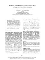
alveolar ridge augmentation of the maxilla (above) and the mandible (below) using autogenous bone grafts from the iliac crestFigure 3
alveolar ridge augmentation of the maxilla (above) and the
mandible (below) using autogenous bone grafts from the iliac
crest.
preoperative panoramic radiographs showing features of dentin dysplasia type IFigure 2
preoperative panoramic radiographs showing features of
dentin dysplasia type I.
initial clinical situationFigure 1
initial clinical situation.
Head & Face Medicine 2007, 3:31 />Page 3 of 5
(page number not for citation purposes)
tured on three-dimensional stereolithographic models of
the mandible and maxilla (figure 5).
After 4 months of socket healing implant surgery was per-
formed under general anaesthesia. The reopening of the
mucoperiostal flaps revealed that the augmented bone
had been resorbed to a significant extend within four
months. By means of the prefabricated templates 10
standard self-tapping implants were inserted in the man-
dible and the maxilla, respectively, according to the prede-
fined planning (figure 6). Bone augmentation around the
dental implants was performed using a mixture (ratio 1:1)
of cancellous bone from the iliac crest and Bio-Oss
®
(par-
ticle size 1–2 mm) (Geistlich, Wolhusen, Switzerland)
held in place by a bioresorbable collagen membrane (Bio-
Mend Extend
®
, Geistlich, Wolhusen, Switzerland). Post-
operative healing was uneventful.
After 4 months of healing, the implants were uncovered
and abutment surgery was performed. All implants were
completely osseointegrated in the new bone. The patient
was provided with a temporary prothesis for two months.
After replacing the healing abutments by definite abut-
ments the final restauration was fabricated and inserted
(figure 7).
Discussion
Dentin dysplasia type I is characterized by primary and
permanent teeth with normal appearance of the crown
but no or only rudimentary root development, incom-
plete or total obliteration of the pulp chamber and peri-
apical radiolucent areas or cysts [1,2]. The abnormal root
morphology is postulated secondary to the abnormal dif-
ferentiation and/or function of the ectomesenchymally
derived odontoblasts [6]. Although various treatment
strategies including conventional endodontic therapy,
periapical curettage or preventive regimen have been pro-
posed to maintain the teeth as long as possible, early exfo-
postoperative clinical situation after completion of the implant treatmentFigure 7
postoperative clinical situation after completion of the
implant treatment.
stereolithographic templates with drill-guide tubes manufac-tured on three-dimensional stereolithographic models of the mandible and maxillaFigure 5
stereolithographic templates with drill-guide tubes manufac-
tured on three-dimensional stereolithographic models of the
mandible and maxilla.
postoperative panoramic radiographs after tooth extraction and bone augmentationFigure 4
postoperative panoramic radiographs after tooth extraction
and bone augmentation.
postoperative panoramic radiographs after implant setting and bone augmentationFigure 6
postoperative panoramic radiographs after implant setting
and bone augmentation.
Head & Face Medicine 2007, 3:31 />Page 4 of 5
(page number not for citation purposes)
liation of the teeth and maxillomandibular atrophy as a
consequence of abnormal root development, periapical
abscesses or cystic formations are characteristics of dentin
dysplasia type I [7].
Successful oral rehabilitation with complete denture after
extraction of all teeth and curettage of cysts has been
described [8].
When implant supported prostheses are planned in
patients affected by dentin dysplasia type I bone regener-
ative therapy is required. Munoz-Guerra et al. reported
successfull treatment of a 24-year old girl after onlay bone
grafting and sinus augmentation [9]. The authors used
cortico-cancellous bone blocks from the iliac crest for
onlay grafting and and a mixture of autologous bone graft
and an autologous platelet concentrate obtained from
platelet-rich plasma for the sinus lift procedure. The teeth
were extracted 4 months after bone augmentation was
performed. No increased and accelerated bone resorption
was observed.
In our patient, extraction of all teeth, cystectomy, bilater-
ally sinus lifting and onlay bone grafting with autogenous
bone grafts were performed as the initial surgical proce-
dure. Already 2 months after bone grafting first signs of
bone resorption were noted.
Resorption of grafted bone is a well known phenomena
that arises during healing and osseointegration processes
and as the result of non physiological loading [10]. Bell et
al. found a 33% resorption rate of mandibular onlay grafts
from the iliac crest during the 4 to 6 months before
implant placement. After implant placement resorption
rate decreased considerably [11]. Several investigations
revealed a high resorption rate of autogenous bone grafts
in the period after grafting and before implant placement
and therefore recommend a mixture of autogenous bone
with allografts [12,13] or stabilizing titanium mesh for
vertical alveolar ridge augmentation [14]. Nevertheless
the presence of a dehiscence defect irrespective of the aug-
mentation treatment used increases the resorption rate
[15]. Bone grafting simultaneous to implant placement
has been published to be a proper strategy as this can
reduce the number of surgical interventions and addition-
ally fix the implant itself [16]. However a staged procedure
is recommended to achieve better implant positioning
after graft consolidation. When iliac bone is used, second
surgeries may be performed at 4 to 6 months [17]. After an
uneventful healing period of 6 month the grafted bone
around the implants will have a prognosis similar to that
of nongrafted bone [18]. The application of autologous
blood plasma enriched with thrombocytes by centrifugal
concentration (platelet-rich plasma: PRP) has been
accredited to enhance the formation of new bone and
improve incorporation and preservation of bone grafts
[19]. Platelet-rich plasma (PRP) is being used to deliver
growth factors in high concentration to sites requiring
osseous grafting. Growth factors released from the plate-
lets include platelet-derived growth factor, transforming
growth factor beta, platelet-derived epidermal growth fac-
tor, platelet-derived angiogenesis factor, insulin-like
growth factor 1, and platelet factor 4. These factors signal
the local mesenchymal and epithelial cells to migrate,
divide, and increase collagen and matrix synthesis. How-
ever there is still lack of scientific evidence to support the
effect of PRP on osteogenic induction and the use of PRP
in combination with bone grafts during augmentation
procedures [20,21]. Although Thor et al. could not dem-
onstrate obvious positive effects of PRP on bone graft
healing the authors observed that the handling of the par-
ticulated bone grafts was improved [19].
In our patient implant placement was performed as a sec-
ond stage procedure. A short period after onlay bone graft-
ing and sinus lifting a high degree bone resorption had
occurred, although healing was uneventfull and no dehis-
cence defect had occured. In this situation presurgical
implant planning using 3D images (SimPlant
®
technol-
ogy) was a helpful tool in this anatomic difficult situation.
We were able to take into account not only the present
bone volume and morphology but also aesthetic consid-
erations regarding the prosthetic treatment. Implant
placement was facilitated by the use of osseous-borne ster-
eolithographic drilling guides. To prevent further exten-
sive secondary bone resorption the principle of guided
bone regeneration was used during the second procedure.
In the present case, despite the hypothesized increased
resorption activity, the secondary performed bone aug-
mentation with a mixture of allogenic materials and
autogenous bone in combination with a resorbable mem-
brane provided a successful longterm result. Munoz-
Guerra et al. recommend a two stage procedure and the
use of autologous cortico-cancellous grafts from the iliac
crest for treatment of their patient with dentin dysplasia
type I [9]. In contrast to our case Munoz-Guerra et al. did
not find an increased affinity for bone resorption in their
patient, but they did not perform tooth extraction and cys-
tectomy before bone augmentation but removed the teeth
4 months after onlay bone grafting and sinuslifting was
performed. Whether this is the crucial difference in treat-
ment strategy or whether patients afflicted by dentin dys-
plasia I posses an increased affinity for bone resorption
has to be discovered by future research.
Conclusion
Oral rehabilitation of patients with dentin dysplasia type
I requires elaborate treatment planning. Surgical implant
planning based on stereolithographic technique is a help-
ful tool in such cases. As we found an increased affinity for
Publish with Bio Med Central and every
scientist can read your work free of charge
"BioMed Central will be the most significant development for
disseminating the results of biomedical research in our lifetime."
Sir Paul Nurse, Cancer Research UK
Your research papers will be:
available free of charge to the entire biomedical community
peer reviewed and published immediately upon acceptance
cited in PubMed and archived on PubMed Central
yours — you keep the copyright
Submit your manuscript here:
/>BioMedcentral
Head & Face Medicine 2007, 3:31 />Page 5 of 5
(page number not for citation purposes)
bone resorption in our patient we recommend guided
bone regeneration using a decelerated biodegradable col-
lageneous membrane and a mixture of autogenous bone
with non resorbable grafting material.
Acknowledgements
We thank our patient and her parents for consenting to publication of this
case.
References
1. O Carroll MK, Duncan WK: Dentin dysplasia type I. Radiologic
and genetic perspectives in a six-generation family. Oral Surg
Oral Med Oral Pathol 1994, 78(3):375-381.
2. Shields ED, Bixler D, el-Kafrawy AM: A proposed classification for
heritable human dentine defects with a description of a new
entity. Arch Oral Biol 1973, 18(4):543-553.
3. O Carroll MK, Duncan WK, Perkins TM: Dentin dysplasia: review
of the literature and a proposed subclassification based on
radiographic findings. Oral Surg Oral Med Oral Pathol 1991,
72(1):119-125.
4. Ommerborn M, Raab W: Allgemeinerkrankungen und Schäden
der Zahnhartsubstanzen. Prophylaxe Impuls 2005, 9:66-73.
5. Pettiette MT, Wright JT, Trope M: Dentinogenesis imperfecta:
endodontic implications. Case report. Oral Surg Oral Med Oral
Pathol Oral Radiol Endod 1998, 86(6):733-737.
6. Melnick M, Levin LS, Brady J: Dentin dysplasia type I: a scanning
electron microscopic analysis of the primary dentition. Oral
Surg Oral Med Oral Pathol 1980, 50(4):335-340.
7. Shankly PE, Mackie IC, Sloan P: Dentinal dysplasia type I: report
of a case. Int J Paediatr Dent 1999, 9(1):37-42.
8. Neumann F, Wurfel F, Mundt T: Dentin dysplasia type I a case
report. Ann Anat 1999, 181(1):138-140.
9. Munoz-Guerra MF, Naval-Gias L, Escorial V, Sastre-Perez J: Dentin
dysplasia type I treated with onlay bone grafting, sinus aug-
mentation, and osseointegrated implants. Implant Dent 2006,
15(3):248-253.
10. Burchardt H: Biology of bone transplantation. Orthop Clin North
Am 1987, 18(2):187-196.
11. Bell RB, Blakey GH, White RP, Hillebrand DG, Molina A: Staged
reconstruction of the severely atrophic mandible with
autogenous bone graft and endosteal implants. J Oral Maxillo-
fac Surg 2002, 60(10):
1135-1141.
12. Pejrone G, Lorenzetti M, Mozzati M, Valente G, Schierano GM: Sinus
floor augmentation with autogenous iliac bone block grafts:
a histological and histomorphometrical report on the two-
step surgical technique. Int J Oral Maxillofac Surg 2002,
31(4):383-388.
13. Szabo G, Huys L, Coulthard P, Maiorana C, Garagiola U, Barabas J,
Nemeth Z, Hrabak K, Suba Z: A prospective multicenter rand-
omized clinical trial of autogenous bone versus beta-trical-
cium phosphate graft alone for bilateral sinus elevation:
histologic and histomorphometric evaluation. Int J Oral Maxil-
lofac Implants 2005, 20(3):371-381.
14. Roccuzzo M, Ramieri G, Bunino M, Berrone S: Autogenous bone
graft alone or associated with titanium mesh for vertical
alveolar ridge augmentation: a controlled clinical trial. Clin
Oral Implants Res 2007, 18(3):286-294.
15. Chen ST, Darby IB, Adams GG, Reynolds EC: A prospective clini-
cal study of bone augmentation techniques at immediate
implants. Clin Oral Implants Res 2005, 16(2):176-184.
16. Penarrocha-Diago M, Gomez-Adrian MD, Garcia-Mira B, Ivorra-Sais
M: Bone grafting simultaneous to implant placement. Pres-
entation of a case. Med Oral Patol Oral Cir Bucal 2005,
10(5):444-447.
17. Triplett RG, Schow SR: Autologous bone grafts and endosseous
implants: complementary techniques. J Oral Maxillofac Surg
1996, 54(4):486-494.
18. Widmark G, Andersson B, Carlsson GE, Lindvall AM, Ivanoff CJ:
Rehabilitation of patients with severely resorbed maxillae by
means of implants with or without bone grafts: a 3- to 5-year
follow-up clinical report. Int J Oral Maxillofac Implants 2001,
16(1):73-79.
19. Thor A, Wannfors K, Sennerby L, Rasmusson L: Reconstruction of
the severely resorbed maxilla with autogenous bone, plate-
let-rich plasma, and implants: 1-year results of a controlled
prospective 5-year study. Clin Implant Dent Relat Res 2005,
7(4):209-220.
20. Arpornmaeklong P, Kochel M, Depprich R, Kubler NR, Wurzler KK:
Influence of platelet-rich plasma (PRP) on osteogenic differ-
entiation of rat bone marrow stromal cells. An in vitro study.
Int J Oral Maxillofac Surg 2004, 33(1):
60-70.
21. Klongnoi B, Rupprecht S, Kessler P, Zimmermann R, Thorwarth M,
Pongsiri S, Neukam FW, Wiltfang J, Schlegel KA: Lack of beneficial
effects of platelet-rich plasma on sinus augmentation using a
fluorohydroxyapatite or autogenous bone: an explorative
study. J Clin Periodontol 2006, 33(7):500-509.