Báo cáo y học: " Isolation and characterization of a virus (CvV-BW1) that infects symbiotic algae of Paramecium bursaria in Lake Biwa, Japan" pptx
Bạn đang xem bản rút gọn của tài liệu. Xem và tải ngay bản đầy đủ của tài liệu tại đây (2.73 MB, 10 trang )
RESEARC H Open Access
Isolation and characterization of a virus
(CvV-BW1) that infects symbiotic algae of
Paramecium bursaria in Lake Biwa, Japan
Ryo Hoshina
1,2
, Mayumi Shimizu
2
, Yoichi Makino
2
, Yoshihiro Haruyama
2
, Shin-ichiro Ueda
2
, Yutaka Kato
2
,
Masahiro Kasahara
2,3
, Bun-ichiro Ono
1,2
, Nobutaka Imamura
2,4*
Abstract
Background: We performed an environmental study of viruses infecting the symbiotic single-celled algae of
Paramecium bursaria (Paramecium bursaria Chlorella virus, PBCV) in Lake Biwa, the largest lake in Japan. The viruses
detected were all Chlorella variabilis virus (CvV = NC64A virus). One of the m, designated CvV-BW1, was subjected to
further characterization.
Results: CvV-BW1 formed small plaques and had a linear DNA genome of 370 kb, as judged by pulsed-field gel
electrophoresis. Restriction analysis indicated that CvV-BW1 DNA belongs to group H, one of the most resistant
groups among CvV DNAs. Based on a phylogenetic tree constructed using the dnapol gene, CvV was classified into
two clades, A and B. CvV-BW1 belonged to clade B, in contrast to all previously identified virus strains of group H
that belonged to clade A.
Conclusions: We conclude that CvV-BW1 composes a distinct species within C. variabilis virus.
Background
Chlorella virus that infects Chlorella-like algae symbiotic
with coelenterate Hydra viridis was first discovered in
1981 and designated HVCV (Hydra viridis Chlorella
virus) [1]. Subsequently, another Chlorella virus that
infects Chlorella-like algae symbiotic with ciliate Para-
mecium bursaria was described (Paramecium bursaria
Chlorella virus [PBCV]) [2]. Studies on HVCV a nd
PBCV have revealed strong host-parasite relationships
[[3] and references therein]: HVCVs do not infect P.
bursaria symbionts, whereas PBCVs do not infect hydra
symbionts; PBCVs collected in the United States infect
algal strain NC64A (representative of U.S. P. bursaria
symbionts) and other U.S. P. bursaria symbionts, but
they do not infect algal strain Pbi (representative of Ger-
man P. bursaria symbionts) or other European P. bur-
saria symbionts; PBCVs collected in Europe infect
European P. bursaria symbionts but do not infect U.S.
P. bursaria symbionts (Fig. 1). Later, another group of
viruses that infect Chlorella-like algae symbiotic with
heliozoon, Acanthocystis turfacea was described [4].
Chlorella viruses studied to date, therefore, can be
divided into four categories: HVCV, NC64A virus, Pbi
virus, and ATCV (Acanthocystis t urfacea Chlorella
virus). Furthermore, none of the Chlorella viruses infect
free-living green algae, and NC64A viruses exhibit a
degree of diversification with regard to, for example,
plaque size, hyaluronan productivity, and DNA methyla-
tion level. Note that viruses attack isolated (or released)
algae but not a lgae inhabiting their hosts (i.e., hydra or
paramecium).
Recent taxonomic studies on P. bursaria symbionts indi-
cated that the algal group “American” containing strain
NC64A and the algal group “European” containing strain
Pbi are genetically distinct from each other, as well as
from any known free-liv ing algae and other symbiotic
algal species [5]. Consequently, each group has been given
a distinct species name, Chlorella variabilis (“American”)
and Micractinium reisseri (“European” ) [6]. Due to the
defects in taxonomy of the host algae, circular virus names
(i.e., Hydra viridis Chlorella virus [HVCV], Paramecium
* Correspondence:
2
Department of Bioscience and Biotechnology, Faculty of Science and
Engineering, Ritsumeikan University, Noji Higashi 1-1-1, Kusatsu, 525-8577
Japan
Full list of author information is available at the end of the article
Hoshina et al. Virology Journal 2010, 7:222
/>© 2010 Hoshina et al; licensee BioMed Central Ltd. This is an Open Access artic le distributed under the terms of t he Creative Co mmons
Attribution License (http://c reativecommons.org/licenses/by/2.0), which permits unrestricted use, distribution, and reproduction in
any medium, provided the original work is properly cited.
bursaria Chlorella [PBCV], and Acanthocystis turfacea
Chlorella virus [ATCV]) and strange names based on host
strains (i.e., NC64A virus and Pbi virus) have been used.
In this report, viruses infecting C. variabilis and M. reisseri
are referred to as C. variabilis virus (CvV) and M. reisseri
virus (MrV), respectively (Fig. 1).
Chlorella variabilis F36-ZK isolated from Japanese
P. bursaria [7] and M. reisseri SW1-ZK isolated from
German P. bursaria [8] are lesser-known hosts in PBCV
studies, although they are well researched strains in phy-
logenetic studies [9,10]. We carried out a screen for
viruses from Lake Biwa and adjacent water environ-
ments using C. variabilis F36-ZK and M. reisseri SW1-
ZK as hosts. Here, we present the results of the environ-
mental study and the results of a biological study of one
strain, CvV-BW1, obtained in the environmental study.
Methods
Algal strains and culture conditions
Chlorella variabilis F36-ZK (NIES-2540) and NC64A
(ATCC 50258) w ere cultured in C liquid medium [11]
with 200 mg L
-1
arginine, while M. reisseri SW1-ZK was
cultured in C liquid medium with 1 g L
-1
casamino acid.
They were maintained under fluorescent illumination
(16 L:8 D, 50 μmol photons m
-2
s
-1
) at 25°C.
Detection of viruses
Water samples were collected from eight sites at Lake
Biwa (the largest lake in Japan) and the adjacent Lake
Yogo. For four sites at Lake Biwa, sampling was carried
out almost every month to observe seasonal variations
in the virus populations. Water samples were centri-
fugedat48,000×g for 30 min, and then virus concen-
trated waters were filtrated through nitrocellulose
membrane (pore size, 0.45 μm). Whether cultures con-
tained the viruses was determined by mixing with
C. variabilis F36-ZK or M. reisseri SW1-ZK liquid cul-
tures on 48-well microplates. The titers (PFU mL
-1
)of
virus-containing cultures were determined by serial
dilution.
Plaque assay and virus isolation
We followed a previously described plaque assay proce-
dure [12] using C medium with 5 g L
-1
glucose and
200 mg L
-1
serine (CGS) in place of modified Bold’ s
basal medium (MBBM). Plaques were observed after
3 days of cultivation. Single plaques were picked up and
transferred to fresh algal lawn plates. Single virus strains
were established by repeating this procedure several
times.
Electric microscopic observation
Chlorella variabilis was incubated for 2 h (25°C) after
adding cultured virus, then fixed with 3% glutaraldehyde
and subsequently with 0.5% osmic acid. Resin-embedded
specimens were cut into ultrathin sections, stained with
3% uranyl acetate, and then observed under an elec tron
microscope at an acceleration voltage of 75 kV.
Another culture was centrifuged at 5000 × g for
5 min, and the resulting supernatant was dropped onto
Veco H-200 mesh (Electron Microscopy Sciences, Hat-
field, PA, USA), stained with 1% uranyl acetate, and
then observed at 75 kV.
SDS-PAGE analysis
Chlorella variabilis-CvV-BW1 culture mixture was first
centrifuged at 12,000 × g for 10 min to remove algal
debris, and the supernatant was centrifuged at 37,000 ×
g for 1 h to precipitate virus particles. Urea was added
to the precipitate at a final concentration of 4 M. After
incubation at 45°C for 1.5 h, the mixture was
centrifuged at 37,000 × g for 10 min to remove the pre-
cipitate. The supernatant was subjected to standard
SDS-PAGE analysis; 4.5% and 7.5% polyacrylamide gels
were used for condensation and separation, respectively.
Electrophoresis was performed at a constant voltage of
200 V using a tank buffer consisting of 0.1% SDS,
192 mM glycine, and 25 mM Tris.
N-terminal amino acid sequence analysis and amino acid
sequence homology search
After SDS-PAGE, proteins in the polyacrylamide gels
were electroblotted onto polyvinylidene fluoride
Figure 1 Schema of PBCV infection of the symbiotic algae of
Paramecium.*Paramecium possessing Chlorella variabilis has been
reported in Japan, China, and Australia as well as the United States.
Hoshina et al. Virology Journal 2010, 7:222
/>Page 2 of 10
membranes (Amersham Biosciences, Piscataway, NJ,
USA) using a Horizeblot apparatus (Atto, Tokyo, Japan)
at a constant current of 0.8 mA cm
-2
for 1 h. Af ter
staining the membrane with 0.1% Ponceau solution,
bands of interest were cut out and subjected to N-term-
inal amino acid sequencing using a PPSQ-21 /23 peptide
sequencer (Shimadzu, Kyoto, Japan ); in the present
study, 15 N-terminal amino acids were examined. Using
the obtained 15 amino acid sequence, a homology
search was carried out using NCBI protein -protein
BLAST />Pulsed-field gel electrophoresis (PFGE)
An equal volume of 1.4% InCert Agarose (45°C; Bio-
Rad, Hercules, CA, USA) was added to a suspension
of Chlorella virus, and the mixture was poured into a
mold and solidified by cooling at room temperature.
An agar block was removed from the mold, soaked in
cell wall-dissolving solution (1 mg mL
-1
proteinase K,
1% lauroyl sarcosinate, 0.01 M Tris-HCl, pH 8.0), and
incubated at 50°C for 16 h. The mixture was dis-
carded, and fresh mixture was supplied and incubated
at 50°C for 24 h. After incubation at 4°C for 2 days in
TE buffer (10 mM Tris-HCl, pH 8.0, containing
0.1 mM EDTA), the gel block was subjected to PFGE
using 1% Seakem GTG agarose (Bio-Rad) and a
CHEF-DRIII system (Bio-Rad). Tank buffer (89 mM
Tris-HCl,pH8.0,containing2mMEDTAand
89 mM boric acid) was used. Electrophoresis was per-
formed at 14°C. Other conditions were as follows:
switching time, 22 to 50 s; total time, 24 h; voltage,
6.6 V cm
-1
. Saccharomyces cerevisiae chromosomes
(Bio-Rad) and l DNA ladde r (Bio-Rad) were used as
size markers.
Extraction of CvV-BW1 DNA
Five units of DNase I was added to the virus particles
(precipitate) described above. The resultant precipitate
was suspended, and the suspension was incubated at
37°C for 1 h. Proteinas e K to at a final concentration of
1mgmL
-1
, EDTA to 0.1 M, and SDS to 0.5% were then
added to the suspension. After incubation at 60°C for
1 h, the mixture was subjected to the standard phenol
extraction procedure [13].
Digestion of CvV-BW1 DNA with restriction enzymes
Restriction enzymes were purchased from Takara Bio
(Otsu, Japan) and/or Nippon Gene (Tokyo, Japan).
Restriction enzymes were used under the conditions
recommended by the manufacturers.
HPLC analysis of methylated nucleotides
CvV-BW1 DNA was mixed with Nuclease P1 (GC Ana-
lysis Standard Kit; Yamasa, Choshi, Japan). The mixture
was incubated at 50°C for 1 h. After digestion, the mix-
ture was subjected to HPLC using a column of ODS-
YMC PACK AQ-312 (6.0 mm in inner diameter and
150 mm in length) (YMC, Kyoto, Japan). HPLC condi-
tions and peak assignment were adopted from Kowalak
et al. [14] and Ushida et al. [15].
Hyaluronan labeling
Hyaluronan labeling was performed according to a
modification of the technique reported by Graves et al.
[16] and Cohen et al. [17]. Chlorella variabilis F36-ZK
was incubated for 2 h (25°C) after adding viruses, of
which 200 μL was centrif uged at 5000 × g for 5 min.
Cells were fixed in phosphate-buffe red saline (PBS)
with 3% paraformaldehyde for 20 min. Centrifugation
and PBS wash were repeated three times. Then, cells
were incubated for 2 h at 37°C with 20 μLofbiotiny-
lated hyaluronic acid binding protein (bHABP, 0.5 mg
mL
-1
;Seikagaku,Tokyo,Japan).Centrifugationand
PBS wash were repeated three times, followed by incu-
bation with 50 mL of CY3-conjugated streptavidin (1.8
mg mL
-1
; ENCO, Petach T ikva, Israel) for 30 min at
37°C. Centrifugation and PBS wash were repeated
three times, and then cells were observed under a
fluorescence microscope with excitation at 510 to 550
nm.
DNA polymerase gene analyses
The DNA polymerase gene (dnapol)regionwas
amplified using the forward primer M37dpo0310F (5′-
CAA TGG TGC AAT TCG TGT TC-3′ )andreverse
primer M37dpo2390R (5′-GTG AAT TTT TCC ATG
GGA TAC TC-3′ ). These primers were designed with
reference to three longer determined sequences of
PBCV-1 (M86836), NY-2A (M86837), and CVK2
(AB011500). A standard three-step PCR protocol was
carried out (annealing temperature of 55°C) using
Takara Ex Taq (Takara Bio) according to the manu-
facturer’ s directions. The PCR product was confirmed
by agarose gel electrophoresis, purified by polyethy-
lene glycol (PEG) precipitation, and then sequenced
directly.
The obtained sequence was compared to those of
Chlorella viruses available in the databases. The align-
ment was performed w ith reference to Zhang et al.
[18], and 663 nucleotide positions (Polymerase
Domain, excluding introns) contributed to phyloge-
netic analysis. Phylogenetic tree was constructed by
the neighbor-joining (NJ) methods of Saito and Nei’ s
evolutionary model using Clustal X ver. 2 [19]. The
significance of each node was tested using 1000 boot-
strap replicates. Evolutionary divergence between
sequences was estimated using the Jukes-Cantor
method in MEGA4 [20].
Hoshina et al. Virology Journal 2010, 7:222
/>Page 3 of 10
Results and discussion
Ecological studies of viruses in Lake Biwa
Using two strains of a lgae, C. variabilis F36- ZK and M.
reisseri SW1-ZK, we surveyed alga e-lytic viruses at nine
sites in Lake Biwa and Lake Yogo, both in Shi ga Prefec-
ture, western Honshu, Japan (Fig. 2), between May and
July 2004. At all sites and at nearly all sampling time
points, we detec ted viruses infecting C. variabilis.None
of the isolated viruses infected M. reisseri in this study,
indicating that all of those obtained were C. variabilis
virus (CvV = NC64A virus).
Since the development of a screening method for virus
sampling [12], both CvV and MrV have been detected
from extensive regions of the world, but MrV h as never
been recorded from East Asia [21,22]. In the present
study, we also found CvVs, but not MrV, from the
water of Lake Biwa. Van Etten [21] indicated that the
factors influencing the distribution patterns of these
viruses are probably latitude and altitude. Based on a
series of taxonomic studies on symbiotic algae, the all
P. bursaria collected so far in Japan have been verified
as C. variabilis-h arbo ring type [6]. The absence of MrV
in Lake Biwa is inevitable if no M. reisseri occur in this
lake.
The results of our ecological studies are summarized
in Table 1. The titers of CvVs were mostly between 0.5
and 50 PFU mL
-1
. This density level is the same or
slightly lower than those reported in previous studies
[e.g., [23,24]]. Exceptionally high values were recorded
in May (85.3 PFU mL
-1
) and June (171.0 PFU mL
-1
)
2004 at Shin-Asahi Windmill Village (site 8). In addi-
tion, no clear seasonal changes in population density
were detected, and the popula tion densities were parti-
cularly low (< 1.5 PFU mL
-1
) in high-temperature waters
(around 30°C) in July 2004 at all the sites except Shin-
Asahi Windmill Village.
Reisser et al. [25] attempted to explain the density of
viruses in natural water environments; the viral density
depends on the P. bursaria population and the probabil-
ity of its burst (i.e., release of symbiotic algae). In 2003
and 2004, a major outbreak of koi herpes virus (KHV)
occurred in Japan. Populations of koi (common carp) in
Lake Biwa were attacked by the viru s from May to June
2004, which caused mass death of the fish. Large num-
bers of koi carcasses washed ashore onto the coastal
area of a sampling point, Shin-Asahi Windmill Village
(site 8). At this time, shallow water around this point
seemed to be under low-oxygen conditions caused by
decomposition of fish carcasses. We detected the highest
virus concentrations at this sampling point at these
times. In contrast, lower densities o f viruses were
Figure 2 Locations of sampling sites. Sites numbered 1 to 4 were
surveyed for seasonal transition.
Table 1 Seasonal transition of Chlorella variabilis viruses concentration (PFU mL
-1
) for nine sampling sites
Sampling date Water temp. (°C) Site 1 Site 2 Site 3 Site 4 Site 5 Site 6 Site 7 Site 8 Site 9
2004 May – 2.67 21.3 21.3 2.67 21.3 2.67 ND 85.3 21.3
June 16.5-19.5 21.3 5.33 10.7 5.33 5.33 – 10.7 171.0 42.7
July 29.3-32.0 ND 0.67 0.67 ND 1.33 – ND 10.7 0.67
Sept. 24.0-25.5 0.67 5.33 10.7 10.7 –––––
Oct. 16.0-16.9 0.67 1.33 10.7 5.33 –––––
Nov. 10.2-12.0 5.33 5.33 5.33 5.33 –––––
Dec. 8.8-11.2 0.67 1.33 21.3 0.33 –––––
2005 Jan. 3.9-6.8 21.3 1.33 0.67 0.67 –––––
Feb. 5.2-8.3 1.33 1.33 10.7 5.33 –––––
Mar. 8.0-9.4 5.33 10.7 21.3 5.33 –––––
Apr. 17.0-18.8 5.33 21.3 21.3 5.33 –––––
June 23.0-24.0 5.33 10.7 42.7 10.7 –––––
Sampling Sites: 1. Karasuma Pen., 2. Kita-Yamada, 3. Yabase Kihan Is., 4. Ohashi Marina, 5. Wani Fishing Port, 6. Aoyagi Beach, 7. Shirahige Beach, 8. Shin-Asahi
Windmill Village, 9. Lake Yogo (also see Fig. 2). ND: Not detected. –: Not determined.
Hoshina et al. Virology Journal 2010, 7:222
/>Page 4 of 10
common in July 2004 at all sampling points (Table 1).
In general, high temperature and strong light prompt
Paramecium to avoid its translatory movement. Low
oxygen levels may have caused bursting of some P.
bursaria cells, with summer heat prompting t he migra-
tion of P. bursaria.
Plaque-forming assay
We performed plaque-forming assay of the viruses, and
all but one plate revealed plaques 3 to 4 mm in dia-
meter . The exceptional plate, for the sample water from
Ohashi Marina (site 4, May 2004), had smaller plaques
(about 1 mm in diameter) in addition to the normal-
sized plaques (Fig. 3). Viruses recovered from one of the
smaller plaques formed smaller plaques on reinfection.
By repeating this procedure several times, we concluded
that we had established a pure clone of smaller plaque-
formi ng virus, which we designated CvV-BW1. We sub-
sequently focused our attention on the biological char-
acteristics of CvV-BW1. We used four independent
clones of normal-sized plaque-forming viruses, CvV-
BW2, -BW3, -BW4, and -BW5, obtained in the same
ecological study. These CvV-BW strains infe ct C. varia-
bilis NC64A but not M. reisseri. Similar to known CvVs,
CvV-BW1 appeared as polyhedral particles about
150 nm in diameter (Fig. 4).
Protein of CvV-BW1
First, we analyzed the protein composition of CvV-BW1
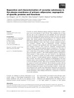
by SDS-PAGE. As shown in Fig. 5, when viral proteins
were not heat-treated (leftmost lane), two major bands,
Figure 3 Plaque formation on the Chlorella variabilis lawn plate
(90 mm petri dish). Both large and small plaques were seen.
Figure 4 Polyhedral particles, attac hing to the external surface of the algal cell wall (TEM, upper panel) and released particles (SEM,
lower panel) of Chlorella variabilis virus. CvV-BW1 is on the left (A and C) and CvV-BW3 is on the right (B and D). Scale bars are 100 nm.
Hoshina et al. Virology Journal 2010, 7:222
/>Page 5 of 10
designated X and Y, were observed. Judging from the
intensity, the proteins in these bands accounted for 80%
of the total viral proteins. By in creasing the temperature
of the heat treatment to 70°C, band X faded, whereas
the intensity of band Y increased. With further increases
in temperature, band Y faded, whereas the intensity of
band Z increased; with heat treatment at 100°C, only
band Z was observed. The sizes of proteins in bands X,
Y, and Z were estimated to be 370 kDa, 105 kDa, and
50 kDa, respectively, compared to size markers. To
identify proteins of these bands, we performed an N-
terminal amino acid sequence analysis. Although we did
not obtain meaningful results for protein of band X,
presumably due to an insufficient amount of protein, we
obtained the same sequence, AGGLSQLVAYGAQDV,
for the proteins recovered from bands Y and Z. The
obtained N-terminal amino acid sequence was comple-
tely identical to those of the majo r capsid proteins of all
PVCVs(NA46AvirusandPbivirus)reportedtodate.
Therefore, we concluded that CvV-BW1 has a major
capsid protein of 50 kDa. We thus contended that band
Y represent dimmer of the 50 kDa major capsid protein.
The assignment of protein of band Z remained to be
established. In addition, CvV-BW1 showed at least nine
distinct bands, which showed no changes in elec tro-
phoretic mobility according to heat treatment condi-
tions. Further studies are required to characterize the
proteins corresponding to these bands.
Size of CvV-BW1 DNA
To estimate the size of CvV-BW1 DNA, we carried out
pulsed-field gel electrophoresis as described in the
Methods section. The result s are shown in Fig. 6.
Compared to Saccharomyces cerevisiae chromosomes
and l DNA ladder, we concluded that the CvV-BW1
DNA is 370 kb in length, assuming that it has a linear
DNA genome. CvV-BW1 DNA was somewhat larger
than those of CvV-BW2, -BW3, and -BW4.
Resistance/susceptibility of CvV-BW1 DNA to restriction
enzymes
CvV has been divided into 16 “ species” based on the
restriction enzyme digestion patterns and various other
characteristics [3]. We attempted to cut the DNA of
CvV-BW strains u sing six widely used restriction
enzymes: HindIII, BamHI, EcoRI, MssI, SfiI, and SwaI
(Fig. 7). The results indicated that CvV-BW1 DNA was
much more resistant to cleavage than the DNAs of
other BW strains. That is, CvV-BW1 DNA was cut only
by MssIandSwaI, while CvV-BW2, -BW3, and -BW5
DNAs were effectively cut by all six enzymes tested.
DNAs of CvV-BW2 and -BW5 showed the same band
pattern, indicating that they are clones of a single
species.
Figure 5 SDS-PAGE analysis of CvV-BW1 virion proteins.From
the left, no heat treatment, heat treatment at 60°C, at 70°C, at 80°C,
at 100°C prior to electrophoresis.
Figure 6 Estimates of virion genome sizes.Fromtheleft,
Saccharomyces cerevisiae chromosomes (Bio-Rad), l DNA ladder
(Bio-Rad), CvV-BW1, CvV-BW2, CvV-BW3, and CvV-BW4.
Hoshina et al. Virology Journal 2010, 7:222
/>Page 6 of 10
An additional 18 restriction enzymes were tested for
CvV-BW1DNA;11oftheenzymesdidnoteffectively
cutCvV-BW1DNA(Fig.8).Theenzymesthatdidnot
effectively cut CvV-BW1 DNA are listed in Table 2
(Enzymes I), while those that cut CvV-BW1 DNA are
shown in Table 3 (Enzymes II). Van Etten et al. [3] clas-
sifiedCvVDNAsinto11restrictiongroups(AtoK)
based on the effects of 13 restriction enzymes. Although
the enzymes they used were not identical to those
applied here, some were common to the two studies.
Judging from the cleavage patterns with the common
enzymes, we concluded that CvV-BW1 DNA belongs to
group H, which is characterized by resistance to EcoRI
but susceptibility to BglII.
Analysis of the enzymes of I and II indicated that the
AT/GC ratio of the recognition sequences was quite dif-
ferent between them; enzymes I were rich (almost 65%)
in GC, whereas enzymes II were rich (75%) in AT. This
result can be rationalized in two ways: CvV-BW1 DNA
isrichinATandpoorinGCorCvV-BW1DNAis
highly modified at G and/or C. Nucleotide sequence
analysis of clones in the CvV-BW1 genome library did
not reveal any evidence that CvV-BW1 DNA was AT-
rich; according to our preliminary genome analysis, the
GC content of CvV-BW1 is in the vicinity of 41.3%.
Figure 7 Restriction enzyme digestion of CvV-BW virion genomes.
Figure 8 Restriction enzyme digestion of the CvV-BW1
genome. For band sizes of the l/HindIII marker, see Fig. 7. A
summary of the effectiveness is shown in Tables 2 and 3.
Table 2 Restriction enzymes that did not effectively cut
CvV-BW1 DNA (Enzymes I)
Restriction enzyme Recognition sequence
BalI TGGCCA
BamHI GGATCC
EcoRI GAATTC
HaeIII RGCGCY
HindIII AAGCTT
HpaI GTTAAC
NcoI CCATGG
NotI GCGGCCGC
PstI CTGCAG
PvuII CAGCTG
SacI GAGCTC
SalI GTCGAC
ScaI AGTACT
SfiI GGCCNNNNNGGCC
Sse8387I CCTGCAGG
Hoshina et al. Virology Journal 2010, 7:222
/>Page 7 of 10
Therefore, we suspected that CvV-BW1 DNA would
have a high incidence of G and/or C modification. To
confirm this speculation, we examined the frequencies
of modified nucleotides in CvV-BW1 DNA; the results
revealed 33.2% 5 mC relative to 5 mC+C and 31.0% 6
mA relative to 6 mA+A.
Production of hyaluronan by CvV-BW1
The best characterized CvV, PBCV-1, encodes hyaluro-
nan synthase (HAS), which functions in the production
of hyaluronan, a polysaccharide covering the outside of
the algal cell wall [26]. Graves et al. [16] showed that
some CvVs produce hyaluronan during infection,
although others do not [27]. Therefore, we examined
whether CvV-BW1 produces hya luronan. Algal cells
showed strong fluorescence 120 min after infection of
CvV-BW1 (stronger than those infected by CvV-BW3)
using the streptavidin-biotin system, indicating the pro-
duction of hyaluronan by CvV-BW1 (Fig. 9).
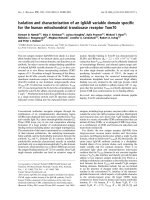
DNA polymerase gene phylogeny of CvV-BW1
The DNA polymerase genes, dnapol, of viruses appear
to have evolved fro m a common ancestral gene, and are
highly conserved within the viral family Phycodnaviridae
[28,29]. Therefore, we attempted to amplify a homolog
from CvV-BW1 via PCR using sequences that are com-
mon to nearly all strains, with PBCV-1, NY-2A, and
CVK2 as primers (Fig. 10). The amplification fragment
of 2060 bp obtained by PCR was then sequenced
(AB572585). Multiple alignme nt with the known PBCV
dnapol sequences indicated that this 2060-bp fragment
contained an intron of 86 bp. Introns of the same length
are present in dnapol of AR-158, NY-2A, NY-2B, and
NYs-1 [18]. In the phylogenetic tree constructed from
the exon regions o f dnapol,CvVwasfoundtobe
divided into two clades, A and B, with a minimum
Table 3 Restriction enzyme that effectively cut CvV-BW1
DNA (Enzymes II)
Restriction enzyme Recognition sequence
BglII AGATCT
DraI TTTAAA
EcoRV GATATC
NdeI CATATG
MssI GTTTAAAC
Sau3AI GATC
SspI ACTAGT
SwaI ATTTAAAT
XbaI TCTAGA
Figure 9 Light (upper) and fluorescence (lower) images of
Chlorella variabilis. A: Noninfected algae. Slight fluorescence
assumed to be intrinsic fluorescence of the chloroplast; B: CvV-BW1-
infected algae; C: CvV-BW3-infected algae.
Figure 10 Domain structure of the dnapo l gene, obtained
sequence, and neighbor-joining tree of PBCVs based on
dnapol gene sequences. Chlorella variabilis virus split into two
lineages, A (101 bp intron group) and B (86 bp intron group).
Numbers at major nodes represent bootstrap probabilities (1000
replicates).
Hoshina et al. Virology Journal 2010, 7:222
/>Page 8 of 10
distance of 0.237 between these clades. As shown in Fig.
10, all CvVs with the 86-bp intron belonged to the same
group that included CvV-BW1 affiliated to clade B,
while all CvVs affiliated to clade A possessed an intron
of 101 bp in their dnapol genes.
Identity of CvV-BW1
Van Etten et al. [ 3] reported that three viral strains, CA-
4A, XZ-4A, and XZ-5C, belong to restriction group H.
Note that these strains all form small plaques (1 mm in
diameter) and are rich in methylated nucleotides (40%
to 45% 5 mC among C+5 mC, and 20% to 30% 6 mA
among A+6 mA). As presented above, CvV-BW1 shares
these properties. However, all the strains belonged to
dnapol clade A (101-bp intron group) (Fig. 10). Mem-
bers of dnapol clade B (86-bp intron group) differ from
CvV-BW1 in some respects. NY-2A belongs to restric-
tion group I, NYs-1 belongs to group F, and NY-2B
belongs to group G. Although the restriction group of
AR158 has not been determined, AR158 does not
encode HAS [30]. Taken together, these findings i ndi-
cate that CvV-BW1 does not belong to any of the 16
CvV “species” defined to date.
Conclusions
We detected C. variabil is virus (NC64A virus) but not
M. reisseri virus ( Pbi virus) in the water of Lake Biwa,
Japan. The highest virus density was recorded in water
under low-oxygen conditions, whereas lower virus densi-
ties were commonly found in the seasons when the lake
waters reached up to around 30°C. These results suggest
that viral de nsity is affected by the population density of
P. bursaria and its burst ratio.
The viral strain CvV-BW1 found in Lake Biwa was
examined in detail with regard to plaque size, electron
microsco pic features, protein composition, genome size,
restriction enzyme digestion, level of DNA methyla tion,
production of hyaluronan, and phylogeny of the DNA
polymerase gene. Taken together, all of these observa-
tions indicate that CvV-BW1 is likely to be a new spe-
cies of C. variabilis virus.
List of abbreviations used
CvV: Chlorella variabilis virus; MrV: Micractinium reisseri virus; PBCV:
Paramecium bursaria Chlorella virus.
Competing interests
The authors declare that they have no competing interests.
Authors’ contributions
MS screened and isolated the viral strains, and then tested hyaluronan
productivity. YK observed viruses by electron microscopy. SiU carried out the
protein analysis. YM examined the viral genome sizes, and then MS and YM
confirmed the results of restriction enzyme digestion. YH examined viral
DNA modification. RH contributed to DNA polymerase gene analyses. RH
and BiO prepared the manuscript. NI initially conceived of this study and RH,
MK, BiO, NI finalized the experimental design. All authors have read and
approved the final manuscript.
Authors’ information
1 Department of Biomedical Science, College of Life Sciences, Ritsumeikan
University, Noji Higashi 1-1-1, Kusatsu, 525-8577 Japan.
2 Department of Bioscience and Biotechnology, Faculty of Science and
Engineering, Ritsumeikan University, Noji Higashi 1-1-1, Kusatsu, 525-8577
Japan.
3 Department of Biotechnology, College of Life Sciences, Ritsumeikan
University, Noji Higashi 1-1-1, Kusatsu, 525-8577 Japan.
4 Department of Pharmacy, College of Pharmaceutical Sciences, Ritsumeikan
University, Noji Higashi 1-1-1, Kusatsu, 525-8577 Japan.
Acknowledgements
We thank Associate Prof. T. Suzaki (Kobe University) for help with electron
microscopy.
Author details
1
Department of Biomedical Science, College of Life Sciences, Ritsumeikan
University, Noji Higashi 1-1-1, Kusatsu, 525-8577 Japan.
2
Department of
Bioscience and Biotechnology, Faculty of Science and Engineering,
Ritsumeikan University, Noji Higashi 1-1-1, Kusatsu, 525-8577 Japan.
3
Department of Biotechnolog y, College of Life Sciences, Ritsumeikan
University, Noji Higashi 1-1-1, Kusatsu, 525-8577 Japan.
4
Department of
Pharmacy, College of Pharmaceutical Sciences, Ritsumeikan University, Noji
Higashi 1-1-1, Kusatsu, 525-8577 Japan.
Received: 20 July 2010 Accepted: 13 September 2010
Published: 13 September 2010
References
1. Meints RH, Van Etten JL, Kuczmarski D, Lee K, Ang B: Viral infection of the
symbiotic Chlorella-like alga present in Hydra viridis. Virology 1981,
113:698-703.
2. Van Etten JL, Meints RH, Kuczmarski D, Burbank DE, Lee K: Viruses of
symbiotic Chlorella-like algae isolated from Paramecium bursaria and
Hydra viridis. Proc Natl Acad Sci USA 1982, 79:3867-3871.
3. Van Etten JL, Lane LC, Meints RH: Viruses and viruslike particles of
eukaryotic algae. Microbiol Rev 1991, 55:586-620.
4. Bubeck JA, Pfitzner AJP: Isolation and characterization of a new type of
chlorovirus that infects an endosymbiotic Chlorella strain of the
heliozoon Acanthocystis turfacea. J Gen Virol 2005, 86:2871-2877.
5. Hoshina R, Imamura N: Multiple origins of the symbioses in Paramecium
bursaria. Protist 2008, 159:53-63.
6. Hoshina R, Iwataki M, Imamura N: Chlorella variabilis and Micractinium
reisseri sp. nov. (Chlorellaceae, Trebouxiophyceae): redescription of the
endosymbiotic green algae of Paramecium bursaria (Peniculia,
Oligohymenophorea) in the 120th year. Phycol Res 2010, 58:188-201.
7. Kamako S-i, Hoshina R, Ueno S, Imamura N: Establishment of axenic
endosymbiotic strains of Japanese Paramecium bursaria and the
utilization of carbohydrate and nitrogen compounds by the isolated
algae. Eur J Protistol 2005, 41:193-202.
8. Hoshina R, Imamura N: Phylogenetically close group I introns with
different positions among Paramecium bursaria photobionts imply a
primitive stage of intron diversification. Mol Biol Evol 2009, 26:1309-1319.
9. Hoshina R, Kamako S-i, Imamura N: Phylogenetic position of
endosymbiotic green algae in Paramecium bursaria Ehrenberg from
Japan. Plant Biol 2004, 6:447-453.
10. Hoshina R, Kato Y, Kamako S-i, Imamura N: Genetic evidence of
“American”
and “European” type symbiotic algae of Paramecium bursaria
Ehrenberg. Plant Biol 2005, 7:526-532.
11. Ichimura T: Sexual cell division and conjugation-papilla formation in
sexual reproduction of Closterium strigosum. In Proceedings of the Seventh
International Seaweed Symposium: August 1971; Hokkaido. Edited by:
Nishizawa K, Arasaki S, Chihara M, Hirose H, Nakamura V, Tsuchiya Y. Tokyo:
University of Tokyo Press; 1971:208-214.
12. Van Etten JL, Burbank DE, Kuczmarski D, Meints RH: Virus infection of
culturable Chlorella-like algae and development of a plaque assay.
Science 1983, 219:994-996.
Hoshina et al. Virology Journal 2010, 7:222
/>Page 9 of 10
13. Sambrook J, Fritsch EF, Maniatis T: Molecular Cloning: A Laboratory
Manual. NY: Cold Spring Harbor Laboratory press, 2 1989.
14. Kowalak JA, Bruenger E, Hashizume T, Peltier JM, Ofengand J, McCloskey JA:
Structural characterization of U*-1915 in domain IV from Escherichia coli
23S ribosomal RNA as 3-methylpseudouridine. Nucleic Acids Res 1996,
24:688-693.
15. Ushida C, Muramatsu T, Mizushima H, Ueda T, Watanabe K, Stetter KO,
Crain PF, McCloskey JA, Kuchino Y: Structural feature of the initiator tRNA
gene from Pyrodictium occultum and the thermal stability of its gene
product, tRNA
i
Met
. Biochimie 1996, 78:847-855.
16. Graves MV, Burbank DE, Roth R, Heuser J, DeAngelis PL, Van Etten JL:
Hyaluronan synthesis in virus PBCV-1-infected Chlorella-like green algae.
Virology 1999, 257:15-23.
17. Cohen M, Klein E, Geiger B, Addadi L: Organization and adhesive
properties of the hyaluronan pericellular coat of chondrocytes and
epithelial cells. Biophys J 2003, 85:1996-2005.
18. Zhang Y, Adams B, Sun L, Burbank DE, Van Etten JL: Intron conservation in
the DNA polymerase gene encoded by Chlorella viruses. Virology 2001,
285:313-321.
19. Larkin MA, Blackshields G, Brown NP, Chenna R, McGettigan PA,
McWilliam H, Valentin F, Wallace IM, Wilm A, Lopez R, Thompson JD,
Gibson TJ, Higgins DG: Clustal W and Clustal X version 2.0. Bioinformatics
2007, 23:2947-2948.
20. Tamura K, Dudley J, Nei M, Kumar S: MEGA4: Molecular Evolutionary
Genetics Analysis (MEGA) software version 4.0. Mol Biol Evol 2007,
24:1596-1599.
21. Van Etten JL: Unusual life styoe of giant chlorella viruses. Annu Rev Genet
2003, 37:153-195.
22. Yamada T, Onimatsu H, Van Etten JL: Chlorella viruses. Adv Virus Res 2006,
66:293-336.
23. Van Etten JL, Van Etten CH, Johnson JK, Burbank DE: A survey for viruses
from fresh water that infect a eukaryotic chlorella-like green alga. Appl
Environ Microbiol 1985, 49:1326-1328.
24. Yamada T, Higashiyama T, Fukuda T: Screening of natural waters for
viruses which infect chlorella cells. Appl Environ Microbiol 1991,
57:3433-3437.
25. Reisser W, Becker B, Klein T: Studies on ultrastructure and host range of a
Chlorella attacking virus. Protoplasma 1986, 135:162-165.
26. DeAngelis PL, Jing W, Graves MV, Burbank DE, Van Etten JL: Hyaluronan
synthase of Chlorella virus PBCV-1. Science 1997, 278:1800-1803.
27. Mohammed Ali AM, Kawasaki T, Yamada T: Genetic rearrangements on
the Chlorovirus genome that switch between hyaluronan synthesis and
chitin synthesis. Virology 2005, 342:102-110.
28. Lee AM, Ivey RG, Meints RH: The DNA polymerase gene of a brown algal
virus: structure and phylogeny. J Phycol 1998, 34:608-615.
29. Schroeder DC, Oke J, Malin G, Wilson WH: Coccolithovirus
(Phycodnaviridae): Characterisation of a new large dsDNA algal virus
that infects Emiliania huxleyi. Arch Virol 2002, 147:1685-1698.
30. Fitzgerald LA, Graves MV, Li X, Feldblyum T, Nierman WC, Van Etten JL:
Sequence and annotation of the 369-kb NY-2A and the 345-kb AR158
viruses that infect Chlorella NC64A. Virology 2007, 358:472-484.
doi:10.1186/1743-422X-7-222
Cite this article as: Hoshina et al.: Isolation and characterization of a
virus (CvV-BW1) that infects symbiotic algae of Paramecium bursaria in
Lake Biwa, Japan. Virology Journal 2010 7:222.
Submit your next manuscript to BioMed Central
and take full advantage of:
• Convenient online submission
• Thorough peer review
• No space constraints or color figure charges
• Immediate publication on acceptance
• Inclusion in PubMed, CAS, Scopus and Google Scholar
• Research which is freely available for redistribution
Submit your manuscript at
www.biomedcentral.com/submit
Hoshina et al. Virology Journal 2010, 7:222
/>Page 10 of 10