Báo cáo y học: "A role for the histone deacetylase HDAC4 in the life-cycle of HIV-1-based vectors" ppsx
Bạn đang xem bản rút gọn của tài liệu. Xem và tải ngay bản đầy đủ của tài liệu tại đây (1.86 MB, 10 trang )
RESEA R C H Open Access
A role for the histone deacetylase HDAC4 in the
life-cycle of HIV-1-based vectors
Johanna A Smith
1
, Jennifer Yeung
1
, Gary D Kao
2
, René Daniel
1,3,4*
Abstract
HIV-1 integration is mediated by the HIV-1 integrase protein, which joins 3′-ends of viral DNA to host cell DNA. To
complete the integration process, HIV-1 DNA has to be joined to host cell DNA also at the 5′-ends. This process is
called post-integration repair (PIR). Integration and PIR involve a number of cellular co-factors. These proteins exhi-
bit different degrees of involveme nt in integration and/or PIR. Some are required for efficient integration or PIR. On
the other hand, some reduce the efficiency of integration. Finally, some are involved in integration site selection.
We have studied the role of the histone deacetylase HDAC4 in these processes. HDAC4 was demonstrated to play
a role in both cellular double-strand DNA break repair and transcriptional regulation. We observed that HDAC4
associates with viral DNA in an integrase-dependent manner. Moreover, infection with HIV-1-based vectors induces
foci of the HDAC4 protein. The related histone deacetylases, HDAC2 and HDAC6, failed to associate with viral DNA
after infection. These data suggest that HDAC4 accumulates at integration sites. Finally, overexpression studies with
HDAC4 mutants suggest that HDAC4 may be required for efficient transduction by HIV-1-based vectors in cells that
are deficient in other DNA repai r proteins. We conclude that HDA C4 is likely involved in PIR.
Introduction
Chromatinundergoesexpansionandcompactioninthe
course of many fundamental cellular processes, including
gene expressi on, differentiation, cell cycle prog ression
and DNA repair. These alterati ons of the chromatin
structure are largely mediated by histone acetylases and
histone deacetylases (HDACs). HDACs deacetylate key
lysine residues of core histones to induce chromatin
compaction. This process usually results in transcrip-
tional repression [1]. Cells contain many HDACs, which
are categorized into four classes, based on sequence
homologies. Class I (homologues of the yeast deacetyl ase
Rpd3) contains HDAC1, HDAC2, HDAC3 and HDAC8
[2-6]. Class II (yeast Hda1 homologues) contains
HDAC4, HDAC5, HDAC6 and HDAC7 [7-12]. Class II
HDACs, unlike Class I, can shuttle in and out of the
nucleus, depending on various signals [13]. Class III con-
tains proteins that are homologous to the yeast deacety-
lase Sir 2 [14,15]. Finally, the Class IV contains enzymes
whicharerelatedtothoseofClassIandClassII,buta
sequence analysis shows they form a distinct class. They
are exemplified by HDAC11 [16].
Although transcriptional repression is apparently an
important function of HDACs, these proteins see m to
play a broader role in regulating cellular processes and
one HDAC, HDAC4, has been found to play a role in cel-
lular double-strand DNA break (DSB) repair. It has been
shown by Kao et al. (2003) that HDAC4 forms nuclear
foci in cells exposed to ionizing radiation, which causes
double-strand DNA breaks [17]. Foci of DNA repair pro-
teins are formed at sites of double-strand DNA breaks,
and the HDAC4 foci overla p with foci of the DNA repair
proteins Rad51 and 53BP1. Silencing of HDAC4 via RNA
interference leads to radiosensitisation of HeLa cells,
underscoring a requirement for HDAC4 in DSB repair.
In addition, HDAC4-deficient cells were shown to loose
the DNA damage-induced G2/M checkpoint. The mole-
cular function of HDAC4 in DSB repair remains to be
fully clarified, although it has been shown very recently
that nuclear tra nslocation of H DAC4 is required and it
may play a ro le in the suppression of promoters of genes
that are activated during G2/M progression [18,19].
It has been shown previously by us and others that
cellular DSB repair proteins are involved in the life-cycle
of retroviruses and retrov iral vectors. We have observed
* Correspondence:
1
Division of Infectious Diseases - Center for Human Virology, Department of
Medicine, Thomas Jefferson University, Philadelphia, PA 19107, USA
Full list of author information is available at the end of the article
Smith et al. Virology Journal 2010, 7:237
/>© 2010 Smith et al; licensee BioMed Central L td. This is a n Open Access a rticle distributed under t he terms of t he Creative Commons
Attribution License ( which pe rmits unrestri cted use, distribution, and reproduction in
any medium, provided the original work is properly cited .
that cellular DSB proteins ar e involved in completing
the integration process. In addition, others suggested
that they are involved in the formation of 2-LTR circles,
and it has been proposed that they might also be
involved in int ranuclear traff icking of the preintegratio n
complex [20-23].
In this study, we have tested the hypothesis that
HDAC4 plays a role in the life-cycle of HIV-1-based
vectors. We show that infection with retroviral vectors
induces, similar to DSBs, nuclear foci of the HDAC4
protein. We show that the formation of these foci is
dependent on active retroviral integrase, and HDAC4,
but not HDAC2 and HDAC6, associates with viral
DNA. Taken together, these data indicate t hat HDAC4
plays a yet undiscovered role at sites of retroviral DNA
integration. In addition, we show that overexpression of
nuclear HD AC4 rescues a defect in retroviral transd uc-
tion that is associated with a deficiency of the cellular
DNA repair protein ATM. We conclude that HDAC4 is
involved in stable transduction by retroviral vectors,
and plays a role in the completion of the integration
process.
Results
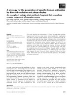
HDAC4, but not HDAC2 or HDAC6, associates with DNA
of an infecting HIV-1-based vector
HeLa cells were infected with a pseudotyped HIV-1-
based vector (containing a lacZ reporter) at an m.o.i. of
0.1 and harvested at the time points indicated (Fig. 1A).
Chromatin immunoprecipitation (ChIP) analysis was
used to identify the association of HDAC4 with viral
DNA. To do so, DNA isolated from infected cells and
the associated proteins were crosslinked, immunopreci-
pitated with the HDAC4 antibody (see Methods), and
associated viral DNA was amplified by real time PCR.
Results are expressed as a n umber of viral DNA ampli-
cons per μl of chromatin immunoprecipitates at each
time p oint. As shown in Fig. 1A, viral DNA was found
to be associated with HDAC4 at 4, 6, 8, and 16 hrs
post-infection. The amount of HDAC4-associated viral
DNA steadily increased from 4 hours, with a peak
reached at 8 hours post-infection. The associated viral
DNA drastically declined at the 16 hour t ime point. To
determine if vector DNA associates with other HDAC
proteins, we have immunoprecipitated lysates from
infected cells with the HDAC2 and HDAC6 antibodies.
Whereas HDAC2 is a Class I HDAC, w e note HDAC6
is a class II HDAC and thus structurally c losely related
to HDAC4. However, as shown in Fig. 1B, we did not
observe any association of these HDACs with viral
DNA. We thus conclude that HDAC4 shows a distinct
preference for associ ation with vector DNA, wh en com-
pared to other HDACs.
Retroviral integration enhances the association of HDAC4
with vector DNA
We note that HDAC4 was reported to associate with
DNA of the avian sarcoma virus, but this association
was detected onl y post-integration [24]. To test t he
hypothesis that integration is requir ed for the associa-
tion of HDAC4 with the DNA o f HIV-1-based vectors,
we have infected HeLa cells and t reated them with the
integrase inhibitor 118-D-24. As shown in Fig. 1C, the
inhibitor decreases the association of HDAC4 with vec-
tor DNA in a dose-dependent manner. However, we
note that the inhibitor effect can be seen only at
8 hours post-infection, when the association of DNA
with HDAC4 is at its peak. In contrast, association at
4 hours post-infection is res istant to the inhib itor treat-
ment. These data suggest that while integration does sti-
mulate the association of vector DNA with HDAC4,
HDAC4 also associates with vector DNA prior to inte-
gration, in an integration-independent manner.
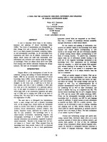
Retroviral integration induces the formation of HDAC4
foci in infected cells
HDAC4 was reported to form foci in irradiated cells.
These foci were a ssociated with the formation of dou-
ble-strand DNA breaks [ 17]. To determine if infection
with HIV-1-based vectors induces the formation of
HDAC4 foci, we have infected HeLa cells at a high mul-
tiplicity of infection (10), fixed infected cells at predeter-
mined time points and st ained with the HDAC4
ant ibody. We observed that in u ninfected cells, HDAC4
is present almost exclusively in the cytoplasm (Fig. 2).
Similarly, we have observed that HDAC4 is predomi-
nantly cytoplasmic at 4 and 6 hours post-infection.
However, we also observed the appearance of HDAC4
foci in infected cells, with the majority of cells contain-
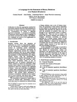
ing foci at 8 hrs p ost-infection (Figs. 2 and 3). Most of
the infected cells contained multiple HDAC4 foci. As
indicated in Fig. 3, the number of foci correlates well
with the multiplicity of infection.
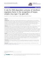
We have observed that integration stimulates the asso-
ciation of HDAC4 with vector DNA and wondered if
integration affects the formation of HDAC4 foci. Thus,
we have infected HeLa cells in the presence and absence
of an integrase inhibitor. We have again detected
HDAC4 foci in cells that were infected with the HIV-1-
based vector in the absence of the inhibitor (Fig. 4).
However, tre atment of infected cells with the inhibitor
significantly reduced (ca. 3.5 fold) the total number of
cells that contained foci (Fig. 4 and Fig. 5). We have
also observed a drop in the average number of foci per
cell among foci-containing cells, although the differen ce
was within the standard deviation due to a wide range
of the n umbers of foci (1 to 8 foci per cell among cells
Smith et al. Virology Journal 2010, 7:237
/>Page 2 of 10
Figure 1 HDAC4 associates with vector DNA in an integrase-dependent manner. (A) ChIP analysis of infected HeLa cells. To establish if
HDAC4 associates with vector DNA, HeLa cells were infected with the HIV-1-based vector at an m.o.i. of 0.1 and ChIP was performed with the
anti-HDAC4 antibody as described in “Experimental Procedures”, followed by real-time PCR to detect vector DNA. Numbers (x-axis) indicate hours
post-infection. Bkgd - background, no antibody added. (B) HDAC2 and HDAC6 association with vector DNA. Lysates of cells infected as
described above (Fig. 1A) were immunoprecipitated with antibodies against HDAC2 and HDAC6, as indicated. Terminology as above, * indicates
samples from A. (C) Effect of an integrase inhibitor on the association of HDAC4 with vector DNA. Cells were infected as in A, except the
integrase inhibitor 118-D-24 was added to samples at the indicated concentrations, together with the vector. Cells were processed as in A.
Smith et al. Virology Journal 2010, 7:237
/>Page 3 of 10
infected with the vector and 1 to 6 foci per cell in cells
infected with the vector and treated with the inhibitor,
Fig. 5B). Treatment with the inhibitor itself had a negli-
gible effect on t he i ntracellular localization of H DAC4
(Figs. 4 and 5). Taken to gether, our result s suggest t hat
although HDAC4 associates with viral DNA even prior
to integration, integration s timulates further accumula-
tion of HDAC4 at integration sites, which are then
marked by the formation of HDAC4 foci.
Effect of HDAC4 knockdown on HIV-1 transduction
We have established a novel interaction between the
cellular HDAC4 protein and HIV-1-based vectors. Our
results suggested that HDAC4 plays a role in the life-
cycle of these vectors. To test this hypothesis, we have
knocked down HDAC4 in HeLa cells using siRNA treat-
ment and determined if HDAC4 is required for stable
integration of HIV-1- vector DNA. As shown in Fig. 6,
HDAC4 had little effect on the efficiency of integration
as me asured by Alu-PCR. In addition, we have infected
siRNA-treated cells with the HIV-1-based vector c arry-
ing an EGFP marker and examined EGFP expression
using flow cytometry. We have not observed a signifi-
cant drop in EGFP expression in HDAC 4 siRNA-treated
cells (data not shown). We conclude that HDAC4 defi-
ciency does not appear to significantly affect the effi-
ciency of integration. Similarly, it appears that HDAC4
is not n ecessary for the last step of the integration pro-
cess, termed post-integration repair (PIR), since PIR fail-
ure r esults in a loss of cells in which integrase-mediated
joining occurred, and thus again manifests as a decrease
in the Alu-PCR signal [25,26].
HDAC4 is involved in PIR in ATM-deficient cells
HDAC4 is a DSB rep air prote in, and it had been
reported by us and others that these proteins are
involved in PIR [23]. However, cellular DSB repair pro-
teins often have overlapping functions and DSB repair
sys tems can partially s ubstitute for each other [27]. It is
thus possible that a los s of the HDAC4 p rotein can be
compensated for by other DSB repair systems or pro-
teins. To test this hypothesis, we have induced a DSB
repair deficiency in HeLa cells by treatment with an
established ATM inhibitor, KU-55933 [28]. The ATM
Figure 2 The HDAC4 protein forms foci following infection with the HIV-1-based vector. HeLa cells were infected with an HIV-1-based
vector as described in “Experimental Procedures”. At indicated time points, samples were fixed and stained with the anti-HDAC4 antibody (top
row). Nuclei were visualized using DAPI staining (middle row). Representative photographs are shown. DIC (differential interference contrast)
shows the cell morphology (bottom row).
Smith et al. Virology Journal 2010, 7:237
/>Page 4 of 10
protein is a major player in cellular DSB repair and was
reported by us and others to be involved in PIR [26-28].
At the same time, in these cells, we have overexpressed
the HDAC4 protein. Since the no rmal HDAC4 p rotein
(denoted h ere as HDAC4-1084) is mainly cytoplasmic,
we have also overexpressed a mutant, which lacks a
nuclear export signal (HDAC4-1061) and is thus present
in the nucleus (Fig. 7, [29]). As expected, the ATM inhi-
bitor reduced the Alu-PCR signal due to the inhibition
of PIR (Fig. 7A). We have observed that in ATM-profi-
cient HeLa cells, the overex pressed HDAC4 proteins do
not appear to affect the efficiency of integration or PIR,
as shown by Alu-PCR (Fig. 7A). However, in HeLa cells
that were treated with the ATM inhibitor, the HDAC4-
1084 reverses the inhibitor effect and upregulates HIV-1
transduction four fo ld. The HDAC4-1061 mutant that is
constitutively present in the nucleus completely reverses
the effect of the ATM inhibitor (Fig. 7A). T o investigate
the possibility that t he differences in the Alu-PCR
signals could be due to variations of the exogenous
HDAC4 express ion levels, we pe rformed a western blot-
ing analysis (Fig. 7B). However HDAC4-1061 and
HDAC4- 1084 levels appear to be the same in our trans-
fected cells. Taken together our results suggest that
HDAC4isinvolvedinPIR,butitsfunctioncanbe
replaced by other DSB protein(s).
Finally, a failure of PIR induces apoptotic death of
infected cells. I f the effect of n uclear HDAC4 on inf ec-
tion ef ficiency is due its role in PIR, it should prevent
PIR-associated cell death. Thus, cells were tre ated and
infected as above (Fig. 7), except at a high m.o.i. (2), and
analyzed by Western blotting for the presence of the 85-
kDa PARP fragment, an apoptotic marker generated by
caspase-medi ated cleavage o f the PARP protein [30]. As
shown in Fig. 8, ATM inhibition and infection stimu-
lated PARP cleavage. However, the apoptosis was
reduced by overexpression of either the full length
HDAC4 (HDAC4-1084) or the truncated mutant
(HDA C4-1061) . This finding is again consistent with an
HDAC4 role in PIR.
Discussion
In this study, we demonstra te that t he histone deacety-
lase HDAC4, a Class II HDAC, associates with DNA of
HIV-1-based vectors and forms foci at s ites of integra-
tion. We also show that overexpression of nuclear
HDAC4 rescues the defect in PIR that is induced by an
ATM deficiency. Our data thus reveals a new cellular
partner, which is involved in the life-cycle of HIV-1-
based vectors. Our finding also supports the hypothesis
that cellular DSB repai r proteins are invo lved in PIR. At
the same time, these proteins clearly have overlapping
functions and can to a degree substitute for each other.
What could be the HDAC4 function in PIR? HDAC4
is a deacetylase, although with relatively low a ctivity
[31]. Histone deacetylation generally results in transcrip-
tional suppression. Thus, one possible function of
HDAC4 could be to suppress transcription at integra-
tion sites, thus allowing access for DNA repair machin-
ery. We note in this context that HIV-1 prefers to
integrate in genes and the likelihood of transcription
interfering with integration is thus very high [32]. Sec-
ond, it is possible that HDAC4 is required for the
recruitment of other DNA repair proteins to PIR sites.
HDAC4 was reported to physically interact with the
53BP1 protein and thus may bring this protein to PIR
sites. It will be a matter of future experiments to distin-
guish between these possibilities.
We also note that HDAC4 associates with vector
DNA prior to integration. These data suggest that
HDAC4 may play a role in steps prior to PIR. However,
since HDAC4 knockdown does not appear to have a
major effect on the transduction efficiency of the vector,
Figure 3 Quantitat ive analysi s of HDAC4 foci in the vect or-
infected cells. Number of HeLa cells containing foci, as well as foci
number per cells were counted in images prepared as described in
Fig. 2 and “Experimental Procedures”.(A) Number of foci-containing
cells. (B) Average number of foci among foci-containing cells.
Numbers (x axis) indicate hours post-infection. Bars indicate
standard deviation. * - only one foci-containing cell was found.
Smith et al. Virology Journal 2010, 7:237
/>Page 5 of 10
it seems likely that HDAC4 is not required for these
steps of the retroviral life-cycle. Nevertheless, it is possi-
ble that HDAC4 affects t he life-cycle in different ways.
One possibility is that HDAC4 has a cell-type-specific
function and affects the retroviral life-cycle differently
depending on cell type. Another possibility is that
HDAC4 affects intracellular or intranuclear trafficking,
which may effect integration s ite selection. Experiments
designed to test these hypotheses are underway in our
laboratory.
What are the practical implications of our data? HIV-
1-based vectors perform integration and PIR in an iden-
tical way to wild-type HIV-1. Thus, proteins which are
required for PIR of HIV-1-based vectors are also
involved in PIR of HIV-1. Since PIR is absolutely
required for HIV- 1 repli cation, proteins involv ed in PIR
are potential targets for ani-HIV-1-therapy. However,
overlapping functions of these proteins suggest that it
will be necessary to inhibit more than one DNA repair
pathway to achieve complete suppression of HIV-1
replication.
Finally, our results indicate that HDAC4 accumulates
at the sites of integration. HDAC4 foci t hus may serv e
as a useful marker for integration, and their numbers
could be used to evaluate the efficacy of HIV-1- inhibi-
tors at the early steps of the HIV-1- life-cycle.
Experimental Procedures
Cells
HeLa cells were maintained in DMEM medium sup ple-
mented with 10% fetal bovine serum and antibiotics
(Penn/Strep).
HIV-1-based vectors
All VSV G-pseudotyped HIV-1 based vectors were pre-
pared as described previously [33,34], and carried either
a lacZ or EGFP reporter gene.
Plasmids and transfections
Plasmids expressing the full-length HDAC4 (amino
acids 1-1084) fused to the EGFP protein (HDAC4-1084)
or the HDAC4 C-terminal truncated mutant, lacking
Figure 4 Effect of an integrase inhibitor on the formation of HDAC4 foci in infected cells. HeLa cells we re infected with the HIV-1-based
vector in the presence and absence of the integrase inhibitor (100 μM), or were treated only with the inhibitor, as indicated. Cells were
processed as in Fig. 2, at 8 hrs post-infection. M - mock, uninfected cells, Inf - cells infected with the HIV-1-based vector, Inf+II - cells infected
with the HIV-1-based vector and treated with the integrase inhibitor (added together with the virus), II - cells treated with the integrase inhibitor
only. Other terminology as in Fig. 2.
Smith et al. Virology Journal 2010, 7:237
/>Page 6 of 10
the nuclear export signal (amino acids 1-1061) fused to
the EGFP protein (HDAC4-1061) have been described,
and were a g enerous gift from Dr. X. J. Yang of McGill
University [12,29]. The PEGFP-C1 control plasmid
expressing the EGFP protein under control of the CMV
promoter was purchased from Clontech (GenBank
Accession # U55763). Plasmids were transfected into
HeLa cells using the Lipofectamine™ 2000 transfection
reagent (Invitrogen, cat # 11668-027) using company
protocols. Cells were infected with the HIV-1-based vec-
tor two days post-transfection.
Chromatin Immunoprecipitation
HeLa cells were infect ed at a multiplici ty of infect ion
(m.o.i.) 0.1 for the indicated time intervals. In some
cases, an integrase inhibitor was added at the time of
infection (118-D-24, NIH AID S Reagent Program). Cells
were then harveste d and ChIP was performed as
described [35], with the anti-HDAC4 antibody (1 μg/
sample, San ta Cruz Biotechnology , cat # sc-11418X) or
anti-HDAC2 antibody (1 μg/sample, Abcam, cat #
ab16032) or anti-HDAC6 antibody (1 μg/sample, Santa
Cruz, cat # sc-11420). Protein-associated vector DNA
was detected by real-time PCR, using primers and
probes detecting HIV-1 LTR. Forward primer: 5′ -
TGTGTGCCCG TCT GTTGT GT-3 ′; Reverse primer: 5′-
CCTGCGTCGAGA GAGCTC-3′. To quantitate the viral
amplicon, a TaqMan dual 5′-6-carboxyfluorescein-and
3′-6-carboxytetramethylrhodamimine-labeled probe was
used: 5′ -(FAM)-CAGTGGCGCCCGAACAGGGA-
(TAMRA)-3′ (Integrated DNA Technologies). Real-time
PCR was performed using a LightCycler 1.5 with soft-
ware 3.5.3 (Roche). Reaction mixtures contained Quanti-
Fast Probe 2× mix (Qiagen), 100 nM probe, and 200 nM
primers. The standard cycling conditions were 95°C - 3
min followed by 50 cycles at 95°C - 3 s and 60°C - 30 s.
Samples were run in triplicate.
Immunofluorescence experiments
HeLa cells were plated at a density of 2 × 10
4
and
grown on 4-well chamber slides. The following day, the
cells w ere infected with the HIV-based vector at m.o.i.
10 for a time course study at 4, 6, and 8 hours. In
another experiment, vector and an integrase inhibitor
(118- D-24, final concentration of 100 μM) or the vector
only had been incubated for 8 hours prior to fixation.
At the indicated time points, cells were washed in PBS
and fixed by adding cold methanol-acetone (1:1 volume)
at room temperature for 2 min. The slides were incu-
bated with the primary antibody in KB buffer overnight
at 4°C. As a control, we used samples incubated in KB
buffer with no primary antibody. The primary antibody
was the rabbit polyclonal anti-HDAC4 (see above),
diluted 1:500 in KB buffer. The seconda ry antibody,
Alexa Fluor 488 donkey anti-rabbit (Invitrogen, cat #
A21206) was used at a 1:1000 dilution. Cells were then
washed with PBS containing 0.1% Triton. Cells were
incubated in the secondary antibodies for 1 hr at room
temperature. Cells were then stained directly with 4 ′,6-
diamidino-2-phenylindole, dihydrochloride (DAPI) (Invi-
trogen, cat # D1306) for 5 min at room temperature.
The stained cells were washed with KB buffer and
mounted with prolo ng gold anti-fade (Invitrogen, cat #
P36930). Images of stained cells were taken using a
Nikon Eclipse TE-2000 S with fluorescence optics at an
objective magnification of 20×.
Quantitation of HDAC4 foci in infected cells
Random images of HeLa cells stained as described above
were taken using Nikon Eclipse TE-2000 S at a magnifi-
cation of 20×. All cells were then counted on a ran-
domly selected slide, both to dete rmine the number of
cells containing foci and number of foci per cell, if the
cell contained foci. This had been performed in dupli-
cate, each time on a different slide.
Figure 5 Quantitative analysis of HDAC4 foci in c ells infected
with the HIV-1-based vector and treated with an integrase
inhibitor. Samples from Fig. 4 were quantitated as described in Fig.
3 and “Experimental Procedures”.(A) Number of foci-containing
cells. (B) Average number of foci among foci-containing cells. Bars
indicate standard deviation. * - only three foci-containing cells were
found. DIC - phase contrast.
Smith et al. Virology Journal 2010, 7:237
/>Page 7 of 10
Figure 6 Effect of HDAC4 knockdown on the efficiency of integration and PIR. (A) HDAC4 was suppressed in HeLa cells using an siRNA
treatment two days in a row (see “Experimental Procedures”). Two days after the first siRNA transfection, cells were infected with the HIV-1-
based vector. DNA was extracted 3 days post-infection and analyzed by Alu-PCR (see “Experimental Procedures”). +Alu - DNA was analyzed using
Alu-PCR, -Alu - a negative control, the Alu primer was left out in the first round of PCR. M - uninfected cells, Ci - cells transfected with control
siRNA and infected with the vector, Hi - cells transfected with HDAC4 siRNA and infected with the vector. (B) HDAC4 levels in cells transfected
with control (Ci) and HDAC4 siRNA (Hi).
Figure 7 Effects of overexp ression of HDAC4 mutants on stable integration in ATM-def icient cells. (A) Control HeLa cells and HeLa cells
overexpressing HDAC4 mutants were infected and treated with the ATM inhibitor. One day post-infection, cells were harvested, DNA extracted
and stable integration analyzed by Alu-PCR. C1 - cells transfected with the control EGFP expressing PEGFP-C1 plasmid and infected with the
vector, 1061 - cells expressing the HDAC4-1061 mutant and infected with the vector, 1084 - cells expressing the HDAC4-1084 protein and
infected with the vector. KU - the ATM inhibitor, KU-55933. +Alu - DNA was analyzed using Alu-PCR, -Alu - a negative control, the Alu primer
was left out in the first round of PCR. (B) Comparison of the levels of overexpressed HDAC4 proteins. Western blotting was performed with an
anti-GFP antibody (sc-9996, Santa Cruz), since HDAC4-1061 and HDAC4-1084 are fused to the GFP protein [29].
Smith et al. Virology Journal 2010, 7:237
/>Page 8 of 10
Alu-PCR
To detect and quantify fully integrated proviral DNA, a
two-step nested Alu-PCR technique was conducted.
Cells w ere infected with the HIV-1-based vector at m.
o.i. 0.1. Three days post-infection genomic DNA was
extracted (Qiagen, cat # 51306). The first round of
Alu-PCR employed a primer targeting the cellular Alu
sequence 5′ - GCCTCCCAAAGTGCTGGGATTACAG
-3′ as well as the primer targeting the HIV-1 LTR/gag
region, 5′ - TTTTGGCGTACTCACCAGTCG - 3′ .
This initial amplification stepused100ngofgenomic
DNA as template. Samples were subjected to 30 PCR
cycles of 95°C - 30 s, 60°C - 45 s, and 72°C - 5 min,
and after the final round, samples were kept a t 72°C
for10min.Productsofthefirstround(4μlofthe50
μl first round reaction) were used in the second, real-
time PCR reaction as described above (see ChIP
experiments).
HDAC4 siRNA-mediated knockdown
A pool of siRNAs targeting HDAC4 (cat # M-003497-
03) and a pool of non-targeting, control siRNA (cat #
D-001206-14-05) were obtained from Dharmacon. A
day after plating 10
5
HeLa cells per 60 mm dish, cells
were transfected with siRNA using Lipofectamine™
RNAiMAX Transfection Reagent (Invitrogen, cat #
13778-075) according to the manufacturer’sprotocol.
The following day, medium was replaced, and cells were
transfected again t he same way. The next day (three
days after cells were plated) cells were infected and
assayed for integration, see Alu-PCR methods above.
HDAC4 levels were measured three days after plating by
western blotting with an anti-HDAC4 antibody (cat #
sc-11418, Santa Cruz Biotechnology).
Detection of apoptosis by western blotting
HeLa cells (transfected with either a control C1,
HDAC4-1061 or HDAC4-1084 plasmid two days prior
to infection, see above) were infected at mo.i. 2. KU-
55933 (Calbiochem, c at # 118500-2 MG) was added at
the time of infection to a final concentration of 10 μM.
One day post-infection, cells were harvested, lysed and
cell l ysates subjected to western blotting with an anti-
PARP antibody (sc-7150, Santa Cruz Biotechnology).
Acknowledgements
This work has been supported by NIH grants CA125272 and CA135214 to R.
D. and CA107956 to G.D.K.
Author details
1
Division of Infectious Diseases - Center for Human Virology, Department of
Medicine, Thomas Jefferson University, Philadelphia, PA 19107, USA.
2
Department of Radiation Oncology, University of Pennsylvania School of
Medicine, Philadelphia, PA 19104, USA.
3
Center for Stem Cell Biology and
Regenerative Medicine, Thomas Jefferson University, Philadelphia, PA 19107,
USA.
4
Kimmel Cancer Center, Immunology Program, Thomas Jefferson
University, Philadelphia, PA 19107, USA.
Authors’ contributions
JAS carried out the HIV-1 transduction experiments and real-time PCR-based
assays. JY carried out the immunofluorescence experiments. RD wrote the
manuscript and participated in western blotting and ChIP experiments GDK
participated in immunofluorescence and transduction experiments. All
authors read and approved the final manuscript.
Competing interests
The authors declare that they have no competing interests.
Received: 1 July 2010 Accepted: 16 September 2010
Published: 16 September 2010
References
1. Hess-Stumpp H: Histone deacetylase inhibitors and cancer: from cell
biology to the clinic. Eur J Cell Biol 2005, 84:109-121.
2. Buggy JJ, Sideris ML, Mak P, Lorimer DD, McIntosh B, Clark JM: Cloning and
characterization of a novel human histone deacetylase, HDAC8. Biochem
J 2000, 350(Pt 1):199-205.
3. Dangond F, Hafler DA, Tong JK, Randall J, Kojima R, Utku N, Gullans SR:
Differential display cloning of a novel human histone deacetylase
(HDAC3) cDNA from PHA-activated immune cells. Biochem Biophys Res
Commun 1998, 242:648-652.
4. Emiliani S, Fischle W, Van Lint C, Al-Abed Y, Verdin E: Characterization of a
human RPD3 ortholog, HDAC3. Proc Natl Acad Sci USA 1998, 95:2795-2800.
5. Hu E, Chen Z, Fredrickson T, Zhu Y, Kirkpatrick R, Zhang GF, Johanson K,
Sung CM, Liu R, Winkler J: Cloning and characterization of a novel human
class I histone deacetylase that functions as a transcription repressor. J
Biol Chem 2000, 275:15254-15264.
6. Yang WM, Inouye C, Zeng Y, Bearss D, Seto E: Transcriptional repression
by YY1 is mediated by interaction with a mammalian homolog of the
yeast global regulator RPD3. Proc Natl Acad Sci USA 1996, 93 :12845-12850.
7. Fischle W, Emiliani S, Hendzel MJ, Nagase T, Nomura N, Voelter W, Verdin E:
A new family of human histone deacetylases related to Saccharomyces
cerevisiae HDA1p. J Biol Chem 1999, 274:11713-11720.
8. Grozinger CM, Hassig CA, Schreiber SL: Three proteins define a class of
human histone deacetylases related to yeast Hda1p. Proc Natl Acad Sci
USA 1999, 96:4868-4873.
Figure 8 EffectsofoverexpressionofHDAC4mutantson
integrase-dependent apoptosis in ATM-deficient cells. Control
HeLa cells and HeLa cells overexpressing HDAC4 mutants were
infected at m.o.i. 2 and treated with the ATM inhibitor (10 μM). One
day post-infection, cells were harvested, lysed and cell lysates
subjected to western blotting with an anti-PARP antibody. M -
uninfected HeLa cells, C1 - cells transfected with the control EGFP
expressing PEGFP-C1 plasmid and infected with the vector, 1061 -
cells expressing the HDAC4-1061 mutant and infected with the
vector, 1084 - cells expressing the HDAC4-1084 protein and infected
with the vector. KU - the ATM inhibitor. N - normal PARP protein, A
- the 85-kDa fragment of PARP, which is an apoptotic marker.
GAPDH served as a loading control.
Smith et al. Virology Journal 2010, 7:237
/>Page 9 of 10
9. Kao HY, Downes M, Ordentlich P, Evans RM: Isolation of a novel histone
deacetylase reveals that class I and class II deacetylases promote SMRT-
mediated repression. Genes Dev 2000, 14:55-66.
10. Miska EA, Karlsson C, Langley E, Nielsen SJ, Pines J, Kouzarides T: HDAC4
deacetylase associates with and represses the MEF2 transcription factor.
EMBO J 1999, 18:5099-5107.
11. Rundlett SE, Carmen AA, Kobayashi R, Bavykin S, Turner BM, Grunstein M:
HDA1 and RPD3 are members of distinct yeast histone deacetylase
complexes that regulate silencing and transcription. Proc Natl Acad Sci
USA 1996, 93:14503-14508.
12. Wang AH, Bertos NR, Vezmar M, Pelletier N, Crosato M, Heng HH, Th’ng J,
Han J, Yang XJ: HDAC4, a human histone deacetylase related to yeast
HDA1, is a transcriptional corepressor. Mol Cell Biol 1999, 19:7816-7827.
13. de Ruijter AJ, van Gennip AH, Caron HN, Kemp S, van Kuilenburg AB:
Histone deacetylases (HDACs): characterization of the classical HDAC
family. Biochem J 2003, 370:737-749.
14. Frye RA: Phylogenetic classification of prokaryotic and eukaryotic Sir2-
like proteins. Biochem Biophys Res Commun 2000, 273:793-798.
15. Landry J, Sutton A, Tafrov ST, Heller RC, Stebbins J, Pillus L, Sternglanz R:
The silencing protein SIR2 and its homologs are NAD-dependent protein
deacetylases. Proc Natl Acad Sci USA 2000, 97:5807-5811.
16. Gregoretti IV, Lee YM, Goodson HV: Molecular evolution of the histone
deacetylase family: functional implications of phylogenetic analysis. J
Mol Biol 2004, 338:17-31.
17. Kao GD, McKenna WG, Guenther MG, Muschel RJ, Lazar MA, Yen TJ:
Histone deacetylase 4 interacts with 53BP1 to mediate the DNA damage
response. J Cell Biol 2003, 160:1017-1027.
18. Basile V, Mantovani R, Imbriano C: DNA damage promotes histone
deacetylase 4 nuclear localization and repression of G2/M promoters,
via p53 C-terminal lysines. J Biol Chem 2006, 281:2347-2357.
19. Geng L, Cuneo KC, Fu A, Tu T, Atadja PW, Hallahan DE: Histone
deacetylase (HDAC) inhibitor LBH589 increases duration of gamma-
H2AX foci and confines HDAC4 to the cytoplasm in irradiated non-small
cell lung cancer. Cancer Res 2006, 66:11298-11304.
20. Daniel R, Katz RA, Skalka AM: A role for DNA-PK in retroviral DNA
integration. Science 1999, 284:644-647.
21. Jeanson L, Subra F, Vaganay S, Hervy M, Marangoni E, Bourhis J,
Mouscadet JF: Effect of Ku80 depletion on the preintegrative steps of
HIV-1 replication in human cells. Virology 2002, 300:100-108.
22. Li L, Olvera JM, Yoder KE, Mitchell RS, Butler SL, Lieber M, Martin SL,
Bushman FD: Role of the non-homologous DNA end joining pathway in
the early steps of retroviral infection. EMBO J 2001, 20:3272-3281.
23. Smith JA, Daniel R: Following the path of the virus: the exploitation of
host DNA repair mechanisms by retroviruses. ACS Chem Biol 2006,
1:217-226.
24. Greger JG, Katz RA, Ishov AM, Maul GG, Skalka AM: The cellular protein
daxx interacts with avian sarcoma virus integrase and viral DNA to
repress viral transcription. J Virol 2005, 79:4610-4618.
25. Daniel R, Kao G, Taganov K, Greger JG, Favorova O, Merkel G, Yen TJ,
Katz RA, Skalka AM: Evidence that the retroviral DNA integration process
triggers an ATR-dependent DNA damage response. Proc Natl Acad Sci
USA 2003, 100:4778-4783.
26. Smith JA, Wang FX, Zhang H, Wu KJ, Williams KJ, Daniel R: Evidence that
the Nijmegen breakage syndrome protein, an early sensor of double-
strand DNA breaks (DSB), is involved in HIV-1 post-integration repair by
recruiting the ataxia telangiectasia-mutated kinase in a process similar
to, but distinct from, cellular DSB repair. Virol J 2008, 5:11.
27. Daniel R, Katz RA, Merkel G, Hittle JC, Yen TJ, Skalka AM: Wortmannin
potentiates integrase-mediated killing of lymphocytes and reduces the
efficiency of stable transduction by retroviruses. Mol Cell Biol 2001,
21:1164-1172.
28. Lau A, Swinbank KM, Ahmed PS, Taylor DL, Jackson SP, Smith GC,
O’Connor MJ: Suppression of HIV-1 infection by a small molecule
inhibitor of the ATM kinase. Nat Cell Biol 2005, 7:493-500.
29. Liu F, Dowling M, Yang XJ, Kao GD: Caspase-mediated specific cleavage
of human histone deacetylase 4. J Biol Chem 2004, 279 :34537-34546.
30. Kim TW, Pettingell WH, Jung YK, Kovacs DM, Tanzi RE: Alternative cleavage
of Alzheimer-associated presenilins during apoptosis by a caspase-3
family protease. Science 1997, 277:373-376.
31. Ficner R: Novel structural insights into class I and II histone deacetylases.
Curr Top Med Chem 2009, 9:235-240.
32. Schroder AR, Shinn P, Chen H, Berry C, Ecker JR, Bushman F: HIV-1
integration in the human genome favors active genes and local
hotspots. Cell 2002, 110:521-529.
33. Naldini L, Blomer U, Gallay P, Ory D, Mulligan R, Gage FH, Verma IM,
Trono D: In vivo gene delivery and stable transduction of nondividing
cells by a lentiviral vector. Science 1996, 272:263-267.
34. Daniel R, Greger JG, Katz RA, Taganov KD, Wu X, Kappes JC, Skalka AM:
Evidence that stable retroviral transduction and cell survival following
DNA integration depend on components of the nonhomologous end
joining repair pathway. J Virol 2004, 78:8573-8581.
35. Smith JA, Ndoye AM, Geary K, Lisanti MP, Igoucheva O, Daniel R: A role for
the Werner syndrome protein in epigenetic inactivation of the
pluripotency factor Oct4. Aging Cell 2010, 9:580-591.
doi:10.1186/1743-422X-7-237
Cite this article as: Smith et al.: A role for the histone deacetylase
HDAC4 in the life-cycle of HIV-1-based vectors. Virology Journal 2010
7:237.
Submit your next manuscript to BioMed Central
and take full advantage of:
• Convenient online submission
• Thorough peer review
• No space constraints or color figure charges
• Immediate publication on acceptance
• Inclusion in PubMed, CAS, Scopus and Google Scholar
• Research which is freely available for redistribution
Submit your manuscript at
www.biomedcentral.com/submit
Smith et al. Virology Journal 2010, 7:237
/>Page 10 of 10