Báo cáo y học: " Measurement of Epstein-Barr virus DNA load using a novel quantification standard containing two EBV DNA targets and SYBR Green I dye" pps
Bạn đang xem bản rút gọn của tài liệu. Xem và tải ngay bản đầy đủ của tài liệu tại đây (486.81 KB, 11 trang )
METH O D O LOG Y Open Access
Measurement of Epstein-Barr virus DNA load
using a novel quantification standard containing
two EBV DNA targets and SYBR Green I dye
Meav-Lang J Lay
1*
, Robyn M Lucas
2
, Mala Ratnamohan
1
, Janette Taylor
1
, Anne-Louise Ponsonby
3
,
Dominic E Dwyer
1
, the Ausimmune Investigator Group (AIG)
Abstract
Background: Reactivation of Epstein-Barr virus (EBV) infection may cause serious, life-threatening complications in
immunocompromised individuals. EBV DNA is often detected in EBV-associated disease states, with viral load
believed to be a reflection of virus activity. Two separate real-time quantitative polymerase chain reaction (QPCR)
assays using SYBR Green I dye and a single quantification standard containing two EBV genes, Epstein-Barr nuclear
antigen-1 (EBNA-1) and BamHI fragment H rightward open reading frame-1 (BHRF-1), were developed to detect
and measure absolute EBV DNA load in patients with various EBV-associated diseases. EBV DNA loads and viral
capsid antigen (VCA) IgG antibody titres were also quantified on a population sample.
Results: EBV DNA was m easurable in ethylenediaminetetraacetic acid (EDTA) whole blood, periph eral blood
mononuclear c ells (PBMCs), plasma and cerebrospinal fluid (CSF) samples. EBV DNA loads were detectable from 8.0 × 10
2
to 1.3 × 10
8
copies/ml in p ost -transplant lymphoproliferative disease (n = 5), 1.5 × 10
3
to 2.0 × 10
5
copies/ml in
infectious mononucleosis (n = 7), 7.5 × 10
4
to 1.1 × 10
5
copies/ml in EBV- associated haemophagocytic syndrome (n = 1),
2.0 × 1 0
2
to 5.6 × 10
3
copies/ml in HIV-infected patients (n = 12), and 2.0 × 10
2
to 9.1 × 10
4
copies/ml in t he population
sample (n = 218). EBNA-1 and BHRF-1 DNA were detected in 1 1.0% and 21.6% of the population sample respectively.
There was a modest correlation between VCA I gG antibody titre and BHRF-1 DNA load (rho = 0 .13, p = 0.05) b ut not
EBNA-1 DNA l oad (rho = 0.11, p = 0.11).
Conclusion: Two sensitive and specific real-time PCR assays using SYBR Green I dye and a single quantification
standard containing two EBV DNA targets, were developed for the detection and measurement of EBV DNA load
in a variety of clinical samples. These assays have application in the in vestigation of EBV-related illnesses in
immunocompromised individuals.
Background
Epstein-Ba rr virus (EBV) causes infectious mononucleo-
sis, an acute but self-limiting disease a ffecting children
and young adults. After primary infection, the virus per-
sists indefini tely in B-lymphocytes [1], only to reactivate
when cellular immunity is impaired. In immunocompro-
mised individuals, EBV-related disorders follo wing virus
reactivation are associated with significant morbidity
and mortality [2]. Up to 15% of trans plant recipients
develop post-transplant lymphoproliferative disease
(PTLD), a heterogeneous group of disorders charac-
terised by EBV transforma tion of lymphocytes [3,4].
Although uncommon, PTLD is aggressive and coupled
with high mortality rates of 50-80% [4]. Also related to
other diseases in immunosuppressed individuals, includ-
ing chronic active EBV, fatal infectious mononucleosis
(IM) and EBV-associated haemophagocytic syndrome
(EBVAHS) [5-7], EBV is linked to several malignancies
such as nasopharyngeal carcinoma (NPC) and Burkitt’s
lymphoma (BL) [5]. In HIV-infected individuals, EBV is
associated with diseases such as oral hairy leukoplakia
and AIDS-related non-Hodgkin’s lymphoma [5,8].
* Correspondence:
1
Virology Department, Centre For Infectious Diseases & Microbiology
Laboratory Services, Institute of Clinical Pathology & Medical Research,
Institute Road, Westmead Hospital, Westmead 2145, New South Wales,
Australia
Full list of author information is available at the end of the article
Lay et al. Virology Journal 2010, 7:252
/>© 2010 Lay et al; licensee BioMed Central Ltd. This is an Open Access article distributed under the terms of the Creative Commons
Attribution License (http://creativecommons.o rg/licenses/by/2.0), which permits unrestricted use, distribution, and reproduction in
any medium, provid ed the original work is properly cited.
Though sometimes detect able in t he immunocompe-
tent [9], EBV DNA is found in greater concentrations in
immuno suppressed populat ions [10-13]. The presence of
circulating EBV DNA does not always correlate with
symptomatic infection, nor does it predict clinical disease
in immunocompetent or immunosuppressed individuals
[2,9]. Nevertheless, although the correlation between
EBV burden and disease status is incompletely under-
stood, several studies have shown an association between
symptomatic infection and elevated DNA loads in clinical
samples [14,15]. Increasing virus burden is also believed
to be a rapid indicator of i mmunopathological changes
preceding and/or underlying the B-lymphocyte driven
changes caused by EBV [16]. Therefore, determining
EBV DNA loads in EBV-related disorders i n immuno-
compromised populations is an important step towards
disease diagnosis, management and treatment [17].
Several methods for quantifying absolute DNA load have
been developed since its first application to EBV diagnos-
tics in 1999 [18-20]. These i nclude semi-quantitative,
quantitative competitive and real-time PCR methods [21],
with each using different means f or amplicon detection;
visualisation on agarose gel, Southern blot analysis and
enzyme immunoassay [21]. Real-time PCR quantification
is generally preferred for its wider dynamic range, speed,
ease of handling, sensitivity and specificity [2,22-25].
Although commercial assays inc orporating probe-based
chemistries are available [26,27], in-house methods
employing high saturating dyes such as SYBR Green I are
more cost-effective and just as sensitive as the widely used
TaqMan PCR [21,28-30].
Here, in an effort to ascertain the relationship between
EBV DNA load and disease, two real-time quantitative
PCR (QPCR) assays using SYBR Green I dye and a sin-
gle quantification standard i ncorporating two separate
EBV genes, Epstein-Barr nuclear antigen-1 (EBNA-1)
and BamHI fragment H rightward open read ing frame-1
(BHRF-1), wer e developed. EBV DNA was measured in
a range of clinical samples, including unfractionated
whole blood, plasma, PBMC and CSF from patients with
EBV-associated disorders or immune dysfunctions. EBV
sero-status was also determined for individuals in a
population sample to assess the correlation between
DNA load and antibody titres.
Methods
Groups with EBV-associated diseases or immune
disorders
A total of 60 clinical samples from 25 individuals with
various EBV-associated diseases or immune disorders
were collected between February 2007 and September
2008. Specimen types included EDTA whole blood,
plasma, PBMC and CSF. Each patient was assigned a
letter (A to Y) and c lassified into one of four groups.
Group 1 consisted of five patients with EBV-related
PTLD following matched-unrelated donor haematopoie-
tic stem cell transplantation, generating 40 samples:
whol e blood (n = 20), plasma (n = 18) and CSF (n = 2).
Group 2 consisted of seven patients with IM, with
plasma (n = 4) or whole blood (n = 3) samples and
Group 3 was based on a single patient with EBVAHS
from whom a whole blood sample was available. Group
4 consisted of PBMC (n = 3) and plasma (n = 9) sam-
ples from 12 HIV-i nfected individuals with HIV RNA
plasma loads greater than 10,000 copies/ml.
Population sample
A fifth group was comprised of 218 individuals from a
population sample for whom whole blood and serum
were collected between 2004 and 2007. This included
46 males and 172 females with a mean age of 39
(SD = 10) and 40 (SD = 9.5) years respectively. These
individuals resided in one of four regions in eastern
Australia including Brisbane (n = 78), Newcastle
(n = 28), Geelong and the western d istricts of Victoria
(n = 45) and Tasmania (n = 67) [31].
Serology testing
EBV-specific antibody detection in the population sample
Quantitative EBV-specific serology was performed on
sera from individuals in Group 5 only. EBV VCA IgG
antibodies titres were determined by an immunofluores-
cence assay (IFA) using FITC conjugated anti-human
IgG prepared in goats (Sigma-Aldrich, Castle Hill, NSW,
Australia). Cells from the B95-8 marmose t cell line pro-
ductively infected with EBV were grown in 27 mls of
RPMI 1640-modifie d (ThermoFisher Scientific, Scoresby,
VIC, Australia) +10% foetal calf serum (FCS) (Thermo-
Fisher Scientific, Scoresby, VIC, Australia) medium
containing 3 mls of 0.4 mM phosphonoacetic acid
(Sigma-Aldrich, Castle Hill, NSW, Australia). Cells were
spotted on 10 well slides (Pathech, Preston, VIC, Austra-
lia) and used as the antigen. Four-fold dilutions of known
EBV positive sera were used as controls. Samples were
diluted using phosphate buffered saline containing 10%
FCS four-fold from 1:10 to an endpoint; samples with a
titre < 1:10 were reported as negative, whilst titres equal
to or greater than 1:10 were defined as positive.
Molecular testing
EBV gene targets, beta-globin and PCR controls
To maximise detection rates and reduce false negative
results, two primer sets targeting the highly conserved
EBV regions, EBNA-1 and BHRF-1, were used for PCR
amplification (Table 1). EBNA-1 is a latent protein
require d for replication and genome maintenance and is
the only v iral protein co nsistently expressed in EBV-
infected cells [32,33]. BHRF-1 is expressed in lytic
Lay et al. Virology Journal 2010, 7:252
/>Page 2 of 11
infection and confers anti-apoptotic properties similar to
Bcl-2 for enhancing cell survival [34]. Groups 2-5 were
evaluated by both PCR targets, while inadequate sample
volume limited testing to EBNA-1 in Group 1. The
beta-globin gene targeting the TAL57 region was used
as a ‘house-keeping’ gene to control for PCR inhibitors
and check for DNA integrity [35]. All samples were sub-
jected to beta-globin PCR prior to EBV QPCR. Contam-
ination was monitored by the use of PCR-grade water
and no template DNA controls.
DNA extraction and molecular assay design
DNA was isolated from 200 μlofEDTAwholeblood,
plasma or CSF using the GenElute™ Mammalian Genomic
DNA Miniprep Kit® (Sigma-Aldrich, Caste Hill, NSW
Australia) according to the manufacturer’s instructions,
andelutedin200μl elution buffer. The QIAamp DNA
mini kit (Qiagen, Donca ster, VIC , Australia) was used to
extract DNA from PBMC in accordance with the manu-
facturer’s instructions. Extracts were aliquoted in single
use volumes to prevent freeze-thaw cycles and stored at
-80°C prior to testing. Each reaction mixture was con-
tained in a PCR-certified colourless 200 μl flat capped
tube (Integrated Sciences, Willoughby, NSW, Australia) to
afinal25μl volume, comprising of 2.0 μl LightCycler® Fas-
tStart DNA Master SYBR Green 1 dye (Roche Diagnostics,
Castle Hill, NSW, Australia) at 10× concentration pre-
combined with the LightCycler® FastStart enzyme, 0.5 μl
of 0.2 mM sense and antisense primers (Invitrogen,
Mount Waverley, VIC, Australia), 0.8 μl of 25 mM MgCl
2
and 5 μl of the DNA eluate. Samples were tested on the
36-well rotor on the Rotor-Gene 6000® analyser (Qiagen,
Doncaster, VIC, Australia). PCR was divided into two
cycles: a first cycle with three repeats at 40 seconds
for each stage, and a second cycle with 40 repeats at
30 seconds per stage. Thermal cycling conditions included
an optimised initial denaturation step followed by 95°C
denaturation, optimised annealing temperatures and
extension at 72°C (Table 1). To ensure complete pr oduct
formation, a final extensionstepat72°Cfor5minutes
concluded the PCR. A melt analysis immediately followed
at between 60°C to 99°C as a c heck for amplicon purity.
For confirmation, EBNA-1 and BHRF-1 products were
electrophoresed in 2% agarose gel containing 1:20 dilution
of SYBR® safe DNA gel stain in 0.5× TBE buffer (Invitro-
gen, Mount Waverley, VIC, Australia).
Cloning of EBNA-1 and BHRF-1 DNA targets into plasmid
vector pGEM and standard curve construction
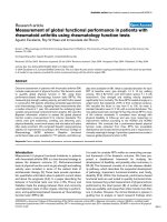
A novel feature of the assay was the design of a quantifica-
tion standard incorporating both EBNA-1 and BHRF-1
DNA targets in a single plasmid (Figure 1). This was done
to minimise the necessity for two separate EBV standards,
thus reducing costs and labour. The EBNA-1 and BHRF-1
DNA targets were linked using randomised primers
(Table 1) and inserted into the pGEM vector, using the
pGEM®-T Easy Vector System II (Promega Corp oration,
Alexandria, NSW, Australia) according to the manufac-
turer’s instructions. The cloned targets were then purified
using the PureYield™ Plasmid MidiPrep System (Promega
Corporation, Alexandria, NSW, Australia), and stored in
single use a liquots. Target copy number was calculated
following double stranded DNA approximation using the
Beckman DU® 530 Life Science UV/Vis spectrophotometer
(Beckman Coulter, Gladesville, NSW, Australia). A new
plasmid aliquot was used fo r standard curve dilution for
Table 1 Oligonucleotides used for EBV QPCR, beta-globin detection, construction of plasmid and PCR thermal cycling
conditions
Target Primer
Name
Oligonucleotide Sequence
5’-3’
Amplicon
Length
GenBank
Accession
(position)
Reference Optimised PCR Thermal
Cycling Conditions
EBNA-1 QP1 GCC GGT GTG TTC GTA TAT GG 213 bp AJ507799
(97174-97386)
Stevens
et al, 1999
95°C initial denaturation for
10 mins; 58°C annealing
QP2 CAA AAC CTC AGC AAA TAT
ATG AG
BHRF-1 EA-1F GGA GAT ACT GTT AGC CCT G 208 bp AJ507799
(42105-42312)
Custom 98°C initial denaturation for
13 mins; 60°C annealing
EA-2R GTG TGT TAT AAA TCT GTT CCA
AG
Plasmid construct
(randomised primers in
bold)
EA-F CTA TAT GTC TGC TTA CTC
CGG CG /G GAG ATA CTG TTA
GCC CTG
554 bp N/A Custom 95°C initial denaturation for
10 mins; 55°C annealing
EB-R CGC CGG AGT AAG CAG ACA
TAT AG /CAA AAC CTC AGC
AAA TAT ATG AG
95°C initial denaturation for
10 mins; 55°C annealing
Beta-Globin TAL57 BG-1F TAG CAA CCT CAA ACA GAC
ACC A
247 bp EU760960
(171-417)
Custom 95°C initial denaturation for
10 mins; 61°C annealing
BG-1R CAG CCT AAG GGT GGG AAA AT
Abbreviations: EBNA-1, Epstein-Barr virus nuclear antigen-1; BHRF-1, BamHI fragment H rightward open reading frame-1; mins, minutes.
Lay et al. Virology Journal 2010, 7:252
/>Page 3 of 11
each PCR run consisting of three replicates starting at 10
1
to 10
6
copies/5 μl. PCR runs were accepted when the stan-
dard curve correlation co-efficient was ≥ 0.99.
Product identification, reproducibility, sensitivity, limit of
detection and specificity
PCR products were identified by an amplification curve,
melt analyses and amplification efficiency generated by the
Rotor-Gene™ 6000 Software 1.7 (Build 90). Positive EBV
DNA samples had a cycle threshold (CT) less than 40, and
melted between 86°C to 87°C with an average amplifica-
tion efficiency of 1.74. PCR products for EBNA-1 DNA
and BHRF-1 DNA were identified on agarose gel by 213
bp and 208 bp bands, respectively. Reproducibility studies
consisting of triplica tes of each standard curve dilution
(10
1
-10
5
copies/5 μl) were performed prior to testing.
Intra-assay variation was determined in three repeat assays
tested within 24 hours on three consecutive days. Inter-
assay variation was assessed using th ree different batches
of the same PCR master mix kit. Sensitivity was deter-
mined by end-point PCR using gel elect rophoresis. To
establish the minimum DNA copy number that could be
reliably detected, ten p lasmid replicates spanning 10
0
to
10
2
copies/5 μl were assayed in three separate runs. Primer
specificity was verified on the Basic Local Alignment
Search Tool on GenBank and by assaying known cytome-
galovirus (CMV), human herpesvirus 6 (HHV6), HIV and
varicella zoster (VZV) positive samples. The EBV QPCR
was evaluated against an external quality assurance pro-
gram (Quality Control for Medical Diagnostics (QCMD),
Glasgow, Scotland, for EBV QPCR
in 2008 and 2009.
Viral load calculation and result interpretation
Viral load calculations were based on DNA extraction
volume and final el ution volumes as well as the number
of replicates tested. Samples were extracted and eluted
in equal quantities, keeping ratios constant. Hence, the
amount of sample used for PCR (5 μl) was multiplied by
a factor of 200 (elution volume) and divided by the
number of replicates to obtain a final measurement
expressed as DNA copies per millilitre (copies/ml) of
sample. This unit of measurement has close correlations
with copie s per microgram of DNA, therefore doe s not
require normalisation to the amount of input DNA [36].
Further more, copies/ml removes unnecessary processing
steps a nd reduces costs, as well as minimising sample
volume for testing. EBV DNA was quantifiable in a
dynamic range spanning six logarithms with the mini-
mum reportable viral load at 2.0 × 10
2
copies/ml of
sample. Samples with no detectable target DNA were
assigned a load of zero and resulted as negative.
Statistical calculations
Data analysis was conducted with SPSS version 17.
Spearman’s (rho) correlation co-efficient was used to
assess the correlations between EBNA-1 and BHRF-1
DNA loads and VCA IgG antibody titres.
Results
Performance of EBV QPCR assays: reproducibility,
sensitivity, detection limit and specificity
The intra-assay and inter-assay co-efficient of variation
for EBNA-1 and BHRF-1 QPCRs are shown in Table 2.
Both EBV targets were detected at levels as low as 2.0 ×
10
2
copies/ml of sample. However, the reliable limit of
detection for both EBNA-1 and BHRF-1 DNA was 2.0 ×
10
3
copies/ml, where the proportion that were detected
(po sitivity ratio) were 97% and 93% respectively. Primers
showed no cross reactivity to other herpesviruses (data
not shown). All samples in both the 2008 and 2009
QCMD programs were correctly identified using the
EBNA-1 primers.
EBV detection and load in EBV-associated disease states
and immunocompromised individuals
Of the 60 samples from 25 immunoco mpromised
patients, 3 0 (50%) samples from 16 (64%) patients h ad
Figure 1 Plasmid vector pGEM showing location of cloned insert.
Lay et al. Virology Journal 2010, 7:252
/>Page 4 of 11
quantifiable viral load using on e or other of the EBV
DNAtargets,EBNA-1orBHRF-1(Table3).EBVDNA
was detected in 100%, 85.7%, 100% and 33.3% of
patients with PTLD, I M, EBVAHS and HIV-infected
individuals (Groups 1 to 4), respectively. EBV DNA
loads were detectable at ran ges from 2.0 × 10
2
to 1.3 ×
10
8
copies/ml in these clinical samples, with the highest
EBV DNA load recorded in an individual with PTLD
(1.3 × 10
8
copies/ml o f sample). High levels were also
seen in individuals with IM (2.0 × 10
5
copies/ml of
sample), EBVAHS (1.1 × 10
5
copies/ml whole blood),
and HIV infection (5.6 × 10
3
copies/ml of sample).
In Group 1 (PTLD), EBV DNA concentrations spanned
six logarithms and were detected in multiple samples from
early to end-stage disease. EBV DNA loads increased
sequentially following transplantation, decreased after
anti-viral therapy in Patients A and C and peaked ten days
prior to death in Patients A to D. EBV DNA loads were
detectable in some samples, but were absent in others. In
Patient D, plasma EBV DNA was qualitative PCR negative
Table 2 Intra- and inter-assay co-efficient of variation for EBNA-1 and BHRF-1 QPCRs
DNA Target
(copies/5 ul)
Mean CT Mean R-G 6000™ Results
(copies/5ul)
Standard Deviation of R-G 6000™ Results
(copies/5ul)
Mean % Variation COV
(%)
Mean R
2
EBNA-1 Intra-Assay Variation (same day)
100,000 18.03 87,329 6,670 12.68% 7.64 0.991
10,000 21.28 11,735 3,092 26.30% 26.34
1,000 25.21 1,057 100 7.00% 9.50
100 29.10 103 38 32.12% 37.25
10 32.91 11 6 46.16% 57.50
EBNA-1 Intra-Assay Variation (different days)
100,000 16.94 89,643 8,164 11.00% 9.11 0.998
10,000 20.31 10,678 1,207 10.00% 11.31
1,000 23.85 1,133 129 16.00% 11.41
100 27.65 102 4 3.00% 3.90
10 31.42 10 2 17.88% 18.95
BHRF-1 Intra-Assay Variation (same day)
100,000 17.23 97,884 9,144 8.08% 9.34 0.994
10,000 20.91 9,852 542 4.45% 5.50
1,000 24.38 1,146 202 16.12% 17.64
100 28.43 94 19 17.70% 20.51
10 31.95 11 5 35.38% 41.91
BHRF-1 Intra-Assay Variation (different days)
100,000 18.05 105,387 4,621 6.02% 4.38 0.997
10,000 21.75 9,779 818 6.23% 8.37
1,000 25.23 1,042 141 11.63% 13.55
100 29.06 89 18 13.76% 19.88
10 32.66 12 2 25.30 16.53
EBNA-1 Inter-Assay Variation
100,000 19.87 101,644 14,058 10.99% 28.35 0.990
10,000 23.75 10,660 1,471 13.65% 13.80
1,000 27.68 1,084 191 18.67% 17.60
100 31.71 111 49 33.58% 43.75
10 35.86 12 9 65.03% 75.85
BHRF-1 Inter-Assay Variation
100,000 17.30 109,065 14,266 10.01% 13.08 0.990
10,000 21.49 9,209 2,154 16.84% 23.39
1,000 25.49 860 251 22.19% 29.15
100 29.01 108 49 35.53% 45.33
10 32.29 15 8 69.41% 57.23
Abbreviations: CT, cycle threshold; Mean % variation, average percentage variation between the calculated (Rotor-Gene results) and the given concentration
(DNA target); COV, co-efficient of variation is the ratio of standard deviation to the mean; R
2
-value, square root of the correlation co-efficient - in quantitation
PCR describes the percentage of the data which matches the hypothesis that the standards conform to a line of best fit.
Lay et al. Virology Journal 2010, 7:252
/>Page 5 of 11
Table 3 EBV DNA loads in various EBV-associated disease states and immunocompromised individuals
Group Patient
ID
Sex/Age Condition Specimen
(Positive/n Tested)
Target Detectable EBV DNA
Load (copies/ml)
Clinical Notes
1. A. M/46y PTLD Plasma (5/6) EBNA-1 Day +32 - 8.0 × 10
2
MUD HSCT for AML; EBV VCA IgG positive
pre-Tx; Plasma collected on Days +32, +39,
+46, +60, +75 and +81 for EBV QPCR; Plasma
EBV (qualitative) PCR positive on Days +75,
+78 and +81; Treatment with Foscarnet and
Rituximab after Day +75; Died of pneumonia
on Day +88
Day +46 - 1.0 × 10
3
Day +60 - 8.8 × 10
3
Day +75 - 1.1 × 10
6
Day +81 - 2.3 × 10
5
CSF (2/2) EBNA-1 Day +75 - 1.3 × 10
6
CSF collected on Days +75 and +78
Day +78 - 2.7 × 10
6
B. M/42y PTLD Whole Blood (1/5) EBNA-1 Day +95 - 2.0 × 10
7
MUD HSCT for AML; Plasma EBV (qualitative)
PCR positive Day +96; Plasma collected on
Day +95 for EBV QPCR; Died on Day +99
due to multi-organ failure
C. F/59y PTLD Plasma (3/6) EBNA-1 Day +45 - 2.2 × 10
5
MUD HSCT for AML; CMV reactivation on Day
+44, Treatment with Foscarnet and
ganciclovir on Day +52; Plasma collected
Days +38, +40, +45, +52 and +59; Died on
Day +66; EBV VCA IgG positive, HHV6 IgG
positive and CMV IgG positive pre-Tx
Day +52 - 9.6 × 10
3
Day +59 - 3.0 × 10
5
Whole Blood (1/8) EBNA-1 Day +46 - 6.6 × 10
4
EDTA collected Days +3, +5, +10, +17, +26,
+31, +33, +46
D. M/48y PTLD Plasma (4/6) EBNA-1 Day +40 - 3.4 × 10
3
MUD HSCT for AML; EBV VCA IgG positive
pre-Tx; Plasma collected Days +28, +33, +40,
+47, +54, +61; Plasma EBV (qualitative) PCR
negative on Day +62; Died Day +72 of multi-
organ failure
Day +47 - 3.6 × 10
4
Day +54 - 3.4 × 10
6
Day +61 - 6.3 × 10
6
Whole Blood (2/2) EBNA-1 Day +62 - 1.3 × 10
8
EDTA collected Days +62 and +63.
Day +63 - 1.8 × 10
7
E. F/57y PTLD Whole Blood (1/5) EBNA-1 9.5 × 10
4
No serology results available however clinical
notes indicate EBV reactivation; Plasma EBV
(qualitative) PCR positive 9-16 days after VL
testing done; negative at 1-7 months
thereafter.
2. F. Unknown IM Plasma (1/1) EBNA-1 3.7 × 10
4
EBV VCA IgM positive
BHRF-1 1.6 × 10
4
G. Unknown IM Plasma (0/1) EBNA-1 0 EBV VCA IgM positive
BHRF-1 0
H. Unknown IM Plasma (1/1) EBNA-1 7.6 × 10
3
EBV VCA IgM positive
BHRF-1 1.5 × 10
3
I. Unknown IM Plasma (1/1) EBNA-1 2.3 × 10
3
EBV VCA IgM positive
BHRF-1 8.7 × 10
4
J. M/17y IM Whole Blood (1/1) EBNA-1 1.0 × 10
5
EBV VCA IgM positive
BHRF-1 1.8 × 10
3
K. F/19y IM Whole Blood (1/1) EBNA-1 2.2 10
3
EBV VCA IgM positive
BHRF-1 5.6 × 10
4
L. F/53y IM Whole Blood (1/1) EBNA-1 2.0 × 10
5
EBV VCA IgM positive; acute glandular fever
BHRF-1 1.8 × 10
4
Lay et al. Virology Journal 2010, 7:252
/>Page 6 of 11
on Day +62 whilst simultaneously QPCR positive in whole
blood. EBV-specific serology results were available for four
patients, and confirmed EBV infection prior to the trans-
plant. Four patients died as a result of PTLD complica-
tions, on average +81.25 days post transplantation. In
Group 2 (IM), EBV DNA was quantifiable from 1.5 × 10
3
to 2.0 × 10
5
copies/ml. One sample was negative for EBV
DNA (Patient G), despite a positive EBV VCA IgM profile.
Group 3 (EBVAHS) EBV DNA load results were similar to
Group 2, however Patient M died as a consequence of the
disease condition. In Group 4 ( HIV), EBV DNA was
detectable in both plasma an d PBMC ranging from 2.0 ×
10
2
to 5.6 × 10
3
copies/ml. However, 50% of these samples
were below 2.0 × 10
3
copies/ml.
EBV detection and load in the population sample
EBNA-1 and BHRF-1 DNA were detected in 11.0% and
21.6% of Group 5 (the p opulation sample), respectively;
22.5% of samples were positive for at least one EBV
DNA target (Table 4). Of the 24 EBNA-1 DNA positive
samples, 91.7% were a lso BHRF-1 DNA positive, and of
the 47 BHRF-1 DNA positive samples, 46.8% were also
EBNA-1 DNA positive. Viral loads (combined targets)
were detectable between 2.0 × 10
2
to 6.2 × 10
4
copies/
ml of whole blood, but 54.2% and 85.1% of samples
were below 2.0 × 10
3
copies/ml for EBNA-1 and BHRF-
1 D NA levels, respectively. All samples with measurable
EBV DNA were EBV VCA IgG antibody positive, which
were found in 95.9% of the population sample. There
was a modest correlation between VCA IgG antibody
titres and BHRF-1 DNA load (Spearman’srho=0.13,
p = 0.05) and a weaker (not statistically significant) cor-
relation between EBNA-1 DNA load and VCA IgG anti-
body titres (Spearman’s rho = 0.11, p = 0.11) (Table 4).
Discussion
With increasing availability of nucleic acid testing
(NAT) methods, measuring EBV DNA in blood has pro-
venvaluableindiagnosingandmonitoringPTLD
[16,21,22,37-41], NPC [42,43], IM [13, 44], EBV infection
in HIV-infected individuals [8,13,45], BL [13] and
chronic active EBV infection [18,46]. In this study, we
Table 3: EBV DNA loads in various EBV-associated disease states and immunocompromised individuals (Continued)
3. M. M/36y EBVAHS Whole Blood (1/1) EBNA-1 7.5 × 10
4
EBV (qualitative) PCR positive; died of EBVAHS
BHRF-1 1.1 × 10
5
4. N. Unknown HIV Plasma (1/1) EBNA-1 0 HIV plasma VL 324, 000 RNA copies/ml
BHRF-1 1.0 × 10
3
O. Unknown HIV Plasma (0/1) EBNA-1, 0 HIV plasma VL 13, 000 RNA copies/ml
BHRF-1 0
P. Unknown HIV Plasma (0/1) EBNA-1, 0 HIV plasma VL 26, 800 RNA copies/ml
BHRF-1 0
Q. Unknown HIV Plasma (0/1) EBNA-1, 0 HIV plasma VL 21, 300 RNA copies/ml
BHRF-1 0
R. Unknown HIV Plasma (0/1) EBNA-1, 0 HIV plasma VL 12, 700 RNA copies/ml
BHRF-1 0
S. Unknown HIV Plasma (0/1) EBNA-1, 0 HIV plasma VL 1, 040, 000 RNA copies/ml
BHRF-1 0
T. Unknown HIV Plasma (0/1) EBNA-1, 0 HIV plasma VL 17, 700 RNA copies/ml
BHRF-1 0
U. Unknown HIV Plasma (0/1) EBNA-1, 0 HIV plasma VL 47, 500 RNA copies/ml
BHRF-1 0
V. Unknown HIV Plasma (1/1) EBNA-1 5.6 × 10
3
HIV plasma VL 16, 400 RNA copies/ml
BHRF-1 3.0 × 10
3
W. Unknown HIV PBMC (1/1) EBNA-1 < 2.0 × 10
2
HIV PBMC VL 12, 800 RNA copies/ml
BHRF-1 < 2.0 × 10
2
X. Unknown HIV PBMC (1/1) EBNA-1 < 2.0 × 10
2
HIV PBMC VL 12, 700 RNA copies/ml
BHRF-1 0
Y. Unknown HIV PBMC (0/1) EBNA-1, 0 HIV PBMC VL 118, 000 RNA copies/ml
BHRF-1
Abbreviations: Y, years; Group 1 (PTLD), post-transplant lymphoproliferative disease; Group 2 (IM), infectious mononucleosis; Group 3 (EBVAHS), Epstein-Barr virus
associated-haemophagocytic syndrome; Group 4 (HIV infection), human immunodeficiency virus; EDTA, ethylenediaminetetraacetic acid; CSF, cerebrospinal fluid;
PBMC, peripheral blood mononuclear cells; EBNA-1, Epstein-Barr virus nuclear antigen-1; BHRF-1, BamHI fragment H rightward open reading frame-1; ml,
millilitres; Bold lettering indicates Day QPCR positive post-transplant; AML, acute myeloid leukaemia; MUD; matched unrelated donor; HSCT, haematopoietic stem
cell transplantation; CMV, cytomegalovirus; VCA, viral capsid antigen; Ig, immunoglobulins; EA-D, early antigen-diffuse; EA-R, early antigen-restricted; VL, viral load.
Lay et al. Virology Journal 2010, 7:252
/>Page 7 of 11
successfully developed two in-house QPCR methods
incorporating a novel single quantification standard con-
taining two EBV DNA targets for measuring viral load
on the Rotor-Gene 6000™ . Substituting SYBR Green I
dye as a fluorescent marker for product accumulation
over fluorogenic probes, this method proved useful for
quantifying EBV DNA concentrations in clinical samples
from individuals with a variety of EB V-associated disor-
ders or immune dysfunctions and in a healthy popula-
tion sample.
Previous studies in PTLD have found that EBV DNA
loads increased with disease progression and decreased
with remission of l ymphoproliferation [47,48]. This pat-
tern was observed in Group 1, where EBV DNA loads
appeared to be correlated with disease status. We found
similar EBV DNA loads to those previously reported,
with most studies showing EBV DNA concentrations
ranging from 5.0 × 10
2
to 2.0 × 10
7
copies/ml in whole
blood, plasma and serum [37,49,50]. EBV DNA was also
detected in CSF at concentrations comparable to plasma,
however detectable CSF EBV DNA has been previously
reported only in association with acquired immunodefi-
ciency syndrome (AIDS)-relate d brain l ymphoma [51].
The significance of EBV DNA in CSF of PTLD remains
to be elucidated.
EBV DNA loads in IM patients were also similar to
those reported in the literature [13, 22,26,44,52], although
some authors described loads as high as 10
6
and 10
7
copies/ml [12,46,53]. In Group 3, EBV DNA loads were
consistent with acute phase EBVAHS [46,54], and corre-
lated with the deterioration of the patient’s disease condi-
tion.Elazaryetalalsofoundthataviralloadranging
from 10
4
-10
5
copies/ml was associated with poor patient
outcome [54]. One study found much higher EBV DNA
loads ( up to 10
7
copies/ml) [55], but this may have been
due to differences in sample type and detection methods.
In Group 4, EBV DNA was detected in 33% of samples
(22% of plasma, 67% of PBMC), compared to 34% to 76%
positivity reported in other studies [8,26]. Notably how-
ever, these studies used whole blood for quantifying EBV
DNA load, which could have increased the probability of
viral DNA detection. As none of the Group 4 patients
were known to have EBV-related disease, low positivity
ratios and viral loads were expected.
Similar to our findings, the literature describes EBV
DNA det ectable from 10
2
to 10
4
copies/ml and positiv-
ity ratios up to 29% in whole blood of healthy indivi-
duals [11-13,26,38,56-59]. However, DNA loads as high
as 5.5 × 10
5
copies/ml of whole blood and a positivity
ratio of 72% have been reported [58]. Differences in t he
results may be attributable to more sensitive methods
associated with nested PCR and dual-labelled probes
[58]. Interestingly, another stu dy showed 100% EBV
DNA positivity in whole blood, although DNA loads
were all below the detection limit of the assay
(2.0 × 10
3
copies/ml) [38].
In the population sample the EBV VCA IgG antibod y
detection rate was consistent with levels of EBV sero-posi-
tivity in Western societies [2]. One study previously showed
a correlation between EBV VCA IgG antibody titres and
EBV viral load (detectable versus non-detectable) [60]. We
similarly found a modest correlation with quantitative
BHRF-1 DNA loads, and a weaker (not statistically signifi-
cant) correlation with E BNA-1 DNA load (see Table 4 ).
We noted some discrepancies in our measure s of EBV
positivity. In one PTLD patient (Patient D), plasma was
qualitative EBV PCR negative whilst simultaneously
reporting an EBV DNA load of 1.3 × 10
8
copies/ml in
whole blood. However, a growing number of studies
have shown that cell-associated EBV is detectable before
plasma EBV DNA and can persist without accompany-
ing p lasma DNA loads [21,48]. In Group 2, Patient G,
despite being EBV VCA IgM antibody positive, was EBV
QPCR negative. As EBV DNA loads can change rapidly
Table 4 EBV DNA load and antibody titre detection rates in the population samples (Group 5, n = 218)
Target Positive
n (%)
Detectable Range Spearman correlation (p)
EBNA-1 DNA
load
BHRF-1 DNA
load
Combined EBV Targets
DNA load
VCA
IgG
EBV EBNA-1 DNA load
(copies/ml)
24 (11.0%) 2.0 × 10
2
- 9.1 × 10
4
1.00
EBV BHRF-1 DNA load
(copies/ml)
47 (21.6%) 2.0 × 10
2
- 3.3 × 10
4
0.63
p < 0.001
1.00
Combined EBV targets DNA
load
(mean of BHRF & EBNA loads
where both
positive) (copies/ml)
49 (22.5%) 2.0 × 10
2
- 6.2 × 10
4
0.73
p < 0.001
0.97
p < 0.001
1.00
Viral capsid antigen IgG
(titres)
209 (95.9%) 1:10 - 1:5120 0.11
p = 0.11
0.13
p = 0.05
0.14
p = 0.04
1.00
Abbreviations: EBV, Epstein-barr virus; EBNA-1, Epstein-barr virus nuclear antigen-1; BHRF-1, BamHI fragment H rightward open reading frame-1; VCA, viral capsid
antigen; IgG, immunoglobulin G; Pos, positive.
Lay et al. Virology Journal 2010, 7:252
/>Page 8 of 11
from being undetectable to being very high in a short
period of time [38], it is possible that sampling occurred
late in the convalescentphasewherelowEBVDNA
positivity ratios of 44% hav e been previously reported
[46]. Other factors contributing to DNA load variation
include differences in sample type, method of extraction
or NAT, and target chosen for PCR amplification.
As specimen type is known to influence DNA loads and
impact on assay performance [36], unfractionated EDTA
whole blood was used f or DNA quantification where
possible. The dynamic changes of EBV DNA are better
reflected in circulating whole blood [38], whic h also
contains all the compartments t hat may harbour virus
[13,21,61]. However, desp ite reports of greater test sensi-
tivity with whole blood [12,36], EBV DNA load has
also been quantified in PBMC [14,16,62-64]. Although
infection is typically associated with cell compartments
[8,12,13], EBV DNA is also found in cell-free blood parti-
tions such as plasma or serum, usually in fragmented, cell-
derived form [12]. In t his study, 2 of 9 plasma samples
from HIV-infected patients had detectable EBV DNA,
compared to 2 out of 3 PBMC samples. As we did not
have simultaneous plasma and PBMC samples from the
same individuals, we were unable to assess the differences
in viral load between these compartments. Further studies
compa ring suitability of different sample types in various
EBV-related diseases and immune disorders are required.
The method of DNA purification is known to affect
viral load measurements. One study showed yield from
manually extracted DNA was 57% higher than that of
robotic systems [65]. Therefore, to improve DNA recov-
ery and maximise PCR sensitivity, samples here were
purified using a commercial silica-based column method
[61,66]. For optimal quantitation result s, an earlier study
showed that DNA should b e subjected to PCR within
one to two weeks post-extraction [67]. Here, delay
between e xtraction and testing could have contributed
to lo w DNA loads and positivity ratios in clinical sam-
ples. Furthermore, DNA from blood samples that had
undergone more than four freeze-thaw cycles were
found to be partially degraded [68]. Since the clinical
samples used here were tested retrospectively, monitor-
ing these conditions were not possible.
EBV DNA loads also vary according to type and size
of gene target [69]. Ryan et al, found assay sensitivity
was dependent on the specific gene segment and that
different targets had varying lower limits of detection
[15]. For EBV, BamHI-W is reportedly 10 times more
sensitive than other targets for PCR, allowing for detec-
tion of viral DNA at trace amounts [8,13,15]. However,
precise quantification of viral genomes is complicated by
the number of reiterated BamHI-W sequences among
EBV strains, which typically ranges between 7 and 11
repeats per genome [15]. To avoid overestimation in
this study, we chose to use the next most sensitive EBV
gene; EBNA-1 [15], and an abundantly expressed gene,
BHRF-1, for QPCR.
Despite targeting highly conserved EBV regions, selec-
tive drop out of amplifiable EBV DNA at the EBNA-1
and BHRF-1 loci was observed in Group 4 (Patients N
and X), and in 25 of 218 (11.5%) whole bloods from the
population sample. Instead of amplifying both EBV
DNA genes, only one target was detected, 93% of which
had viral loads less than 2.0 × 10
3
copies/ml. As beta-
globin was detected in all samples, PCR inhibitors and/
or defective nucleic acid purification methods were
excluded [70]. Alternatively, selective drop out may have
been due to low viral load and/or sampling error [71].
Since load determination is reliant on the amount of
EBV gen omes pipetted into a reaction and assumes viral
homogeneity, QPCR results, particularly at low viral
load levels are prone to random sampling error. This
phenomenon is well documented in DNA quantification
and results in less reliable viral load mea surements
[70,71]. Ther efore, samples reporting low levels of target
nucleic acid may not be reproduci ble in repeated assays
from the same or different specimens [72].
Currently, there are no standardised methods for mea-
suring EBV DNA, complicating inter-laboratory compar-
isons in multicentre studies of EBV-related diseases.
Standardisation is difficult as PCR assay conditions vary
between laboratories, leading to variations in the accu-
racy and reproducibility of viral loa d quantification [21].
Although there appears to be a strong concordance
between laboratories for qualitative EBV DNA estimates,
there continues to be ma rked inconsistency in quantita-
tive results [73]. It has been suggested that the use of
unfractionated whole blood [26] or an international cali-
bration standard could be the first step towards standar-
disation [73]. However, instrumentation, chemistries,
gene targets and other test-related aspects remain
diverse. One solution for enabling inter-laboratory com-
parisons is the distribution of proficiency panels su ch as
QCMD. Such programs have already been used for
assessing methods for the detection and quantification
of EBV and other viruses [27,74,75].
Conclusion
This is the first reported study that uses the SYBR Green
I dye on the R otor-gene 6000™ with a novel quantifica-
tion standard containing two EBV targets for measuring
EBV DNA load. The assays proved successful in the
quantification of EBV genomes in clinical cases and
should be considered as a cost effective and sensitive
PCR alternative to probe-based assays. This approach
can be modified to detect and quantify other latent
Lay et al. Virology Journal 2010, 7:252
/>Page 9 of 11
herpesviruses such as HHV6, CMV, and VZ V. This pro-
cedure is suitable for robot ics and automation, and
would be a useful addition in larger laboratories.
Acknowledgements
The Ausimmune Investigator Group includes C Chapman, A Coulthard, K
Dear, T Dwyer, T Kilpatrick, R Lucas, T McMichael, MP Pender, A-L Ponsonby,
B Taylor, P Valery, I van der Mei and D Williams. The Ausimmune Study is
funded by the National Multiple Sclerosis Society of the USA, the National
Health & Medical Research Council (Project Grant 316901) and Multiple
Sclerosis Research Australia. We also acknowledge the work of the
Ausimmune Study research nurses who undertook sample collection: S
Agland, B Alexander, M Davis, Z Dunlop, A Wright, R Scott, J Selvidge, M
Steele, K Turner, B Wood and the study project officers, H Rodgers and C
Jozwick. Clinical samples were kindly provided by N Gilroy, D Gottlieb, P
Ferguson, F Kwok and I Kay. We would also like to thank B Wang of the
Westmead Millennium Institute for assisting with the cloning work, B
O’Toole for statistical analyses, D Patel for assistance with the serology and C
Toi for laboratory guidance and review of the manuscript.
Author details
1
Virology Department, Centre For Infectious Diseases & Microbiology
Laboratory Services, Institute of Clinical Pathology & Medical Research,
Institute Road, Westmead Hospital, Westmead 2145, New South Wales,
Australia.
2
National Centre for Epidemiology and Population Health, The
Australian National University, Canberra, ACT, 0200 Australia.
3
Murdoch
Childrens Research Institute, 9th Floor AP Building, The Royal Children’s
Hospital, Flemington Road, Parkville, Victoria 3052, Australia.
Authors’ contributions
MLL developed the assays, carried out all of the DNA work, assisted in the
data analysis and result interpretation, and writing of the manuscript. On
behalf of the Ausimmune Investigator group, RML supplied the whole blood
and serum from the population sample, and was involved in the data
analysis. VMR aided in primer design and JT performed the serology testing.
MLL, DED, VMR, RML and ALP were involved in the design and conception
of the study. All authors have read, reviewed and approved the final
manuscript.
Competing interests
The authors declare that they have no competing interests.
Received: 26 May 2010 Accepted: 22 September 2010
Published: 22 September 2010
References
1. Steven NM: Epstein-Barr virus latent infection in vivo. Rev Med Virol 1997,
7(2):97-106.
2. Hess RD: Routine Epstein-Barr virus diagnostics from the laboratory
perspective: still challenging after 35 years. J Clin Microbiol 2004,
42(8):3381-7.
3. Tsai DE, et al: EBV PCR in the diagnosis and monitoring of posttransplant
lymphoproliferative disorder: results of a two-arm prospective trial. Am J
Transplant 2008, 8(5):1016-24.
4. Loginov R, et al: Monitoring of EBV-DNAemia by quantitative real-time
PCR after adult liver transplantation. J Clin Virol 2006, 37(2):104-108.
5. Timbury MC, Edmond E: Herpesviruses. J Clin Pathol 1979, 32(9):859-81.
6. Cohen JI: Epstein-Barr virus infection. N Engl J Med 2000, 343(7):481-92.
7. Kimura H, et al: Viral load in Epstein-Barr virus-associated
hemophagocytic syndrome. Microbiol Immunol 2002, 46(8):579-82.
8. Stevens SJ, et al: High Epstein-Barr virus (EBV) DNA loads in HIV-infected
patients: correlation with antiretroviral therapy and quantitative EBV
serology. AIDS 2002, 16(7):993-1001.
9. Espy MJ, et al: Comparison of three methods for extraction of viral
nucleic acids from blood cultures. J Clin Microbiol 1995, 33(1):41-4.
10. Leung E, et al: Dynamic EBV gene loads in renal, hepatic, and
cardiothoracic transplant recipients as determined by real-time PCR light
cycler. Transpl Infect Dis 2004, 6(4):156-64.
11. Bai X, et al: Quantitative polymerase chain reaction for human
herpesvirus diagnosis and measurement of Epstein-Barr virus burden in
posttransplant lymphoproliferative disorder. Clin Chem 1997,
43(10):1843-9.
12. Kozic S, et al: Evaluation of a commercial real-time PCR assay for
quantitation of Epstein-Barr virus DNA in different groups of patients.
J Virol Methods 2006, 135(2):263-8, Epub 2006 May 2.
13. Stevens SJ, Pronk I, Middeldorp JM: Toward standardization of Epstein-
Barr virus DNA load monitoring: unfractionated whole blood as
preferred clinical specimen. J Clin Microbiol 2001, 39(4):1211-6.
14. Leung E, et al: Use of real-time PCR to measure Epstein-Barr virus
genomes in whole blood. J Immunol Methods 2002, 270(2):259-67.
15. Ryan JL, et al: Epstein-Barr virus quantitation by real-time PCR targeting
multiple gene segments: a novel approach to screen for the virus in
paraffin-embedded tissue and plasma. J Mol Diagn 2004, 6(4):378-85.
16. Rowe DT, et al: Use of quantitative competitive PCR to measure Epstein-
Barr virus genome load in the peripheral blood of pediatric transplant
patients with lymphoproliferative disorders. J Clin Microbiol 1997,
35(6):1612-5.
17. Fan H, Gulley ML: Epstein-Barr viral load measurement as a marker of
EBV-related disease. Mol Diagn 2001, 6(4):279-89.
18. Kimura H, et al: Quantitative analysis of Epstein-Barr virus load by using a
real-time PCR assay. J Clin Microbiol 1999, 37(1):132-6.
19. Lo YM, et al: Quantitative and temporal correlation between circulating
cell-free Epstein-Barr virus DNA and tumor recurrence in
nasopharyngeal carcinoma. Cancer Res 1999, 59(21):5452-5.
20. Lo YM, et al: Quantitative analysis of cell-free Epstein-Barr virus DNA in
plasma of patients with nasopharyngeal carcinoma. Cancer Res 1999,
59(6):1188-91.
21. Stevens SJ, et al: Role of Epstein-Barr virus DNA load monitoring in
prevention and early detection of post-transplant lymphoproliferative
disease. Leuk Lymphoma 2002, 43(4):831-40.
22. Niesters HGM, et al: Development of a real-time quantitative assay for
detection of Epstein-Barr virus. J Clin Microbiol 2000, 38(2):712-5.
23. Xu S, et al: A comparison of quantitative-competitive and realtime PCR
assays using an identical target sequence to detect Epstein-Barr virus
viral load in the peripheral blood. J Virol Methods 2006, 137(2):205-12.
24. Brengel-Pesce K, et al: Routine use of real-time quantitative PCR for
laboratory diagnosis of Epstein-Barr virus infections. J Med Virol 2002,
66(3):360-9.
25. Mackay IM, Arden KE, Nitsche A: Real-time PCR in virology. Nucleic Acids
Res 2002, 30(6):1292-305.
26. Fafi-Kremer S, et al: Evaluation of the Epstein-Barr virus R-gene
quantification kit in whole blood with different extraction methods and
PCR platforms. J Mol Diagn 2008, 10(1):78-84.
27. Ruiz G, et al: Comparison of commercial real-time PCR assays for
quantification of Epstein-Barr virus DNA. J Clin Microbiol 2005,
43(5):2053-7.
28. Papin JF, Vahrson W, Dittmer DP: SYBR green-based real-time quantitative
PCR assay for detection of West Nile Virus circumvents false-negative
results due to strain variability. J Clin Microbiol 2004, 42(4):1511-8.
29. Karlsen F, Steen HB, Nesland JM: SYBR green I DNA staining increases the
detection sensitivity of viruses by polymerase chain reaction. J Virol
Methods 1995, 55(1):153-6.
30. Hilscher C, Vahrson W, Dittmer DP: Faster quantitative real-time PCR
protocols may lose sensitivity and show increased variability. Nucleic
Acids Res 2005, 33(21):e182.
31. Lucas RM, et al: Associations between silicone skin cast score, cumulative
sun exposure, and other factors in the ausimmune study: a multicenter
Australian study. Cancer Epidemiol Biomarkers Prev 2009, 18(11):2887-94.
32. Humme S, et al: The EBV nuclear antigen 1 (EBNA1) enhances B cell
immortalization several thousandfold. Proc Natl Acad Sci USA 2003,
100(19):10989-94.
33. Lee MA, Diamond ME, Yates JL: Genetic evidence that EBNA-1 is needed
for efficient, stable latent infection by Epstein-Barr virus. J Virol 1999,
73(4):2974-82.
34. Henderson S, et al: Epstein-Barr virus-coded BHRF1 protein, a viral
homologue of Bcl-2, protects human B cells from programmed cell
death. Proc Natl Acad Sci USA 1993, 90(18):8479-83.
Lay et al. Virology Journal 2010, 7:252
/>Page 10 of 11
35. Huang Y, et al: Proper developmental control of human globin genes
reproduced by transgenic mice containing a 160-kb BAC carrying the
human beta-globin locus. Blood Cells Mol Dis 2000, 26(6):598-610.
36. Hakim H, et al: Comparison of various blood compartments and
reporting units for the detection and quantification of Epstein-Barr virus
in peripheral blood. J Clin Microbiol 2007, 45(7):2151-5.
37. van Esser JW, et al: Molecular quantification of viral load in plasma allows
for fast and accurate prediction of response to therapy of Epstein-Barr
virus-associated lymphoproliferative disease after allogeneic stem cell
transplantation. Br J Haematol 2001, 113(3):814-21.
38. Stevens SJ, et al: Frequent monitoring of Epstein-Barr virus DNA load in
unfractionated whole blood is essential for early detection of
posttransplant lymphoproliferative disease in high-risk patients. Blood
2001, 97(5):1165-71.
39. Lucas KG, et al: Semiquantitative Epstein-Barr virus (EBV) polymerase
chain reaction for the determination of patients at risk for EBV-induced
lymphoproliferative disease after stem cell transplantation. Blood 1998,
91(10):3654-61.
40. Kimura H, et al: Measuring Epstein-Barr virus (EBV) load: the significance
and application for each EBV-associated disease. Rev Med Virol 2008,
18(5):305-19.
41. Wheless SA, et al: Post-transplantation lymphoproliferative disease:
Epstein-Barr virus DNA levels, HLA-A3, and survival. Am J Respir Crit Care
Med 2008, 178(10) :1060-5.
42. Chan KC, et al: Molecular characterization of circulating EBV DNA in the
plasma of nasopharyngeal carcinoma and lymphoma patients. Cancer
Res 2003, 63(9):2028-32.
43. Lin JC, et al : Quantification of plasma Epstein-Barr virus DNA in patients
with advanced nasopharyngeal carcinoma. N Engl J Med 2004,
350(24):2461-70.
44. Bauer CC, et al : Serum Epstein-Barr virus DNA load in primary Epstein-
Barr virus infection. J Med Virol 2005, 75(1):54-8.
45. Dehee A, et al: Quantification of Epstein-Barr v irus load in peripheral
blood of human immunodeficiency virus-infected patients using real-
time PCR. J Med Virol 2001,
65(3):543-52.
46. Yamamoto M, et al: Detection and quantification of virus DNA in plasma
of patients with Epstein-Barr virus-associated diseases. J Clin Microbiol
1995, 33(7):1765-8.
47. Wagner HJ, et al: Patients at risk for development of posttransplant
lymphoproliferative disorder: plasma versus peripheral blood
mononuclear cells as material for quantification of Epstein-Barr viral
load by using real-time quantitative polymerase chain reaction.
Transplantation 2001, 72(6):1012-9.
48. Rowe DT, et al: Epstein-Barr virus load monitoring: its role in the
prevention and management of post-transplant lymphoproliferative
disease. Transpl Infect Dis 2001, 3(2):79-87.
49. Rosselet A, et al: Associations of serum EBV DNA and gammopathy with
post-transplant lymphoproliferative disease. Clin Transplant 2009,
23(1):74-82.
50. Wada K, et al: Simultaneous quantification of Epstein-Barr virus,
cytomegalovirus, and human herpesvirus 6 DNA in samples from
transplant recipients by multiplex real-time PCR assay. J Clin Microbiol
2007, 45(5):1426-32.
51. Bossolasco S, et al: Epstein-Barr virus DNA load in cerebrospinal fluid and
plasma of patients with AIDS-related lymphoma. J Neurovirol 2002,
8(5):432-8.
52. Stevens SJ, et al: Monitoring of epstein-barr virus DNA load in peripheral
blood by quantitative competitive PCR. J Clin Microbiol 1999, 37(9):2852-7.
53. van Laar JA, et al: Epstein-Barr viral load assessment in
immunocompetent patients with fulminant infectious mononucleosis.
Arch Intern Med 2002, 162(7):837-9.
54. Elazary AS, et al: Severe Epstein-Barr virus-associated hemophagocytic
syndrome in six adult patients. J Clin Virol 2007, 40(2):156-9.
55. Teramura T, et al: Quantitative analysis of cell-free Epstein-Barr virus
genome copy number in patients with EBV-associated hemophagocytic
lymphohistiocytosis. Leuk Lymphoma 2002, 43(1):173-9.
56. Compston LI, et al: Multiplex real-time PCR for the detection and
quantification of latent and persistent viral genomes in cellular or
plasma blood fractions. J Virol Methods 2008, 151(1):47-54.
57. Kullberg-Lindh C,
et al: Comparison of serum and whole blood levels of
cytomegalovirus and Epstein-Barr virus DNA. Transpl Infect Dis 2008,
10(5):308-15.
58. Hudnall SD, et al : Herpesvirus prevalence and viral load in healthy blood
donors by quantitative real-time polymerase chain reaction. Transfusion
2008, 48(6):1180-7.
59. Engelmann I, et al: Rapid quantitative PCR assays for the simultaneous
detection of herpes simplex virus, varicella zoster virus, cytomegalovirus,
Epstein-Barr virus, and human herpesvirus 6 DNA in blood and other
clinical specimens. J Med Virol 2008, 80(3):467-77.
60. Besson C, et al: Positive correlation between Epstein-Barr virus viral load
and anti-viral capsid immunoglobulin G titers determined for Hodgkin’s
lymphoma patients and their relatives. J Clin Microbiol 2006, 44(1):47-50.
61. Fafi-Kremer S, et al: Assessment of automated DNA extraction coupled
with real-time PCR for measuring Epstein-Barr virus load in whole blood,
peripheral mononuclear cells and plasma. J Clin Virol 2004, 30(2):157-64.
62. Jabs WJ, et al: Normalized quantification by real-time PCR of Epstein-Barr
virus load in patients at risk for posttransplant lymphoproliferative
disorders. J Clin Microbiol 2001, 39(2):564-9.
63. Wadowsky RM, et al: Measurement of Epstein-Barr virus DNA loads in
whole blood and plasma by TaqMan PCR and in peripheral blood
lymphocytes by competitive PCR. J Clin Microbiol 2003, 41(11):5245-9.
64. Baldanti F, et al: Kinetics of Epstein-Barr virus DNA load in different blood
compartments of pediatric recipients of T-cell-depleted HLA-
haploidentical stem cell transplantation. J Clin Microbiol 2008,
46(11):3672-7.
65. Riemann K, et al: Comparison of manual and automated nucleic acid
extraction from whole-blood samples. J Clin Lab Anal 2007, 21(4):244-8.
66. Schuurman T, et al: Reduced PCR sensitivity due to impaired DNA
recovery with the MagNA Pure LC total nucleic acid isolation kit. J Clin
Microbiol 2005, 43(9):4616-22.
67. Malcomson RD, et al: The scope of quantitative polymerase chain
reaction assays in clinical molecular pathology. Clin Mol Pathol
1995,
48(4):M178-M183.
68. Lahiri DK, Schnabel B: DNA isolation by a rapid method from human
blood samples: effects of MgCl2, EDTA, storage time, and temperature
on DNA yield and quality. Biochem Genet 1993, 31(7-8):321-8.
69. Stevens SJ, et al: Diagnostic value of measuring Epstein-Barr virus (EBV)
DNA load and carcinoma-specific viral mRNA in relation to anti-EBV
immunoglobulin A (IgA) and IgG antibody levels in blood of
nasopharyngeal carcinoma patients from Indonesia. J Clin Microbiol 2005,
43(7):3066-73.
70. Gulley ML, Fan H, Elmore SH: Validation of Roche LightCycler Epstein-Barr
virus quantification reagents in a clinical laboratory setting. J Mol Diagn
2006, 8(5):589-97.
71. Diaco D: Practical Considerations for the Design of Quantitative PCR
Assays. In PCR Strategies. Edited by: G DH, S JJ, Innis MA. Academic Press
Limited, London; 1995:84-107.
72. Smith TF, et al: Quantitative real-time polymerase chain reaction for
evaluating DNAemia due to cytomegalovirus, Epstein-Barr virus, and BK
virus in solid-organ transplant recipients. Clin Infect Dis 2007,
45(8):1056-61.
73. Hayden RT, et al: Multicenter comparison of different real-time PCR
assays for quantitative detection of Epstein-Barr virus. J Clin Microbiol
2008, 46(1):157-63.
74. Perandin F, et al: Comparison of commercial and in-house Real-time PCR
assays for quantification of Epstein-Barr virus (EBV) DNA in plasma. BMC
Microbiol 2007, 7:22.
75. Pillet S, Bourlet T, Pozzetto B: Comparative evaluation of a commercially
available automated system for extraction of viral DNA from whole
blood: application to monitoring of epstein-barr virus and
cytomegalovirus load. J Clin Microbiol 2009, 47(11):3753-5.
doi:10.1186/1743-422X-7-252
Cite this article as: Lay et al.: Measurement of Epstein-Barr virus DNA
load using a novel quantification standard containing two EBV DNA
targets and SYBR Green I dye. Virology Journal 2010 7:252.
Lay et al. Virology Journal 2010, 7:252
/>Page 11 of 11