Báo cáo y học: " Development and evaluation of an immunochromatographic strip test based on the recombinant UL51 protein for detecting antibody against duck enteritis virus" ppsx
Bạn đang xem bản rút gọn của tài liệu. Xem và tải ngay bản đầy đủ của tài liệu tại đây (731.22 KB, 8 trang )
RESEARC H Open Access
Development and evaluation of an
immunochromatographic strip test based on the
recombinant UL51 protein for detecting antibody
against duck enteritis virus
Chanjuan Shen
1
, Anchun Cheng
1,2,3*
, Mingshu Wang
1,2,3*
, Kunfeng Sun
1
, Renyong Jia
2
, Tao Sun
1
, Na Zhang
1
,
Dekang Zhu
1,2
, Qihui Luo
2
, Yi Zhou
2
, Xiaoyue Chen
1,2,3
Abstract
Background: Duck enteritis virus (DEV) infection causes substantial economic losses to the worldwide duck-
producing areas. The monitorin g of DEV-specific antibodies is a key to evaluate the effect of DEV vaccine and
develop rational immunization programs. Thus, in this study, an immunochromatographic strip (ICS) test was
developed for detecting DEV serum antibodies.
Results: The ICS test is based on membrane chromatography, and uses both the purified recombinant UL51
protein conjugated with colloidal gold and goat anti-rabbit IgG conjugated with colloidal gold as tracers, the
purified recombinant UL51 protein as the capture reagent at the test line, and rabbit IgG as the capture reagent at
the control line. The specificity of the ICS was evaluated by sera against DEV, Duck hepatitis virus (DHV), Riemerella
anatipestifer (RA), Duck E. coli, Muscovy duck parvovirus (MPV), or Duck Influenza viruses (DIV). Only sera against
DEV sho wed the strong positive results. In order to determine the sensitivity of the ICS, anti-DEV serum diluted
serially was tested, and the minimum detection limit of 1:128 was obtained. The ICS components, which are
provided in a sealed package, require no refrigeration and are stable for 12 months. To evaluate the effect of the
ICS, 110 duck serum samples collected from several non-immune duck flocks wer e simultaneously tested by the
ICS test, enzyme-linked immunosorbent assay (ELISA) and neutralization test (NT). The results showed that the
sensitivity of the ICS test was almost consistent with ELISA and much higher than NT, has low cost, and is rapid
(15 min) and easy to perform with no requirement of specialized equipment, reagent or technicians.
Conclusions: In this work, we successfully developed a simple and rapid ICS test for detecting DEV serum
antibodies for the first time. The ICS test was high specific and sensitive for the rapid detection of anti-DEV
antibodies, and has great potential to be used for the serological surveillance of DEV infection in the field.
Background
Duck viral enteritis (DVE) is an acute contagious disease
of various types of waterfowl (ducks, geese, and swans)
caused by duck enteritis virus (DEV), which is a mem-
ber of the subfamily Alpha-herpesviridae [1]. The dis-
ease affects waterfowl of all ages. Cases of the disease
were recorded in domestic ducks in Holland as early as
1923 [2]. In China, the first outbreak of DVE was in
1957 [3]. To date, only a serotype of DEV has been
characterized. In duck-producing areas of the world
where the disease has been reported, DVE has resulted
in significant economic losses in domestic and wild
waterfowls due to h igh mortality, condemnations and
decreased egg production [1]. Several studies have indi-
cated that DVE is difficult to monitor and control,
because DEV establishes an asymptomatic carrier state
in b oth farmed and wild waterfowl and it is only detect-
able during the intermittent shedding period of the
virus [1,4].
* Correspondence: ;
1
Avian Diseases Research Center, College of Veterinary Medicine of Sichuan
Agricultural University, Ya’an, Sichuan, 625014, China
Full list of author information is available at the end of the article
Shen et al. Virology Journal 2010, 7:268
/>© 2010 Shen et al; licensee BioMed Central Ltd. This is an Open A ccess art icle di stributed under the terms of the Creative Commons
Attribution License (http://cre ativecommons.org/licenses/by/2.0 ), which permits unrestricted use, distribution, and reproduction in
any medium, provided the original work is properly cited.
Vaccination has been used as a preventive measure
and also for controlling DVE disease outbreaks. Clinical
and laboratory tests have confirmed that the attenuated
DEV vaccine is an effective biological agents for the pre-
vention and control of DVE, and the monitoring of
DEV-specific antibodies is a key to evaluate the effect of
the attenuated DEV vaccine and develop the rational
immunization programs [5,6]. Rapid and simple test is
needed for routine field practice to monitor whether the
vaccines have induced antibody to DEV. Generally, the
detection of anti-DEV antibodies in the serum samples
of ducks usually relies on conventional techniques, such
as the neutralization test (NT) [7,8], enzyme-linked
immunosorbent assay (ELISA) [9-11], agar gel diffusion
test, Dot-ELISA assay, and passive hemagglutination
assay [12]. However, the time consuming process,
requiring special instrumentations and professional skills
would inevitably inhibit these immunoassay techniques
from benefiting the poultry farms in field applications.
In contrast with these immunoassay methods, immuno-
chromatographic strip (ICS) tests combine chromatogra-
phy technology with conventional immunoassay to offer
an economic, simple and rapid approach for protein
analysis and clinical diagnosis, which is especially suita-
ble for a wide variety of field applications eve n without
the use of instruments [13,14]. It has been widely used
as an in-field diagnosis tool to detect antibodies [15,16]
or antigens [17,18].
The DEV UL51 protein, a conserved t egument pro-
tein, is one of 78 putative proteins encoded by the gen-
ome of DEV [19-21], and may be involved in virion
maturation, similar to other alpha-herpesvir uses UL51
proteins described previously [22-24]. Thus, in the pre-
sent study, based on a recombinant DEV UL51 protein
[19], we developed an ICS test for the field detection of
DEV serum antibo dy, and compared the new assay with
standard diagnostic tests, ELISA and NT.
Results
Preparation and purification of the recombinant UL51
protein
By the fermenter cultivation, a large number of bacter-
ial cells containing the recombinant UL51 protein were
harvest. The recombinant protein obtained was ana-
lyzed by SDS-PAGE and western blotting. Coomassie
blue staining showed that the UL51 fusion protein was
expressed with a molecular mass of approximately 34
kDa (Figure 1a). Western blotting using positive rabbit
anti-DEV antiserum as the first antibody demonstrated
that the recombinant UL51 protein reacted strongly
and specifically with the antiserum raised against DEV
(Figure 1b), suggesting that the purified recombinant
UL51 protein was suitable as the capture reagent of
the I CS.
Specificity, sensitivity and stability of the ICS test
All of the 5 healthy ducks serum samples and 25 standard
serum samples positive for other non-DEV pathogens were
found negative for anti-DEV antibodies with the ICS test
(Figure 2). The results were similar with the blank control
which had only one red band at control line (Figure 2).
Two bands are seen when 5 standard serum samples posi-
tive for DEV was tested (Figure 2). Similar result patterns
were reproduced in repeat experiments (data not shown).
The s ensitivity of the ICS was tested with anti-DEV serum
diluted serially. Two red bands developed at the test line
and control line with a highest dilution of 1:128 (Figure 3).
The same results were repeated for 3 times with different
personnel. This indicates that the I CS test has a high sensi-
tivity for detecting s mall amount of anti-DEV antibo dies.
With strips being respectively stored for 3, 6, 9, and
12 months at room temperature (about 25°C), all test
results were the same from 3 to 12 months, with all
known DEV-positive sera being positive and all known
DEV-negative sera being negative. False positives were
not detected (data not shown).
Comparison with ELISA and NT
The high sensitivity of the ICS test was also evidenced
from th e analysis of 110 field serum sa mples (Table 1).
Among the 110 serum samples, 41 samples (37.27%)
were positively determined by ICS tests; the percentage
Figure 1 SDS-PAGE and western blotting a nalysis of
recombinant UL51 protein. (a) SDS-PAGE analysis of recombinant
UL51 protein. Lane M, molecular mass markers (in kDa); lane 1,
extract from I mL fermentation cultures of E. coli BL21 (DE3)
containing pET28a-UL51 recombinant plasmid; lane 2, the purified
recombinant UL51 protein by washing inclusion bodies thrice; lane
3, refolding of inclusion bodies of the purified recombinant UL51
protein by dialyzing. (b) Western blotting analysis of recombinant
UL51 protein. Lane 1, western blotting of the purified recombinant
UL51 protein, with rabbit anti-DEV antibody and horseradish
peroxidase (HRP)-labeled sheep anti-rabbit IgG as the first and
second antibody, respectively. The arrowhead indicates the position
of recombinant UL51 protein (about 34 kDa).
Shen et al. Virology Journal 2010, 7:268
/>Page 2 of 8
of positive sera was comparable to the rate of 42.73%
analyzed by the highly sensitive ELISA (P ≥ 0.05), and
was notably higher than the 22.73% characterized by NT
(P ≤ 0.05). Further analyse s revealed that 35 of 41 posi-
tive sera samples determined by ICS tests were also
positively analyzed by ELISA, while 57 of 69 negative
sera were negatively confirmed by ELISA. The ratio of
positive and ne gative consistency for the two methods
was 85.37 and 82.61%, respectively (Table 2), with no
significant difference in terms of sensitivity between the
methods. Compared with NT, 8 of 41 positive ser a
determined by ICS tests were positively characterized by
NT and 52 of 69 negative sera analyzed by ICS tests
failed to show positive in NT assays (Table 2). Notably,
while very few NT-posit ive sera were overlooked by ICS
tests, many ICS-positive sera, which were confirmed by
ELISA, were missed by NT. This suggests that the sensi-
tivity of the ICS test was almost consistent with ELISA
and much higher than NT. Importantly, the detection of
anti-DEV IgG using the ICS test o nly took about 15
min; the s ame serum required a couple of hours wit h
the ELISA assay and more than 3 days with NT.
Discussion
As far as we know, the antigen and a specific antibody
to it are the two most important components of any
serologically diagnostic assay. Generally, because the
complex construction of the purified vi rus may incorpo-
rate various host cell proteins, antibodies against
expressed protein produced during an immune reaction
are more specific than those against purified virus [25].
Moreover, our studies showed that large quantities of
recombinant DEV UL51 protein can be produced by
large-scale fermentation and purified quickly, but the
whole DEV virus cannot be easily produced and purified
[26]. Furthermore, in the recent years, ICS test based on
a certain reco mbinant p rotein [13,15,27], has been
widely used for detecting the corresponding anti-virus
antibodies. So, the recombinant DEV UL51 protein
described in this study, which may be substituted for
the whole DEV virus, will no doubt be suitable for
ongoing use in the ICS as described above, and will
have widespread application in both diagnostic and
research work.
In the past few decades, vari ous classical serological
methods have been used for detecting antibodies against
DEV. The ELISA, which is considered currently the
commercial standard for detecting antibody to DEV,
uses the purified DEV virions as coating antigen, and is
sensitiveandspecifictoantibodyagainstDEV,accord-
ing to the described previously [10,9]. It can detect large
quantities of serum samples with a high s ensitivity;
Figure 2 Specificity of the immunochromatographic strip (ICS) test. The positive sera against Duck enteritis virus (DEV), Duck hepatitis virus
(DHV), Riemerella anatipestifer (RA), Duck E. coli, Muscovy duck parvovirus (MPV), or Duck Influenza viruses (DIV), and sera from healthy ducks
were simultaneously tested by the ICS. Similar result patterns were reproduced in repeat experiments (data not shown).
Figure 3 Sensitivity of the immunoc hromatograp hic strip (ICS) test. Reference positive sera against DEV at different dilutions (from 1:2 to
1:512) were used to analyze DEV-specific antibodies by the ICS test. Data are presented as the mean dilution of each serum at a single assay.
Shen et al. Virology Journal 2010, 7:268
/>Page 3 of 8
however, the ELISA using the whole virus as coating
antigen to detect antibodies usually leads to false posi-
tives, owing to the complex components of the purified
virus, which may incorporate various host cell proteins.
Furthermore, ELISA usually requires laboratory o pera-
tion, skilled technicians, a special instrument, and takes
about 3.5 h to complete the measurement, making it dif-
ficult for use in the rapid and on-site detection of anti-
DEV antibodies. The NT using duck embr yo fibroblasts
[7], one of the gold standard tests, usually detects anti-
bodies against DEV. This test is very specific, but it has
lower sensitivity and commonly takes about 3-5 days to
obtain results, and is not suitable for testing large quan-
tities of serum samples. Other methods for detecting
anti-DEV antibodies, such as agar gel precipitin test,
Dot-ELISA assay, and passive hemagglutination assay
[12], are either less sensitive and time-consuming assay,
or require special equipments and complex procedures.
Therefore, the development of this new, simple and
powerful ICS test for the rapid and on-site detection of
DEV-specific antibodies is significant.
In this paper, a simple and rapid ICS test based on
recombinant UL51 protein has been successfully devel-
oped, which could rapidly detect duck IgG antibodies
against the UL51 of DEV, both qualitatively and quanti-
tatively, if using serially diluted duck serum, without
cross-reaction with antibodies against other tested
viruses. In comparison with the commercial standard
assay, ELISA, the sensitivity of the ICS test was
comparable to the highly sensitive ELISA. Simulta-
neously, compared with the gold standard assay, NT,
the sensitivity of the ICS test was significantly higher
than the NT. Unlike these commonly used assays, the
ICS test for the detection of DEV-specific antibodies
does not require any equipment or skilled technicians
and can be conveniently performed on the duck farm by
a d uck farmer. Importantly, the detection of DEV-speci-
fic antibodies by the ICS test only takes about 15 min,
which is much faster than the time required for the
ELISA and NT assays, and the results can be read
directly by the naked eye. Therefore, the ICS test is a
high specific and sensitive assay for the rapid and repro-
ducible detection of DEV spe cific antibodies, which is
easy to operate and low in cost. It could be adapted for
on-site surveillance in duck flocks.
Outbreaks of DEV throughout the world have resulted
in significant economic losses in the duck breeding
industry. Effective vaccination to induce immune
responses to DEV is expected to control the spread of
DVE. Therefore, the epidemiological surveillance of
DVE a nd vaccine-induced immune responses require a
sensitive and specific assay that can be conveniently
operated to rapidly detect antibodies against DEV. The
ICS test has been shown to rapidly detect antibodies to
DEV. Its application may economically benefit duc k
farmer by monitoring the antibody levels of vaccinated
duck flocks, and investigating the epidemiology of DEV
in unvaccinated duck flocks.
Conclusions
In summary, we successfully developed a simple and
rapid ICS test for detecting DEV serum antibodies for
the first time. Compared with the ELISA and NT, the
ICS test was able to detect anti-DEV ant ibodies in natu-
rally infected duck sera with hig h sensitivity and specifi-
city. The ICS components, which are provided in a
sealed package, require no refrigeration and are stable
for 12 months. This ICS test is convenient, rapid and
easy to perform, with no requirement of specia lized
equipment, reagent or technicians. Thus, it has great
potential to be used for the serological surveillance of
DEV infection in the field.
Methods
Large-scale preparation and purification of the
recombinant UL51 protein
Strain and ex pression vector: A recombinant expression
plasmid pET28a-UL51 was successfully constructed as
described previously [19]. Then, the pET28a-UL51 plas-
mid was transformed into E. coli strain BL21 (DE3)
(obtained from the Key Laboratory of Animal Disease
and Human Health of Sichuan Province). The bacterial
cells transformed with the pET28a-UL51 plasmid were
Table 1 Comparison of the percentages of anti-DEV
positive sera among ICS, ELISA and NT
a
Method
ICS ELISA NT
Positive serum 41 47 25
Negative serum 69 63 85
Ratio of positive
b
37.27% 42.73% 22.73%
a
The total of 110 duck sera samples were simult aneously analyzed by the ICS,
ELISA and NT assays.
b
The percentiles of anti-DEV positive sera were analyzed by the Chi-square
test.
Table 2 Comparison of consistency ratios among ICS,
ELISA
a
and NT
b
ELISA NT
Positive Negative Positive Negative
ICS
Positive 35 6 8 33
Negative 12 57 17 52
a
The consistency ratio of the positive number of ICS to the positive number
of ELISA is 85.37% and that of the negative number of ICS to the negative
number of ELISA is 82.61%.
b
The consistency ratio of the positive number of ICS to the positive number
of NT is 19.51% and that of the negative number of ICS to the negative
number of NT is 75.36%.
Shen et al. Virology Journal 2010, 7:268
/>Page 4 of 8
grown in Luria-Bertaini (LB) agar medium containing 50
μg/mL kanamycin, and were incubated overnight at 37°
C. 200 m L LB medium containing 50 μg/mL kanamycin
was inoculated with a freshly grown colony containing
the pET28a-UL51 plasmid, and was incubated for 16 h
at 37°C as the seed culture.
Fermentation: A twenty liter fermenter (B.Braun,
BIOSTATRB, Germany) containing 10 L of LB medium
containing 50 μg/mL kanamycin and 1 mL antifoam
was inoculated with 2% v/v seed culture (200 mL). 10 L
fermentation culture was grown at 640 rpm, 37°C, pH
7.0, and 50% dissolved oxygen (DO) for 2-3 h, until bac-
terial cells reached the mid-log phase of growth (A
550
nm
= 0.5-1.0). Then the recombinant UL51 protein
expression was induced by the addition of 0.4 mmol/L
isopropyl-1-thio-b -D-galactoside (IPTG) for 3 h at the
same conditions. 1 mL bacterial cultures was taken at 3
h after induction, and the induced bacterial cells were
pelleted by centrifugation at 8,000 rpm for 5 min, resus-
pended in 50 μL of 1 × SDS loading buffer, boiled for 5
min, and analyzed by SDS -PAGE. Then large quantities
of bacterial cells were harvested by centrifuging at 8,000
rpm for 10 min and stored at -20°C.
Purification and solution of inclusion bodies: The
harvested bacterial cell paste (50.6 g) was resuspended
thoroughly in 240 mL of TE buffer (20 mmol/L Tris-
HCl, 5 mmol/L EDTA, pH 8.0). The suspension was
sonicated for 30-spulses, at least ten time s, at 1 min
intervals, using a microtip (Branson Ult rasonic Cor-
poration). The pellets of the inclusion bodies were col-
lected by centrifugation at 10,000 rpm for 10 min at 4°
C, were resuspended in 120 mL washing buffer (10
mmol/L PBS, 2 mol/L urea, 1% TritonX-100 (v/v), pH
7.4) under constant stirring for 10 min, then followed
by centrifugation at 10,000 rpm for 10 min at 4°C, and
the above steps repeated twice to release the trapped
protein. Finally, the purified inclusion bodies were dis-
solved in denaturing buffer (10 mmol/L PBS and 8
mol/L urea, pH 7.4) for 1 h at 4°C, and were analyzed
by SDS-PAGE.
Renaturation of inclusion bodies: The inclusion
bodies were dialyzed in different concentrations of
urea buffer solution (6 mol/L, 4 mol/L, 3 mol/L, 2
mol/L, 1 mol/L and 0 mol/L urea in 10 mmol/L PBS,
pH 7.4) to refold before determination of the protein
content by the Bradford protein assay [28]. The fusion
protein solution was adjusted to the concentration of 2
mg/mL, divided into small aliquots, and was analyzed
by SDS-PAGE. Rabbit anti-DEV antiserum (obtained
from our laboratory) and horseradish peroxidase
(HRP)-labeled sheep anti-rabbit IgG were used as the
first and second antibody, respectively, for western
blotting. The remaining protein solution was stored at
-20°C for later use.
Preparation and assembly of ICS
An ICS test for detecting DEV-specific antibodies was
developed. A sandwich immunoreaction was performed
on the ICS [16,29,30]. Briefly, the ICS assembly consists of
a sample pad, a conjugate pad, a nitrocellulose membrane,
and an absorption pad. Both the recombinant UL51 pro-
tein conjugated with colloidal gold and the goat anti-rabbit
IgG conjugated with colloidal gold (provided by Shanghai
Goldbio Tech Co., Ltd) were sprayed onto a glass fiber
pad. The pad was then dried at 37°C overnight. Purified
recombinant UL51 protein, whose optimal concentration
was determined as 2 mg/mL, was micro-sprayed onto a
nitrocellulose membrane at 1 μL/cm at a position that
would become the capture test band (T) of the completed
strip. The purified rabbit IgG, whose optimal concentra-
tion was determined as 1 mg/mL, was micro-sprayed onto
the same nitrocellulose membrane at 1 μL/cm at a posi-
tion that would become the control band (C); the m em-
brane was dried at 37°C overnight. The conjugate pad was
cut into strips 5 mm long and 5 mm wide. The nitrocellu-
lose membrane was sliced into strips 25 mm long and 5
mm wide. One end of the conjugate pad was attach ed to
the sample pad and the other end overlapped the mem-
brane. An absorption pad (cellulose membrane) was
attached to the end of the membrane to remove excess
reaction mixture. The sample pad, conjugate pad, immobi-
lized nitrocellulose membrane, and absorption pad were
glued together on a plastic backing plate (60 mm × 5
mm), as shown in Figure 4a. Each strip was housed in a
plastic case that was then stored in a desiccated plastic bag
(Shanghai Goldbio Tech Co., Ltd).
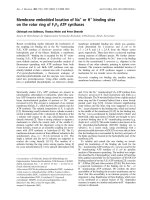
Principle of ICS test
The principle of the ICS test is based on the following
theory. If the tested duck serum contains the antibody
against DEV, the antibody will be absorbed from the
sample pad, which will interact with the recombinant
UL5 1 protein on the conjugat e pad to form an antigen-
antibody complex. The complex will migrate into the
nitrocellulose membrane by capillary action and, subse-
quently, react with the immobilized recombinant UL51
protein on the testing line (T), generating a red band,
the density of which will be in proportion to the con-
centration of antibody against DEV. Nonreactive goat
anti-rabbit IgG on the conjugate pad will run over the
test line, and then reacts with the rabbit IgG at the con-
trolline(C)ofthestriptoformthesecondvisiblered
band. Thus, after approximately 100 μLoftheduck
serum specimen was added to the sample chamber and
let stand for 15 min, the results were considered positive
(if the red band was present at both the test line and the
control line) (Figure 4b), negative (if the red band
appeared only at the control line) (Figure 4b), or invalid
(if no red band developed at either lines or only one
Shen et al. Virology Journal 2010, 7:268
/>Page 5 of 8
band appeared at the test line). Evaluation of the test-
strip results ca n be performed with the nake d eye a nd
total assay time is less than 15 min.
Specificity, sensitivity and stability of the ICS test
The spe cificity of the ICS test was evaluated with stan-
dard ne gative serum s amples from 5 healthy ducks, 25
standard serum samples positive for non-DEV patho-
gens (the pathogens for Duck hepatitis virus (DHV),
Riemerella anatipestifer (RA), Duck E. coli, Muscovy
duck parvovirus (MPV), or Duck Influenza viruses
(DIV)), 5 standard serum samples positive for DEV. All
the standard serum samples were supplied by our
laboratory.
The sensitivity of the ICS was tested with serially
diluted anti-DEV serum. The standard serum sample
was diluted 8 times with 10 mmol/L PBS from 1:2 to
1:512. The diluted sera were tested with this ICS. The
same procedure was repeated three times with different
operators.
The stability of the ICS was determined with the stan-
dard positive serum and the standard negative serum.
At each sa mple time, 8 strips that had been respectively
stored for 3, 6, 9, and 12 months at room temperature
(about 25°C), were tested.
ELISA
An ELISA for the detect ion IgG antibody against DEV in
serum was pe rformed as previously described [10,9]. In
brief, the DEV CHv strain (obtained from our laboratory)
virions abundantly propagated in duck embryo fibro-
blasts (DEF) was purified by differential velocity centrifu-
gation and sucrose density gradient centrifugation.
Round-bottomed 96 well polystyrene plates (Nunc Maxi-
Sorp) were coated overnight with the prepared highly
purified DEV vir ions (100 μL/well) at 4°C in a humidity
chamber. The plates were washed three times with PBS-
T buffer (10 mmol/L PBS containing 0.05% Tween-20),
non-specific protein binding sites were blocked with
blocking buffer (10 mmol/L PBS containing 1% fetal calf
serum) for 60 min at 37°C, and the plates were then
washed three f urther times with PBS-T buffer. A 10-fold
dilution series of serum, diluted with PBS, were added
and the plates incubated for 60 min at 25°C following by
washing, 50 μL of HRP-labeled goat anti-duck IgG (KPL)
(1:4000 dilution with PBS containing 1% bovine serum
albumin) was added. Following incubation for 60 min at
25°C, the plates were washed and 100 μL3,3
’
,5,5’ -etra-
methylbenzidine (TMB) substrate solution (KPL) was
added along with 0.01% of H
2
O
2
in 0.05 mol/L citric acid
buffer (pH5.0). After 15 min, the reaction was terminated
by adding 50 μLof0.5mol/Lsulfuricacidsolution.The
absorbance was read at 450 nm on a 96-well plate reader
(Model 460, Bio-Rad). The results were expressed as
serum antibody titer defined as the log10 of the dilution
that generated an optical density (OD) equal to two stan-
dard deviations (SD) above the mean background OD of
negative control duck sera at 450 nm.
Figure 4 Schematic diagram of the immunochromatographic strip (ICS) and interpretation of the results detected by the ICS.(a)
Schematic diagram of the ICS. The sample pad, conjugate pad, immobilized nitrocellulose membrane, and absorption pad were glued together
on a plastic backing plate. At the test band (T) and control band (C), the purified recombinant UL51 protein of DEV and the rabbit IgG were
immobilized, respectively. (b) Interpretation of the results detected by the ICS. A negative result and a positive result were showed in the picture,
respectively.
Shen et al. Virology Journal 2010, 7:268
/>Page 6 of 8
NT
The NT was performed as previously described [7,8].
Briefly, the serum was heated at 56°C for 30 min to
inactivate complement and dilut ed by means of serial
two-fold dilutions in MEM. Then, the diluted sera were
equally mixed with a 200 TCID
50
dose of DEV CHv
strain at 37°C for 1 h. The mixtures were inoculated
into the DEF cultured in 24-well plates (Corning Incor-
porated). The cytopathic effect (CPE) was observed, and
the neutralizing antibody titer of the serum wa s calcu-
lated using the Reed-Muench formula.
The analysis of 110 field serum samples
Using the ICS, 110 sera that had been collected from
several non-immune duck flocks in Sichuan province,
were tested. They were also tested for antibody against
DEV using the ELISA and NT following the above
instructions.
Statistical analysis
The percentiles of anti-DEV positive sera were statisti-
cally analyzed by Chi-square test and a P value of ≤ 0.05
was considered significantly.
Acknowledgements
The research were supported by grants from the National Natural Science
Foundation of China (No.30771598), Changjiang Scholars and Innovative
Research Team in University (PCSIRT0848), the earmarked fund for Modern
Agro-industry Technology Research System (nycytx-45-12 ) and the
Cultivation Fund of the Key Scientific and Technical Innovation Project, the
Ministry of Education of China (No.706050).
Author details
1
Avian Diseases Research Center, College of Veterinary Medicine of Sichuan
Agricultural University, Ya’an, Sichuan, 625014, China.
2
Key Laboratory of
Animal Diseases and Human Health of Sichuan Province, Ya’an, Sichuan,
625014, China.
3
Epizootic Diseases Institute of Sichuan Agricultural University,
Ya’an, Sichuan, 625014, China.
Authors’ contributions
CJS carried out most of the experiments and drafted the manuscript. ACC,
MSW, KFS, RYJ, TS, NZ, DKZ, QHL, YZ, and XYC helped in experiments and
drafted the manuscript. All authors read and approved the final manuscript.
Competing interests
The authors declare that they have no competing interests.
Received: 23 June 2010 Accepted: 14 October 2010
Published: 14 October 2010
References
1. Sandhu TS, Metwally SA: Duck Virus Enteritis (Duck Plague). In Diseases of
poultry. Edited by: Saif YM. Singapore: Blackwell Publishing; , 12
2008:384-393.
2. Baudet AE: Mortality in ducks in the Netherlands caused by a filterable
virus. Fowl plague 1923, 50:455-459.
3. Huang YX: Study on duck plague-like disease. J South China Agric Univ
1959, 1:1-12.
4. Burgess EC, Ossa J, Yuill TM: Duck plague: a carrier state in waterfowl.
Avian Dis 1979, 23:940-949.
5. Lin W, Lam KM, Clark WE: Active and Passive Immunization of Ducks
against Duck Viral Enteritis. Avian Dis 1984, 28:968-977.
6. Islam MA, Samad MA, Rahman MB, Hossain MT, Akter S: Assessment of
Immunologic Responses in Khaki Cambell Ducks Vaccinated Against
Duck Plague. Int J Poult Sci 2005, 4:36-38.
7. Dardiri AH, Hess WR: The incidence of neutralizing antibodies to duck
plague virus in serums from domestic ducks and wild waterfowl in the
United States of America. Proc Annu Meet US Anim Health Assoc 1967,
71:225-237.
8. Wolf K, Burke CN, Quimby MC: Duck viral enteritis: microtiter plate
isolation and neutralization test using the duck embryo fibroblast cell
line. Avian Dis 1974, 18:427-434.
9. Qi X, Cheng A, Wang M, Yang X, Jia R, Chen X: Development of an
indirect-ELISA kit for detection of antibodies against duck plague virus.
Vet Sci Chin 2007, 37:690-694.
10. Yang X, Qi X, Cheng A, Wang M, Zhu D, Jia R, Chen X: Intestinal mucosal
immune response in ducklings following oral immunisation with an
attenuated Duck enteritis virus vaccine. Vet J 2009.
11. Qi X, Yang X, Cheng A, Wang M, Zhu D, Jia R, Luo Q, Chen X: Intestinal
mucosal immune response against virulent duck enteritis virus infection
in ducklings. Res Vet Sci 2009, 87:218-225.
12. Malmarugan S, Sulochana S: Comparison of dot-ELISA passive
haemagglutination test for the detection of antibodies to duckplague.
Indian Vet J 2002, 79:648-651.
13. Peng D, Hu S, Hua Y, Xiao Y, Li Z, Wang X, Bi D: Comparison of a new
gold-immunochromatographic assay for the detection of antibodies
against avian influenza virus with hemagglutination inhibition and agar
gel immunodiffusion assays. Vet Immunol Immunopathol 2007, 117:17-25.
14. Mao X, Baloda M, Gurung AS, Lin Y, Liu G: Multiplex electrochemical
immunoassay using gold nanoparticle probes and
immunochromatographic strips. Electrochemistry Communications 2008,
10:1636-1640.
15. Cui S, Chen C, Tong G: A simple and rapid immunochromatographic strip
test for monitoring antibodies to H5 subtype Avian Influenza Virus. J
Virol Methods 2008, 152:102-105.
16. Cui S, Zhou S, Chen C, Qi T, Zhang C, Oh J: A simple and rapid
immunochromatographic strip test for detecting antibody to porcine
reproductive and respiratory syndrome virus. J Virol Methods 2008,
152:38-42.
17. Kameyama K, Sakoda Y, Tamai K, Igarashi H, Tajima M, Mochizuki T,
Namba Y, Kida H: Development of an immunochromatographic test kit
for rapid detection of bovine viral diarrhea virus antigen. J Virol Methods
2006, 138:140-146.
18. Tsuda Y, Sakoda Y, Sakabe S, Mochizuki T, Namba Y, Kida H: Development
of an immunochromatographic kit for rapid diagnosis of H5 avian
influenza virus infection. Microbiol Immunol 2007, 51:903-907.
19. Shen C, Cheng A, Wang M, Guo Y, Zhao L, Wen M, Xie W, Xin H, Zhu D:
Identification and characterization of the duck enteritis virus UL51 gene.
Arch Virol 2009, 154:1061-1069.
20. Shen C, Guo Y, Cheng A, Wang M, Zhou Y, Lin D, Xin H, Zhang N:
Characterization of subcellular localization of duck enteritis virus UL51
protein. Virol J 2009, 6:92.
21. Li Y, Huang B, Ma X, Wu J, Li F, Ai W, Song M, Yang H: Molecular
characterization of the genome of duck enteritis virus. Virology 2009,
391:151-161.
22. Klupp BG, Granzow H, Klopfleisch R, Fuchs W, Kopp M, Lenk M,
Mettenleiter TC: Functional analysis of the pseudorabies virus UL51
protein. J Virol 2005, 79:3831-3840.
23. Nozawa N, Kawaguchi Y, Tanaka M, Kato A, Kato A, Kimura H, Nishiyama Y:
Herpes simplex virus type 1 UL51 protein is involved in maturation and
egress of virus particles. J Virol 2005, 79:6947-6956.
24. Koshizuka T, Kawaguchi Y, Nozawa N, Mori I, Nishiyama Y: Herpes simplex
virus protein UL11 but not UL51 is associated with lipid rafts. Virus Genes
2007, 35:571-575.
25. Jia R, Cheng A, Wang M, Qi X, Zhu D, Ge H, Luo Q, Liu F, Guo Y, Chen X:
Development and evaluation of an antigen-capture ELISA for detection
of the UL24 antigen of the duck enteritis virus, based on a polyclonal
antibody against the UL24 expression protein. J Virol Methods 2009,
161:38-43.
26. Guo Y, Cheng A, Wang M, Zhou Y: Purification of anatid herpesvirus 1
particles by tangential-flow ultrafiltration and sucrose gradient
ultracentrifugation. J Virol Methods 2009, 161(1):1-6.
Shen et al. Virology Journal 2010, 7:268
/>Page 7 of 8
27. Yang J, Hua Q, Chen H, Lv J, Qin Z, Jin M, Tao H, Zeng S, Ruan Z, Chen B,
Zhou X: Development and evaluation of an immunochromatographic
strip for the detection of serum antibodies against bluetongue virus. J
Virol Methods 2010, 163:68-73.
28. Bradford MM: A rapid and sensitive method for the quantitation of
microgram quantities of protein utilizing the principle of protein-dye
binding. Anal Biochem 1976, 72:248-254.
29. Tanaka R, Yuhi T, Nagatani N, Endo T, Kerman K, Takamura Y, Tamiya E: A
novel enhancement assay for immunochromatographic test strips using
gold nanoparticles. Anal Bioanal Chem 2006, 385:1414-1420.
30. Liu G, Lin YY, Wang J, Wu H, Wai CM, Lin Y: Disposable electrochemical
immunosensor diagnosis device based on nanoparticle probe and
immunochromatographic strip. Anal Chem 2007, 79:7644-7653.
doi:10.1186/1743-422X-7-268
Cite this article as: Shen et al.: Development and evaluation of an
immunochromatographic strip test based on the recombinant UL51
protein for detecting antibody against duck enteritis virus. Virology
Journal 2010 7:268.
Submit your next manuscript to BioMed Central
and take full advantage of:
• Convenient online submission
• Thorough peer review
• No space constraints or color figure charges
• Immediate publication on acceptance
• Inclusion in PubMed, CAS, Scopus and Google Scholar
• Research which is freely available for redistribution
Submit your manuscript at
www.biomedcentral.com/submit
Shen et al. Virology Journal 2010, 7:268
/>Page 8 of 8