Báo cáo y học: " Genital herpes evaluation by quantitative TaqMan PCR: correlating single detection and quantity of HSV-2 DNA in cervicovaginal lavage fluids with cross-sectional and longitudinal clinical data" doc
Bạn đang xem bản rút gọn của tài liệu. Xem và tải ngay bản đầy đủ của tài liệu tại đây (286.18 KB, 8 trang )
RESEARC H Open Access
Genital herpes evaluation by quantitative TaqMan
PCR: correlating single detection and quantity of
HSV-2 DNA in cervicovaginal lavage fluids with
cross-sectional and longitudinal clinical data
Bulbulgul Aumakhan
1*
, Andrew Hardick
2
, Thomas C Quinn
2,3
, Oliver Laeyendecker
2,3
, Stephen J Gange
1
,
Chris Beyrer
1
, Christopher Cox
1
, Kathryn Anastos
4
, Mardge Cohen
5
, Ruth M Greenblatt
6
, Daniel J Merenstein
7
,
Howard Minkoff
8
, Marek Nowicki
9
, Charlotte A Gaydos
2
Abstract
Objective: To evaluate the utility of a single quantitative PCR (qPCR) measurement of HSV (HSV-1&2) DNA in
cervicovaginal lavage (CVL) specimens collected from women with predominantly chronic HSV-2 infection in
assessing genital HSV shedding and the clinical course of genital herpes (GH) within a cohort with semiannual
schedule of follow up and collection of specimens.
Methods: Two previously described methods used for detection of HSV DNA in mucocutaneous swab samples
were adapted for quantification of HSV DNA in CVLs. Single CVL specimens from 509 women were tested.
Presence and quantity of CVL HSV DNA were explored in relation to observed cross-sectional and longitudinal
clinical data.
Results: The PCR assay was sensitive and reproducible with a limit of quantification of ~50 copies per milliliter of
CVL. Overall, 7% of the samples were positive for HSV-2 DNA with median log
10
HSV-2 DNA copy number of 3.9
(IQR: 2.6-5.7). No HSV-1 was detected. Presence and quantity of HSV-2 DNA in CVL directly correlated with the
clinical signs and symptoms of presence of active symptomatic disease with frequent recurrences.
Conclusion: Single qPCR measurement of HSV DNA in CVL fluids of women with chronic HSV-2 infection provided
useful information for assessing GH in the setting of infrequent sampling of specimens. Observed positive
correlation of the presence and quantity of HSV-2 DNA with the presence of active and more severe course of
HSV-2 infection may have clinical significance in the evaluation and management of HSV-2 infected patients.
Introduction
Genital herpes (GH) is a common chronic sexually
transmitted infection w orldwide with substantial mor-
bidity [1,2] caused mainly by Herpes S implex Virus
Type 2 (HSV-2) and sometime s by HSV-1. Women, in
particular, are disproportionately affected. GH is also
commonly found among Human Immunodeficiency
Virus (HIV) infected individuals in whom it is associated
with increased HIV replication [3,4].
The majority of HSV-2 infected individuals is ‘asymp-
tomatic’ or unaware of infection [5,6]. Those with symp-
tomatic HSV-2 can experience recurrent episodes of
genital lesions that appear to diminish in severity and
frequency over time [7-9]. Most individuals with chronic
HSV-2 have mild or asymptomatic infection.
Cell culture isolation of HSV is the preferred diagnos-
tic test, usually used in conjunction with symptomatic
primary or first clinical episode. However, its sensitivity
for recurrent or healing lesions is low. More recently,
PCR- based methods have been actively investig ated for
the detection of HSV DNA in mucocutaneous lesions
and have shown to be superior to viral culture [10-12].
* Correspondence:
1
Johns Hopkins Bloomberg School of Public Health, Baltimore, MD, USA
Full list of author information is available at the end of the article
Aumakhan et al. Virology Journal 2010, 7:328
/>© 2010 Aumakhan et al; licensee BioMed Central Ltd. This is an Open Access article distributed under the terms of the Creative
Commons Attribution License ( which permits unrestricted use, distribution, and
reproduction in any medium, provided the original work is properly cited.
PCR has also been shown to be more sensitive in detect-
ing asymptomatic shedding or shedding episodes in the
absence of clinically obvious lesions [13-16]. Neverthe-
less, the potential utility of broad based application of
PCR based techniques in the evaluation and manage-
ment of HSV-2 infected patients, especially of those
with longstanding and/or asymptomatic GH, is less clear
given the plausibility of reduced genital shedding over
time. In ad dition, the essential goal of most PCR assays
was detection, i.e. determining the presence or absence
of HSV target nucleic acid sequences in the s ample.
However, for pathogenesis studies and clinical manage-
ment purposes, including prognosis or determining opti-
mal drug regimens, quantification of actual viral load
may be useful. Data on the usefulness of quantification
of HSV DNA in genital secretions, perhaps due to mild
nature of most HSV-2 infections, is limited and
restricted mainly to evaluating clinical and virologic effi-
cacy of antiviral compounds and defining the threshold
of HSV infectivity as a potential factor in the transmissi-
bility of infection [17-22]. Nevertheless, available evi-
dence suggests that HSV-2 viral titer in genital
secretion s can be a useful means for disease monitoring
purposes. A study by Filen et al., for example, found
that first e pisodes of GH were associated w ith signifi-
cantly higher viral loads compared to recurrent or atypi-
cal cases [9]. Yet, other studies doubt the usefulness of
monitoring HSV loads in clinical samples [21,23]. Some
of the challenges in ascertaining these issues are related
to intermittent nature and wide variability in the fre-
quency and amount of HSV shedding observed among
infected individuals. Many investigators use repeated
and frequent sampling up to multiple times a day to
overcome these challenges [24]. However, for practical
reasons, not all research and clinical settings can easily
implement such an approach and, hence, the clinical
usefulness of quantitative PCR (qPCR) methods, espe-
cially for those with established chronic GH and in the
setting of infrequent sampling of specimens, is unclear.
Therefore, using quantitative PCR technique, we
aimed to explore the usefulness of assessing genital HSV
infection by single qPCR measurement of HSV DNA in
cervicovaginal lavage (CVL) specimens of women with
mostly longstanding HSV-2 infection within the setting
of a research cohort with semiannual scheduling of fol-
low up and specimen sampling. The presence and quan-
tity of CVL HSV DNA were explored in relation to
observed cross-sectional and longitudinal clinical data.
Methods
Study population and specimens
The study population consisted of HIV infected and
uninfected participants of Women’s Interagency HIV
Study (WIHS), a multicenter cohort study of HIV in
womenacrosssixsitesintheUS(LosAngeles,CA;
Washington, DC; San Francisco, CA; New York City/
Bronx, NY; Brooklyn, NY; and Chicago, IL). WIHS
enrolled 2059 HIV infected and 569 high risk HIV unin-
fected women between October 1994 and November
1995 [25]. At enrollment, over 90% of WIHS partici-
pants were seropositive for HSV-1 and more than 80%
of HIV infected women seropositive for H SV-2. HSV
serostatus was determined by HSV type specific antibo-
dies by glycoprotein G-based enzyme immunoassay (gG-
EIA, Gull Laboratories, Salt Lake City, Utah). Negative
and equivocal results were confirmed by Western Blot
[26]. Gynecological examination included assessment for
genital tract infections and genital tract dysplasia as pre-
viously described [27]. Self-reports of GH sores and
observationsofpresenceoflesions, sites of the lesions
and whether the lesi ons w ere observed at multiple
(three or more) locations were collected at each study
visit. CVL specimens were collected by flushing the cer-
vix with 10 ml sterile norm al saline aspirated from the
posterior vaginal fornix. The specimens were then trans-
ported to the processing lab oratory on ice within 24-26
hour s and 1 ml aliquots were stored at a central reposi-
tory at -70°C. Whole unspun and unfractionated CVL
was used for this study.
Total of 509 single CVL samples from 509 women
were retrieved from repository for testing. Ten samples
each from dual positive ( HIV+/HSV+), HIV only (HIV
+/HSV-), HSV only (HIV-/HSV+) positive women and
40 samples from dual negative (HIV-/HSV-) women
were retrieved from the baseline visit to use in the assay
validation. The rest were selected based on the following
criteria: 1) had known baseline HSV serology status; 2)
had information on self reported history of GH sores,
physical and gynecological exams; 3) had at least one
follow-up visit since the baseline; and 4) had sufficient
volume of more than 5 ml CVL available to preserve the
specimens. To assess the correlation of the initial or
‘baseline’ CVL HSV DNA titer with the number of sub-
sequent lesion recurrences, we identified eligible samples
from wome n who had multiple (> 1) recurrent episodes
of lesion outbreaks (referred thereafter as lesion-episode)
observed d uring the follow up. For these women, CVL
sample from the earliest available lesion-episode was
retrieved for testing and considered as a ‘baseline’ epi-
sode. Since the earliest available lesion-episode is differ-
ent for each woman, the visits from which samples were
pulled ranged from 1 to 24 with the median visit num-
ber of 3 (IQR: 1-8).
Extraction of HSV DNA
CVL fluids were thawed at room temperature. DNA was
extracted by QIAamp DNA blood minikit from 200 μl
of whole CVL (Qiagen, Valencia, California) using the
Aumakhan et al. Virology Journal 2010, 7:328
/>Page 2 of 8
Blood and Body Fluid Spin Protocol. The DNA was
eluted into 55 μl of Q iagen AE buf fer. Each extraction
included positive control HSV isolates (HSV-1 strain
GHSV-UL46andHSV-2strainMS,ATCC,Manassas,
VA) a nd RNase- and DNase-free water as the negative
control.
Preparation of HSV DNA standards
Ten-fold serial dilutions were prepared with commercial
HSV-1&2 quantified DNA (ABI Advanced Technologies,
Inc., Columbia, Maryland) to generate a standard curve.
The DNA stocks were serially diluted with RNase- and
DNase- free water and/or with C VL fluids pooled from
HIV (+) and HIV (-) wome n whose CVLs were negative
for HSV-1&2 DNA. To avoid repeated freeze-thaw of
the DNA stock which could negatively affect the repro-
ducibility of the assay, single use panels of serial dilu-
tions were prepared immediately upon receipt of the
DNAstockandstoredat-20°Cuntil further use. Stan-
dards were analyzed in duplicates and used to generate
a standard curve as well as a positive control for each
PCR run.
Primers, probes and target sequence for amplification
Primers were adapted from two different sources. The
forward primer (GbTypF: 5’-CGC ATC AAG ACC ACC
TCC TC-3’ ) was as described by Corey L. et al. [28].
The reverse primer (HSV1&2-R: 5’-AGC TTG CGG
GCC TCG TT-3’ ) and probes (HSV1-probe: 5 ’-CGG
CCC AAC ATA TCG TTG ACA TGG C-3’ and HSV2-
probe: 5’-CGC CCC AGC ATG TCG TTC ACG T-3’)
were as described by Namvar et al. [29]. The probe
region differs by 5 nucleotides and was previo usly
shown to differen tiate between HSV-1 and HSV-2 with-
out cross-reactivity [29]. Probes were labeled at the 5’-
end with FAM or VIC and at the 3’- end with TAMRA.
Primers allowed amplification of a highly specific 155-
nucleotide region of gB envelope gene homologous for
HSV-1&2 which represented the summed extension of
overlapping target sequences used by the two groups.
TaqMan PCR
The fina l 50 μl PCR reaction mix contained 25 μlof2×
TaqMan universal master mix (P E Applied Biosystems,
Foster City, CA), 900 nM of each primer, 100 nM of
each probe and 10 μlofsampleDNA.PCRwasper-
formed using an ABI 7900 HT sequence detection sys-
tem (PE Applied Biosystems, Foster City, CA) with the
following cycling conditions: incubation for 2 min at 50°
C (uracil-N-glycosylase digestion) and denaturation at
95°C for 10 min followed by 45 cycles of 15 s denatura-
tion at 95°C and 60 s anne aling/extension at 58°C. Spe-
cimens were blinded to clinical i nformation and run in
duplicate. A sample was considered posi tive if the
detected quantity was above or equal to assay limit of
quantification in both replicates.
Statistical analysis
Assay performance was evaluated using within and
between assay measures of efficiency (slope of standard
curve), linearity (R-square) and reproducibility (mean
threshold (Ct) values, standard deviation (SD) and coef-
ficient of variation (CV)) from standard curve data.
Limit of detection (LoD) and limit of quantification
(LoQ) were estimated using the delta method to approx-
imate the relative precision of the estimated concentra-
tion as previously described [30]. Values of HSV-2 DNA
were log
10
transformed for analyses. Proportions with
detectable HSV DNA by clinical markers of genital HSV
infection were compared using chi-square and median
quantities by Wilcoxon rank-sum tests. The markers
were HSV-2 seropositivity, self report of GH lesions, the
presence of any lesions and/or lesions clinically sus-
pected as herpetic. To assess whether there is any rela-
tionship between the initial ‘ baseline’ HSV-2 viral load
and subsequent clinical course of GH, the correlation
between the CVL HSV-2 DNA titer and the total num-
ber of lesion-recurrences observed during the subse-
quent follow up was explored. Duration of subsequent
follow up was determined by the total number of follow
up visits observed since the detection of HSV DNA in
CVL. Ratio of frequency of subsequent lesion-episodes
on duration of follow up was used to account for vary-
ing lengths of follow up among women. Pearson’sror
Spearman’s rho were used to estimate the c orrelations
of interest. P-values of < 0.1 were considered significant.
Statistica l analyses were ca rried out using STATA 1 0.1
software (STATA Corporation, College Station, Texas).
Graphs were created using GraphPad Prism Software, v.
5.03 (GraphPad Software, La Jolla, California).
Results
Assay performance
For HSV-1, the Ct values ranged from 21.62 for log
10
5to
35.68 for log
10
1 with an average slope of -3.22 (range:
-3.17 to -3.27). For HSV-2, the corresponding Ct values
ranged from 23.74 to 38.50 with average slope of -3.33
(range: -3.23 to -3.49) indicating high efficiencies for
both HSV types. The intra-assay CV (Ct) values for five
dilutions of HSV-1&2 ranged from 0.02% to 4.26%. The
inter-assay CV (Ct) ranged from 0.1% to 1.3%. R-square
values for all runs were ≥ 0.99. Standards were stable
with consist ent Ct values for all concentrations in multi-
ple runs performed over the course of 6 months. No sig-
nificant differences were observed in Ct values between
water and CVL diluted standards (≤ 1-2 Ct difference).
HIV status did not influence the test performance. Data
for HSV-2 are shown in Table 1.
Aumakhan et al. Virology Journal 2010, 7:328
/>Page 3 of 8
Limit of detection and limit of quantification
One to 1.5 copies per reaction were detected 50% of the
time and 10 copies w ere detected in 100% of the runs.
Thus, the LoD was considered as 1-2 copies/assay or
20-40 copies/ml. The LoQ for HSV-2 was ~2.3 copies
per reaction corresponding to ~47 copies per mL of
CVL and the LoQ for HSV-1 was ~6.4 copies per reac-
tion or ~127 copies per mL of CVL. The higher LoQ
observed for HSV-1 was due to slightly lower precision
of the estimates in the linear regression compared to
HSV-2. At least 6 replicates for each concentration from
multiple runs were used to estimate LoQ.
Study population
The study population consis ted of 379 (74%) dually
infected (HIV+/HSV+), 22 (4%) HIV only (HIV+/HSV-),
68 (13%) HSV only (HIV-/HSV+) and 40 (8%) neither
HIV nor HSV (HIV-/HSV-) infected individuals (Table
2). Median baseline age of women was 35 years. HSV
seropositive women were predominan tly African Ameri-
can and significan tly older a s opposed to seronegative
women. Intravenous drug use and heterosexual risk
were the commonly identified routes of HIV exposure
among HSV seropositive women. Median follow up o f
women was 24 visits (IQR: 14-24).
HSV-2 DNA detection by HIV/HSV status
Overall, 35 (7%) individuals were positive for genital
HSV- 2 DNA with a median log
10
DNA copy number of
3.9 (IQR: 2.6 - 5.7). No HSV-1 was detected. Log
transformed values of HSV-2 DNA were normally dis-
tributed (Shapiro-Wilk normality test p = 0. 406). By
HIV/HSV status, HIV+/HSV+ group had 27 women
with detectable HSV-2 DNA (n = 379, 7%, median log
10
HSV-2 DNA = 4.4, IQR: 2.6-5.9), HIV-/HSV+ group - 4
(n = 68, 6%, median log
10
HSV-2DNA=2.8,IQR:2.1-
4.0) and HIV-/HSV- group - 4 women p ositive for
HSV- 2 DNA (n = 40, 10%, median log
10
HSV-2 DNA =
3.7, IQR: 3.4-5.2). The number of HIV+/HSV- women
was small (n = 22) an d none had detectable HSV-2
DNA.
HSV-2 DNA detection by clinical markers GH infection
Detection was highest for lesions clinically suspected as
herpetic, 27% (p < 0.001) and 8% for presence of any
lesions (Table 3). About 6% of those with lesions not
identified as herpetic were positive for CVL HSV-2
DNA.
’Baseline’ CVL HSV-2 viral load and subsequent clinical
course
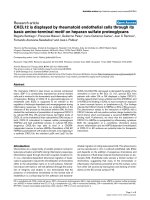
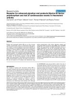
Positive correlation was observed b etween the CVL
HSV-2 DNA load and the frequency of lesion-episodes
observed during the subsequent follow up (Pearson r =
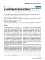
0.48, p = 0.005, Figure 1). Strength of the correlation
did not change when the ratio of lesion-episodes fre-
quency on the duration of follow up was used to
account for varying lengths of follow up (Pearson r =
0.50, p = 0.004, Fig ure 2). There was also no cor relation
between the length of subsequent follow up and
Table 1 Assay Reproducibility (HSV-2)
Intra-assay Inter-assay
# copies per assay Run Ct
1
Ct
2
mean Ct Ct SD CV (Ct) mean Ct Ct SD CV (Ct)
150,000 1 24.11 23.99 24.05 0.08 0.34 24.05 0.11 0.46
2 24.18 24.13 24.15 0.04 0.15
3 24.12 23.74 23.93 0.27 1.13
15,000 1 27.34 27.28 27.31 0.04 0.14 27.25 0.16 0.57
2 27.46 27.25 27.36 0.15 0.55
3 26.97 27.16 27.07 0.13 0.50
1,500 1 30.67 30.56 30.62 0.08 0.25 30.76 0.14 0.45
2 30.97 30.81 30.89 0.12 0.38
3 30.81 30.74 30.77 0.05 0.16
150 1 34.82 33.90 34.36 0.66 1.91 34.30 0.25 0.74
2 34.50 34.52 34.51 0.01 0.03
3 34.09 33.94 34.02 0.10 0.31
15 1 37.09 37.44 37.27 0.25 0.67 37.41 0.15 0.41
2 37.83 37.32 37.57 0.36 0.96
3 38.50 36.25 37.38 1.59 4.26
Aumakhan et al. Virology Journal 2010, 7:328
/>Page 4 of 8
quantity of HSV-2 DNA (Spearman’srho=-0.003,p=
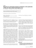
0.852) . Although there were no differences in the detec-
tion status, quantitatively, women with positive history
of GH sores and lesions identified at three or more loca-
tions tended to have higher median quantities of HSV-2
DNA that were statistically significant at alpha level of
0.1 (Figure 3). Women with clinically suspected herpetic
lesions had slightly higher median HSV-2 DNA titer but
the difference did not reach statistical significance of p
< 0.1. No quantitative differences by other markers were
observed.
HIV and HSV detection
Detailed analysis of the association between HIV and
HSV detection including multivariate regression was
described elsewhere (Aumakhan B, Gange SJ, Beyrer C,
Gaydos CA, Minkoff H, Merenstein DJ, Cohen M, Ana-
stos K, Greenblatt RM, Nowicki M, Quinn TC: Quanti-
tative and qualitative correlates of ce rvicovaginal HSV-2
shedding among HIV infected women in Women’ s
Table 2 Demographic and risk characteristics of the 509 women by HIV/HSV serostatus
Definition HIV+/HSV+
N = 379 (74%)
HIV+/HSV-
N = 22 (4%)
HIV-/HSV+
N= 68 (13%)
HIV-/HSV-
N = 40 (8%)
Median age at baseline, years (IQR) 38 (33-42) 36 (28-40) 34 (28-40) 26(22-30)
Race n (%)
African American 225 (59%) 6 (27%) 45 (66%) 13 (33%)
Hispanic 79 (21%) 3 (14%) 15 (22%) 7 (18%)
White 55 (15%) 11 (50%) 6 (9%) 18 (45%)
Other 20 (5%) 2 (9%) 2 (3%) 2 (5%)
Risk exposure
Intravenous drug use 135 (36%) 6 (27%) 16 (24%) 8 (20%)
Heterosexual risk 149 (39%) 12 (55%) 11 (16%) 10 (25%)
Transfusion risk 10 (3%) 0 (0%) 1 (1%) 1 (3%)
No identified risk 85(22%) 4 (18%) 40 (59%) 21 (53%)
Lifetime # of sex partners
0-1 16 (4%) 1 (5%) 3 (4%) 1 (3%)
2-4 51 (13%) 5 (23%) 13 (19%) 6 (15%)
5-9 81 (21%) 8 (36%) 19 (28%) 10 (25%)
10-50 122 (32%) 6 (27%) 19 (28%) 21 (53%)
> 50 108 (28%) 2 (9%) 14 (21%) 2 (5%)
missing 1 (0.3%) –– –
Table 3 CVL HSV-2 DNA detection by markers of genital
herpes
Definition Status
(+/-)
a
HSV-2 DNA (+),
n/N (%)
p-value
HSV-2 serostatus + 28/356 (8%) 0.251
- 7/153 (5%)
Self report of GH sores + 4/42 (10%) 0.518
- 31/467 (7%)
Any lesions
b
+ 30/394 (8%) 0.296
- 5/114 (4%)
Herpetic lesions
c
+ 7/26 (27%) 0.001
- 28/481 (6%)
a
(+) condition present; (-) condition absent
b
any genital lesions vs. no any lesions
c
lesions clinically suspected as herpetic vs. no lesions or lesions not
suspected as herpetic
Figure 1 Correlation between the frequency of subsequent
lesion-episodes and CVL HSV-2 DNA titer, Pearson r = 0.48, p
= 0.005.
Aumakhan et al. Virology Journal 2010, 7:328
/>Page 5 of 8
Interagency HIV Study, submitted). Briefly, trend for
reduced detection of HSV-2 D NA with higher CD+ T
cell counts was observed (p-value for trend = 0.08). No
significant associations were observed with HIV viral
load and use of antiretroviral therapy.
Discussion
We explored the correlation of the presence and quan-
tity of HSV-2 DNA in cervicovaginal fluids collected
from women with predominantly established genital
herpes infection with clinical manifestations observed at
the visit (cross-sectionally) and over the course of follow
up (longitudinally) using real time PCR technique. The
PCR assay adapted two previously reported methods
[28,29] used for detection and typing of HSV DNA in
mucocutaneous swab samples to quantification of HSV
DNA in CVL samples. The combination of primers and
probes from two different sources was a result of preli-
minary review of primers and probes from reported
methods during which it was determined that the target
sequences of these two methods overlapped resulting in a
final amplicon of 155-nucleotide region of glycoprotein B
gene highly specific for HSV-1 and HSV-2 differentiation.
TwotypespecificforwardprimersusedbyNamvaretal.
[29] were conveniently replaced by one common type
primer described by Corey et al. [28] and the assay was
implemented using the absolute quantification guidelines
recommended by the manufacturer (ABI 7900 HT SDS,
PE Applied Biosystems, Foster City, CA).
Overall, we found a 7% HSV-2 DNA detection rate in
the test ed samples. Despite test ing for HSV-1, no HSV-1
DNA was detected. Herpetic lesions had the most corre-
lation with the probability of detectable HSV-2 DNA in
CVL with 27% positivity rate. Although this finding may
not be surprising, the main point of this observation is
the extent of this correlation in this particular population
and what to expect if broader categories, such as pre-
sence of any lesions, are used. The latter was associated
with 8% positivity rate.
Four HIV-/HSV- women tested positive for HSV-2
DNA in CVL suggesting that they may have had pri-
mary genital HSV-2 infection. Three of them had multi-
ple lesion-episodes observed during the subsequent
follow up. However, only one had active lesion at the
time of sampling and one reported positive history of
genital sores in the past 6 months. The individual with
the active lesion had the highest viral load with log10
HSV-2 DNA copy number of 6.4. Follow up measure-
ment of serum anti-HSV-2 Ig G will be needed to con-
firm any subsequent seroconversion.
An interesting finding is the significant correlation
observed between the ‘ baseline’ CVL HSV-2 DNA load
and the frequency of subsequent lesion recurrences
observed during the follow up, which suggests that hi gh
HSV-2 load could be associated with frequent reactiva-
tions. Absence of the correlation between the length of
subsequent follow up and HSV-2 DNA titer suggests that
this association was not due to varying lengths of follow
up. Trend towards higher median HSV-2 DNA titer with
the presence of lesions at multiple locations could indi-
cate that H SV-2 viral load plays a role in the severity of
GH clinical expression. Although only 10% of women
with self reported positive history of GH sores had
detectable HSV-2 DNA, they tended to have higher
HSV-2 DNA copy numbers compared to women without
such history, which suggests that more readily recogniz-
able lesions may harbor high levels of infectious virus.
Figure 2 Correlation between the ratio of the frequency of
subsequent lesion-episodes on duration of follow up and CVL
HSV-2 DNA titer, Pearson r = 0.50, p = 0.004.
Figure 3 Median CVL HSV-2 DNA titer by presence of lesions
at 3 or more locations, presence of self reported history of
genital herpes sores and presence of herpetic lesions.
Aumakhan et al. Virology Journal 2010, 7:328
/>Page 6 of 8
Additional studies with a larger number of positive end-
points will be needed to validate these results. Neverthe-
less, findings of this study were the basis for classifying
HSV-2 infected women into groups of gradient degree of
GH clinical activity (determined by the presence/absence
of active symptomatic disease with multiple recurrences)
in another study by our group, in which we observed
direct dose dependent association between classic mar-
kers of HIV disease progression (CD4+ T cell count, HIV
RNA load) and a degree of HSV-2 clinical activity, which
lends additional support to these results [31].
Our 7% of HSV-2 DNA detection rate in CVL may seem
low compared to some other reports that measured genital
HSV shedding using CVL specimens [32-34]. This may
have been due to differences in the methods of CVL col-
lection employed, sampling frequency or the population
characteristics in which these assays were utilized. It is
also lower than estimates of HSV shedding reported in
previous WIHS study by Augenbraun et al. [35]. However,
direct comparison between this and the previous study
may not be cogent as studies used differed selection
criteria for enrolling participants as well as different
specimen types and sampling strategies.
Despite this limitation, the study has several unique
strengths. First, although HSV-2 shedding was measured
at single time point, we used rich longitudinal clinical
data accumulated by WIHS over many years to link our
PCR results with the observed clinical course of GH in
these women. Second, as many studies explore HSV-2
using frequent sampling such as daily or even multiple
sampling in a day [36,37], these results point to poten-
tial feasibili ty of studies of HSV-2 natural history in the
settings with a less frequent sampling schedule and col-
lection of data.
In summary, single qPCR measurement of HSV DNA
in CVL specimens among w omen with chronic HSV-2
infection can provide useful information for assessing
genital herpes in the setting of infrequent sampling of
specimens. Observed positive correlation of the presence
and quantity of HSV-2 DNA with active symptomatic
disease w ith frequent reactivations suggests that HSV-2
quantification could be a useful tool in evaluating HSV-
2 infected patients with chronic genital herpes and may
guide better antiviral therapy.
Acknowledgements
Data in this manuscript were collected by the Women’s Interagency HIV
Study (WIHS) Collaborative Study Group with centers (Principal Investigators)
at New York City/Bronx Consortium (Kathryn Anastos); Brooklyn, NY (Howard
Minkoff); Washington DC Metropolitan Consortium (Mary Young); The
Connie Wofsy Study Consortium of Northern California (Ruth Greenblatt); Los
Angeles County/Southern California Consortium (Alexandra Levine); Chicago
Consortium (Mardge Cohen); Data Coordinating Center (Stephen Gange).
The WIHS is funded by the National Institute of Allergy and Infectious
Diseases (UO1-AI-35004, UO1-AI-31834, UO1-AI-34994, UO1-AI-34989, UO1-AI-
34993, and UO1-AI-42590) and by the National Institute of Child Health and
Human Development (UO1-HD-32632). The study is co- funded by the
National Cancer Institute, the National Institute on Drug Abuse, and the
National Institute on Deafness and Other Communication Disorders. Funding
is also provided by the National Center for Research Resources (UCSF-CTSI
Grant Number UL1 RR024131). The contents of this publication are solely the
responsibility of the authors and do not necessarily represent the official
views of the National Institutes of Health.
Author details
1
Johns Hopkins Bloomberg School of Public Health, Baltimore, MD, USA.
2
Johns Hopkins University School of Medicine, Baltimore, MD, USA.
3
Laboratory of Immunoregulation, National Institute of Alle rgy and Infectious
Diseases, National Institutes of Health, Bethesda, MD, USA.
4
Albert Einstein
College of Medicine, Bronx, NY, USA.
5
Cook County Medical Center, Chicago,
IL, USA.
6
University of California, San Francisco School of Medicine, San
Francisco, CA, USA.
7
Georgetown University Medical Center, Washington, D.
C., USA.
8
Maimonides Medical Center and SUNY Downstate, Brooklyn, NY,
USA.
9
University of Southern California, Los Angeles, CA, USA.
Authors’ contributions
BA, TCQ, SJG, CB, CAG conceived and designed the study. BA, AH, OL, CAG
performed the assay design and experiments. BA, SJG, CC carried out
statistical analysis. KA, MC, RMG, DJM, HM, MN, SJG contributed samples and
data. BA wrote initial draft of the manuscript. All authors read and approved
the final manuscript.
Competing interests
The authors declare that they have no competing interests.
Received: 6 September 2010 Accepted: 18 November 2010
Published: 18 November 2010
References
1. Smith JS, Robinson NJ: Age-specific prevalence of infection with herpes
simplex virus types 2 and 1: a global review. J Infect Dis 2002, 186(Suppl
1):S3-28.
2. Xu F, Sternberg MR, Kottiri BJ, McQuillan GM, Lee FK, Nahmias AJ,
Berman SM, Markowitz LE: Trends in herpes simplex virus type 1 and
type 2 seroprevalence in the United States. JAMA 2006, 296(8):964-973.
3. Mole L, Ripich S, Margolis D, Holodniy M: The impact of active herpes
simplex virus infection on human immunodeficiency virus load. J Infect
Dis 1997, 176(3):766-770.
4. Schacker T, Zeh J, Hu H, Shaughnessy M, Corey L: Changes in plasma
human immunodeficiency virus type 1 RNA associated with herpes
simplex virus reactivation and suppression. J Infect Dis 2002,
186(12):1718-1725.
5. Ashley RL: Sorting out the new HSV type specific antibody tests. Sex
Transm Infect 2001, 77(4):232-237.
6. Ashley RL, Wald A: Genital herpes: review of the epidemic and potential
use of type-specific serology. Clin Microbiol Rev 1999, 12(1):1-8.
7. Corey L, Adams HG, Brown ZA, Holmes KK: Genital herpes simplex virus
infections: clinical manifestations, course, and complications. Ann Intern
Med 1983, 98(6):958-972.
8. Benedetti J, Corey L, Ashley R: Recurrence rates in genital herpes after
symptomatic first-episode infection. Ann Intern Med 1994, 121(11):847-854.
9. Filen F, Strand A, Allard A, Blomberg J, Herrmann B: Duplex real-time
polymerase chain reaction assay for detection and quantification of
herpes simplex virus type 1 and herpes simplex virus type 2 in genital
and cutaneous lesions. Sex Transm Dis 2004, 31(6):331-336.
10. W a ld A, Huang ML, C ar rell D, Selke S, Corey L : Polymerase chain reaction for
detection of he rpes simplex virus (HSV) DNA on m ucosal surfaces: c omparison
with HSV i solation in cell cu lture. JInfectDis2003, 188(9):1345-1351.
11. Hobson A, Wald A, Wright N, Corey L: Evaluation of a quantitative
competitive PCR assay for measuring herpes simplex virus DNA content
in genital tract secretions. J Clin Microbiol 1997, 35(3):548-552.
12. van Doornum GJ, Guldemeester J, Osterhaus AD, Niesters HG: Diagnosing
herpesvirus infections by real-time amplification and rapid culture. J Clin
Microbiol 2003, 41(2):576-580.
13. Cone RW, Hobson AC, Palmer J, Remington M, Corey L: Extended duration
of herpes simplex virus DNA in genital lesions detected by the
polymerase chain reaction. J Infect Dis 1991, 164(4):757-760.
Aumakhan et al. Virology Journal 2010, 7:328
/>Page 7 of 8
14. Cone RW, Hobson AC, Brown Z, Ashley R, Berry S, Winter C, Corey L:
Frequent detection of genital herpes simplex virus DNA by polymerase
chain reaction among pregnant women. JAMA 1994, 272(10):792-796.
15. Slomka MJ, Emery L, Munday PE, Moulsdale M, Brown DW: A comparison
of PCR with virus isolation and direct antigen detection for diagnosis
and typing of genital herpes. J Med Virol 1998, 55(2):177-183.
16. Safrin S, Shaw H, Bolan G, Cuan J, Chiang CS: Comparison of virus culture
and the polymerase chain reaction for diagnosis of mucocutaneous
herpes simplex virus infection. Sex Transm Dis 1997, 24(3):176-180.
17. Fuchs J, Celum C, Wang J, Hughes J, Sanchez J, Cowan F, Reid S, Delany-
Moretlwe S, Corey L, Wald A, HIV Prevention Trials Network 039 Protocol
Team: Clinical and virologic efficacy of herpes simplex virus type 2
suppression by acyclovir in a multicontinent clinical trial. J Infect Dis
2010, 201(8):1164-1168.
18. Gupta R, Wald A, Krantz E, Selke S, Warren T, Vargas-Cortes M, Miller G,
Corey L: Valacyclovir and acyclovir for suppression of shedding of
herpes simplex virus in the genital tract. J Infect Dis 2004,
190(8):1374-1381.
19. Koelle DM, Wald A: Herpes simplex virus: the importance of
asymptomatic shedding. J Antimicrob Chemother 2000, 45(Suppl T3):1-8.
20. Leone P, Warren T, Hamed K, Fife K, Wald A: Famciclovir reduces viral
mucosal shedding in HSV-seropositive persons. Sex Transm Dis 2007,
34(11):900-907.
21. Sacks SL, Griffiths PD, Corey L, Cohen C, Cunningham A, Dusheiko GM,
Self S, Spruance S, Stanberry LR, Wald A, Whitley RJ: Introduction: Is viral
shedding a surrogate marker for transmission of genital herpes? Antiviral
Res 2004, 63(Suppl 1):S3-9.
22. Wald A, Corey L, Cone R, Hobson A, Davis G, Zeh J: Frequent genital
herpes simplex virus 2 shedding in immunocompetent women. Effect of
acyclovir treatment. J Clin Invest 1997, 99(5):1092-1097.
23. Tang JW, Lin M, Chiu L, Koay ES: Viral loads of herpes simplex virus in
clinical samples–a 5-year retrospective analysis. J Med Virol 2010,
82(11):1911-1916.
24. Mark KE, Wald A, Magaret AS, Selke S, Olin L, Huang ML, Corey L: Rapidly
cleared episodes of herpes simplex virus reactivation in
immunocompetent adults. J Infect Dis 2008, 198(8):1141-1149.
25. Barkan SE, Melnick SL, Preston-Martin S, Weber K, Kalish LA, Miotti P,
Young M, Greenblatt R, Sacks H, Feldman J: The Women’s Interagency HIV
Study. WIHS Collaborative Study Group. Epidemiology 1998, 9(2):117-125.
26. Ameli N, Bacchetti P, Morrow RA, Hessol NA, Wilkin T, Young M, Cohen M,
Minkoff H, Gange SJ, Greenblatt RM: Herpes simplex virus infection in
women in the WIHS: epidemiology and effect of antiretroviral therapy
on clinical manifestations. AIDS 2006, 20(7)
:1051-1058.
27. Greenblatt RM, Bacchetti P, Barkan S, Augenbraun M, Silver S, Delapenha R,
Garcia P, Mathur U, Miotti P, Burns D: Lower genital tract infections
among HIV-infected and high-risk uninfected women: findings of the
Women’s Interagency HIV Study (WIHS). Sex Transm Dis 1999,
26(3):143-151.
28. Corey L, Huang ML, Selke S, Wald A: Differentiation of herpes simplex
virus types 1 and 2 in clinical samples by a real-time taqman PCR assay.
J Med Virol 2005, 76(3):350-355.
29. Namvar L, Olofsson S, Bergstrom T, Lindh M: Detection and typing of
Herpes Simplex virus (HSV) in mucocutaneous samples by TaqMan PCR
targeting a gB segment homologous for HSV types 1 and 2. J Clin
Microbiol 2005, 43(5):2058-2064.
30. Cox C: Limits of quantification for laboratory assays. Appl Statist 54:63-75.
31. Aumakhan B, Gaydos CA, Quinn TC, Beyrer C, Benning L, Minkoff H,
Merenstein DJ, Cohen M, Greenblatt R, Nowicki M, Anastos K, Gange SJ:
Clinical reactivations of herpes simplex virus type 2 infection and
human immunodeficiency virus disease progression markers. PLoS One
2010, 5(4):e9973.
32. Aryee EA, Bailey RL, Natividad-Sancho A, Kaye S, Holland MJ: Detection,
quantification and genotyping of Herpes Simplex Virus in cervicovaginal
secretions by real-time PCR: a cross sectional survey [abstract]. Virol J
2005, 2:61.
33. Legoff J, Bouhlal H, Gresenguet G, Weiss H, Khonde N, Hocini H, Desire N,
Si-Mohamed A, de Dieu Longo J, Chemin C, Frost E, Pepin J, Malkin JE,
Mayaud P, Belec L: Real-time PCR quantification of genital shedding of
herpes simplex virus (HSV) and human immunodeficiency virus (HIV) in
women coinfected with HSV and HIV [abstract]. J Clin Microbiol 2006,
44:423-432.
34. Nagot N, Foulongne V, Becquart P, Mayaud P, Konate I, Ouedraogo A,
Defer MC, Weiss H, Van de Perre P, Segondy M: Longitudinal assessment
of HIV-1 and HSV-2 shedding in the genital tract of West African
women. J Acquir Immune Defic Syndr 2005, 39(5):632-634.
35. Augenbraun M, Corey L, Reichelderfer P, Wright DJ, Burns D, Koelle DM,
Robison E, Cohen M, Women’s Health Studies 002 Study Group: Herpes
simplex virus shedding and plasma human immunodeficiency virus RNA
levels in coinfected women. Clin Infect Dis 2001, 33(6):885-890.
36. Tata S, Johnston C, Huang ML, Selke S, Magaret A, Corey L, Wald A:
Overlapping reactivations of herpes simplex virus type 2 in the genital
and perianal mucosa. J Infect Dis 2010, 201(4):499-504.
37. Mark KE, Wald A, Magaret AS, Selke S, Olin L, Huang ML, Corey L: Rapidly
cleared episodes of herpes simplex virus reactivation in
immunocompetent adults. J Infect Dis 2008, 198(8):1141-1149.
doi:10.1186/1743-422X-7-328
Cite this article as: Aumakhan et al.: Genital herpes evaluation by
quantitative TaqMan PCR: correlating single detection and quantity of
HSV-2 DNA in cervicovaginal lavage fluids with cross-sectional and
longitudinal clinical data. Virology Journal 2010 7:328.
Submit your next manuscript to BioMed Central
and take full advantage of:
• Convenient online submission
• Thorough peer review
• No space constraints or color figure charges
• Immediate publication on acceptance
• Inclusion in PubMed, CAS, Scopus and Google Scholar
• Research which is freely available for redistribution
Submit your manuscript at
www.biomedcentral.com/submit
Aumakhan et al. Virology Journal 2010, 7:328
/>Page 8 of 8