Báo cáo y học: " Quantitative PCR used to Assess HIV-1 Integration and 2-LTR Circle Formation in Human Macrophages" potx
Bạn đang xem bản rút gọn của tài liệu. Xem và tải ngay bản đầy đủ của tài liệu tại đây (318.4 KB, 6 trang )
RESEA R C H Open Access
Quantitative PCR used to Assess HIV-1 Integration
and 2-LTR Circle Formation in Human
Macrophages, Peripheral Blood Lymphocytes and
a CD4+ Cell Line
Brian Friedrich
†
, Guangyu Li
†
, Natallia Dziuba, Monique R Ferguson
*
Abstract
Background: Integration is an intermediate step in the HIV life cycle and is defined as the insertion of HIV-1
proviral DNA into the host chromosome. If integratio n does not occur when HIV-1 cDNA enters the nucleus, it
circularizes upon itself and forms a 2-LTR circle. Monitoring the level of integrated HIV-1 cDNA in different primary
cell subsets is very important, particularly regarding the effect of HAART in HIV-1 infected individuals. Because of
limitations of prior HIV-1 integration assays, there is limited data on the level of integration and 2-LTR circle
formation in primary cell subsets, particularly in human monocyte-derived macrophages and peripheral blood
lymphocytes (PBL).
Results: In this study, we utilized a well-defined, sensitive two-step quantitative real-time PCR method to detect
HIV-1 integration as well as conventional real-time PCR to detect 2-LTR circle formation in human macrophages
and PBL isolated from six different healthy donors, as well as U373 CD4
+
cells by infecting with HIV-1
SX
(R5) or
dual-tropic isolate HIV-1
89.6
(R5/X4) virus strains. We used the FDA-approved integrase inhibitor, raltegravir, to
determine quantitative differences of integrated HIV viral cDNA in HIV-1 infected cells with and without raltegravir
treatment. Our results show that integration and 2-LTR circle formation can be assessed in primary macrophages,
PBL, and a CD4+ cell line by this method. Specifically, our results demonstrate that this two-step real-time PCR
method can distinguish between HIV-1 integrated viral cDNA and non-integrated nuclear HIV-1 2-LTR circles
caused by impaired integration with raltegravir-treatment. This further confirms that only integrated HIV-1 cDNA
can be specifically amplified and quantified by two-step PCR without non-spe cifically detecting non-i ntegrated viral
cDNA.
Conclusion: These results consistently demonstrate that the well-established real-time PCR assays used are robust,
sensitive and quantitative for the detection of HIV-1 integration and 2-LTR circle formation in physiologically
relevant human macrophages and PBL using lab-adapted virus strains, instead of pseudovirus. With two-step real-
time PCR, we show that unintegrated, nuclear HIV-1 cDNA is not detected in raltegravir-treated cells, while specific
for only integrated HIV-1 cDNA in non-treated cells. These methods could be applied as a useful tool in further
monitoring specific therapy in HIV-1 infected individuals.
Background
Human immunodeficiency virus type 1 (HIV-1) is
known to infect several primary cell types, predomi-
nantly CD4
+
T lymphocytes and macrophages. HIV-1
infection results in a gradual decline in the number of
CD4
+
T cells, leading to the development of AIDS.
Macrophages are also of particular importance for the
pathogenesis of HIV-1, as the cells are likely to be the
major cell type involved in mucosal transmission of
HIV-1 [1-3]. In addition, macrophages appear to be
more resistant to the cytopathic effects of HIV-1
* Correspondence:
† Contributed equally
Department of Internal Medicine, Division of Infectious Diseases, University
of Texas Medical Branch, Galveston, Texas 77555-0435, USA
Friedrich et al. Virology Journal 2010, 7:354
/>© 2010 Friedrich et al; licensee BioMed Central Ltd. This is an Open Access ar ticle distributed under the terms of the Creative
Commons Attribution License ( which permits unrestricted use, distribution, and
reproduction in any medium, provided the original work is properly cited.
infection, so they are thought to play a crucial role in
viral persistence, latency, and dissemination [4,5].
Early steps of HIV-1 infection include viral entry by
binding to the main receptor CD4 and either of two co-
receptor s CCR5 or CXCR4. Upon membrane fusion, the
viral core is released into the cytoplasm. Once inside the
cell, reverse transcripta se converts viral RNA into DNA
which is then transported into the nucleus and inte-
grates into the host chromosome. Integration, the inter-
mediate step of the HIV-1 lifecycle, is dependent on
viral integrase activity for eff ici ently spreading infec tion
[6-10]. If HIV-1 cDNA enters the nucleus but does not
integrate into the host cell chromosome, then the viral
cDNA circularizes to form a 2-LTR circle [11,12].
Advent of more sensitive assays for HIV-1 integration
can enhance our knowledge of how cellular factors play
a role in HIV-1 integration [13,14]. Previous methods to
quantify integrated viral DNA include one-step amplifi-
cation [15], nested linker primer PCR (LP-PCR) [16],
virus-specific primer with tag sequence [17], and real-
time nested PCR using Alu-specific primers [18,19].
Liszewski et al. described the limitations of each assay
and recently showed that this two-step Alu-gag PCR
method has high sensitivity as well as robust quantita-
tion [18]. Since this two-step Alu-gag PCR assay i s well-
defined and high ly sensitive and specific, we used this
assay for detecting and quantifying integration in our
cell subsets. Additionally, while the previous studies uti-
lized pseudotyped virus in t heir assays, we used clinical,
lab-adapted HIV-1 strains to measure the level of inte-
grated DNA in human macrophages, peripheral blood
lymphocytes (PBL) and U373 CD4
+
cell lines. We also
employed the use of the FDA-approved integrase inhibi-
tor, raltegravir. Because raltegravir prevents HIV-1 inte-
gration and causes formation of HIV-1 2 -LTR circles,
this allowed us to quantitatively assess the dif ferences
between integrated HIV-1 proviral DNA and uninte-
grated HIV-1 cDNA in HIV-1 infected cells.
Results
HIV-1 Integration in U373 cells
To verify Alu-gag two-step PCR could be used to detect
HIV-1 integration system, HIV-1
SX
, a CCR5-tropic virus
strain, was used to infect U373-MAGI-CCR5 cells (MOI
= 0.1) with or without ral tegravir treatment (Merck &
CO. Inc., Whitehouse Station, NJ). FDA-approved ralte-
gravir blocks HIV-1 integration by preventing strand
transfer, and thus preventing HIV-1 from successfully
inserting its viral cDNA into the host chromosome
[20,21]. Forty-eight hours post-infection, cellular geno-
mic DNA was isolated from U373 cells for detection of
HIV-1 i ntegration; meanwhile, b-galactosidase activity
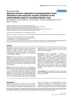
was analyzed for determination of HIV-1 infection. As
shown in Figure 1A b-galactosidase activity in raltegrav ir
treated cells with HIV-1
SX
infection was not seen, similar
to control condition (non-infected/non-treated cells).
However, there was a 6-fold increase in HIV-1
SX
infected
cells without raltegravir treatment. In Figure 1B, HIV-1
integration was shown to be significantly different
between infected cells with and without raltegravir trea t-
ment (P < 0.01), indicating detection of integration in
HIV-infected cells in the absence of raltegravir treatment.
Figure 1C shows that raltegravir-treated cells prevent
integration and is the only treatment causing formation
of 2LTR circles. This also confirms specificity for HIV-1
integration because non-integrated HIV-1 cDNA is not
amplified by these real-time PCR probes. Three indepen-
dent experiments were performed, and the data were
consistent each time, proving to be a reproducible and
reliable method for detection of integration in U373 cells.
HIV-1 Integration in human PBL
In order to assess integration in primary cell subsets,
PBL were isolated from human blood, and infected with
dual-tropic virus strain HIV-1
89.6
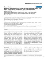
.AsshowninFigure
2A, virus production (HIV-1 p24 measured by ELISA)
in infected PBL was significantly lower (more than 7-
fold) with ralte gravir treatment compared to those with-
out raltegravir (P < 0.01). The integration data (Figure
2B) was highly consistent with p24 data, show ing HIV-
1
89.6
integration as significantly higher (more than 6-
fold) in in fected cells without ralte gravir compared to
raltegravir-treated cells (P < 0.01). In addition, Figure
2C shows that raltegra vir treatment does increase 2LTR
circle formation. These data are representative of six
experiments in PBL.
Figure 1 Quantitation of HIV-1 integration and 2-LTR circle
formation in CD4+ U373 cells. U373-MAGI-CCR5 cells were plated
in 6-well plates with or without raltegravir treatment 24 h prior to
infection and during infection (MOI = 0.1). Two days after infection,
(A) b-galactosidase activity (expressed as RLU = Relative Light Units)
was analyzed for determination of HIV-1
SX
infection; (B) cellular
genomic DNA was extracted from U373 cells 48 h after infection
and HIV-1
SX
integration was detected using two-step quantitative
PCR, and (C) 2-LTR circle formation was measured by real-time PCR.
(**p < 0.01)
Friedrich et al. Virology Journal 2010, 7:354
/>Page 2 of 6
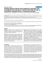
HIV-1 Integration in human macrophages
Human monocyte-derived macrophages were isolated
from human blood, and infected with HIV-1
SX
.As
shown in Figure 3A, virus production in infected macro-
phages was approximately 5-fold higher in cells without
raltegravir treatment as compared to those with ralte-
gravir treatment (P < 0.01). Similarly to other cell types,
macrophages treated with raltegravir show a significant
decrease in viral cDNA integration in to the genome
when compared with the cells without raltegravir treat-
ment (P < 0.01), as shown in Figure 3B. Figure 3C
shows that raltegravir-treatment increases 2LTR circle
formation. For all cell systems used in this study, there
was no cytotoxicity observed in raltegravir-treated cells
(data not shown).
Taken together, these resultssuggestthatthistwo-
step quantitative PCR method can be used effectively to
quantitate HIV-1 integration in primary human macro-
phages and PBL, as well as our CD4
+
U373 cell line.
Conclusions
We used the antiretroviral integrase inhibitor, raltegravir,
to distinguish between integrated and non-integrated
HIV-1 cDNA in infected primary PBL, macrophages, and
ahumanCD4
+
cell line. We detected HIV-1 integration
by utilizing a well-defined two-step quantitative PCR
method [19], which has proven to be a specific and sensi-
tive approach in different cell subsets based on our
reproducible results. In both raltegravir-treated and non-
treated cells, viral RNA is reverse transcribed into viral
cDNA and transported into the nucleus. In non-treated
cells, viral cDNA integrates into the host chromo some,
as detected by two-step real-time PCR; whereas in ralte-
gravir-treated cells, viral cDNA forms 2-LTR circles pre-
venting it from integrating into the host chromosome, as
shown by conventional real-time PCR. Yu et al. have
used this method to show that patients on HAART have
decreased levels of integrated HIV-1 proviral DNA as
compared to patients off HAART [22]. Thus, this method
may be considered for the routine analysis of HIV-1
DNA integration to evaluate t he integrati on efficiency of
retroviral vectors in different cell subsets.
Our study extends the previo us work performed by
others [18,19] to detect integration in primary human
cell subsets - PBL and macrophages using this two-step
PCR technique. This is important because macrophages
and PBL are crucial for HIV-1 infection, latency, and
persistence [4,5]. As such , we infect human macro-
phages or PBLs derived from six different healthy
donors, as we ll as inf ect a CD4+ cell line, and consis-
tently demonstrate similar results u sing two different
virus strains. By using these primary cell subsets, we
show that this method can be useful in precisely moni-
toring the level of integration in laborato ry settings and
perhaps in HIV-infected patients to conclusively deter-
mine if it is affected by specific antiretroviral therapy.
Thus, by using raltegravir as a c ontrol, we demonstrate
that two-step PCR is specific in detecting only inte-
grated HIV-1 cDNA and not other HIV-1 cDNA in the
nucleus or cell. Additionally, we utilized lab-adapted R5-
and dual-tropic strains of HIV-1 instead of pseudovirus
to more closely mimic natural infection. Furthermore,
this approach could reveal if HIV-1 integration persists
within specific cellular subsets in patients on highly
active antiretroviral therapy (HAART).
Figure 2 Quantitation of HIV-1 integration and 2-LTR circle
formation in human PBL. PBL were plated in 6-well plates with or
without raltegravir treatment 24 h prior to infection with HIV-1
89.6
,
during infection and 48 h after infection (MOI = 0.1). (A) Seven days
after infection, supernatant was assessed for p24 level of each
group by p24 capture ELISA; (B) Six days after infection, cellular
genomic DNA was extracted from PBLs and HIV-1 integration was
measured by two-step quantitative PCR, and (C) 2-LTR circle
formation was measured by real-time PCR. (**p < 0.01)
Figure 3 Quantitation of HIV-1 integration and 2-LTR circle
formation in human macrophages. Macrophages were plated in
6-well plates with or without raltegravir treatment 24 h prior to
infection, during infection and 48 h after infection (MOI = 0.1).
(A)Seven days after infection, supernatant was assessed for p24 level
of each group by p24 capture ELISA; (B) Six days after infection,
cellular genomic DNA was extracted from macrophages, and HIV-
1
SX
integration was measured by two-step quantitative PCR, and
(C) 2-LTR circle formation was measured by real-time PCR. (**p < 0.01)
Friedrich et al. Virology Journal 2010, 7:354
/>Page 3 of 6
Methods
U373 cells
U373-MAGI-CCR5 cells (contributed by Drs. Michael
Emerman and Adam Geballe), are modified U373 glio-
blastoma cells that are used for HIV infection experi-
ments. U373-MAGI-CCR5 cells express b-galactosidase
under the control of HIV LTR, which is trans-activated
by HIV Tat protein in relation to the level of virus repli-
cation [23,24]. In addition, these cells express CD4 and
human chemokine receptor CCR5 on its surface, which
allow infection by primary HIV R5 strains [24]. U373
cells were propagated in 90% DMEM supplemented
with 10% fetal bovine serum, 0.2 mg/ml G418, 0.1 mg/
ml hygromycin B, and 1.0 μg/ml puromycin. For infec-
tion experiments, U373 cells were maintained in 90%
DMEM, 10% fetal bovine serum, a nd 1% penicillin/
streptomycin.
Preparation of human PBL
PBL were isolated from PBMC obtained from six differ-
ent healthy human buffy coats prepared by the Univer-
sity of Texas Medical Branch (UTMB) Blood Bank in
Galveston, TX. After the initial 24 h incubation of
PBMC on 10 cm petri dishes, supernatant (containing
PBL) was transferred to 50 ml tube and cells were iso-
lated by centrifugation. Cells then were resuspended in
stimulation media (RPMI 1640 m edia with 20% Fetal
calf serum (FCS); 1% Penicillin/Streptomycin; 5 μg/ml
phytohemagglutinin) and incubated at 37°C with 5%
CO
2
for 72 h. PBL were then collected by centrifugation
and resuspended in growth media (RPMI 1640 with 1%
L-glutamine; 1% Penicillin/Streptomycin; 20% FCS; 20
units/ml IL-2).
Preparation of human macrophages
Primary human macrophages were purified from healthy
human PBMC (from the same six blood donors as
human PBL isolation) by adherence to plastic tissue cul-
ture dishes as described previously [25]. Briefly, PBMC
were purified by Ficoll-Hypaque centrifugation from
buffy coats of healthy HIV-negative blood donors pre-
pared by the UTMB Blood Bank. Primary monocyte-
derived macrophages were obtained by adherence for 7
days to plastic petri dishes initially coated with human
AB serum [26]. During differentiation, macrophages
were cultured in Iscove’s modified Dulbecco’smedium
supplemented with 20% FCS; 1% L-glutamine and 1%
Penicillin/Streptomycin.
Viruses and infection
HIV-1
SX
, which is a chimeric M-tropic virus (R5)
encoding the majority of the HIV-1
JRFL
envelope protein
in an HIV-1
NL4-3
backbone, and dual-tropic (R5/X4)
HIV-1
89.6
, which is a HIV-1 laboratory adapted strain
originally isolated from infected individuals, were pur-
chased from the Vir ology Core Facility, Center for AIDS
Research at Baylor College of Medicine, Houston, TX.
HIV-1
SX
stock containing 69.681 ng/ml of HIV p24
with 65,325 TCID50/ml was used to infect macropha ges
and U373 cell s. HIV-1
89.6
stock containing 49.977 ng/ml
of HIV p2 4 with 261,300 TCID50/ml was us ed to infect
PBL. HIV-1 stocks were titrated, and for all experi-
ments, the inoculum was 7 ng of p24 per 1.5 × 10
5
cells
(MOI 0.1). Raltegravir (Merck & Co., Inc., Whitehouse
Station, NJ) is a well-characterized, FDA-approved HIV-
1 integrase inhibitor. It had been previously tested in
our lab and showed no visual cytopathic effects or any
cytotoxicity at 20 μM (data not shown). U373-MAGI
cells, primary macrophages, and PBL were plated in
6-well plates at 1.5 × 10
5
cells per well 24 h prior to
infection. Each of these cell subsets was plated into
three 6-well plates. In the first plate, cells were infected
with HIV-1 only; the second plate w as treated with
raltegravir (20 μM) 24 h prior to HIV-1 infection and
during infectio n; the third plate contained non-infected/
non-treated cells serving as a negative control. After 4 h
incubation of virus inoculum (0.5 ml/well) at 37°C, fresh
medium (1.5 ml) was added to each well. For macro-
phages and PBL, genomic DNA was extracted 6 days
post-infection using DNeasy Blood and Tissue Kit (QIA-
GEN, Alameda, CA) according to the manufacturer’ s
instructions. To assess infection, supernatant was
harvested for HIV p24 levels in each group by a p24
capture ELISA kit (Immuno Diagnostics, Inc, Woburn,
MA) according to the manufacturer’ s instructions.
Since the HIV replication kinetics are more rap id in
U373-MAGI cells than in primary macrophages and
PBL, genomic DNA was extracted from U373 cells 48 h
post-infection. To as sess infection of HIV-1SX in U373
cells, the cells were lyse d and analyzed for b-galactosi-
dase activity using the Beta-Glo Assay System (Promega,
Madison, WI) and a Dynex MLX Luminometer.
PCR
For the pre-amplification of genomic DNA from macro-
phages, PBL, and U373 cells the following primers were
used: Alu forward, 5’-GCC TCC CAA AGT GCT GGG
ATT ACA G-3’;andHIV-1gag reverse, 5’-GCT CTC
GCA CCC ATC TCT CTC C-3’ [18,19]. The PCR solu-
tion contained 1× TaqMan Universal Ma ster Mix, No
AmpErase UNG (Applied Biosystems, Carlsbad, CA),
100 nM Alu forward primer, and 600 nM gag reverse
primer, and 5 μl of DNA for every 15 μlofPCRsolu-
tion. The Thermocycler (Applied Biosystems GeneAmp
PCR system 2700) was programmed to perform a 2 min
hot start at 94°C, followed by 30 steps of denaturation
Friedrich et al. Virology Journal 2010, 7:354
/>Page 4 of 6
at 93°C for 30 seconds, annealing at 50°C for 1 minute,
and extension at 70°C for 1 minute 40 seconds.
Quantitative real-time PCR
For quantitation of HIV-1 integration, a second round
real-time quantitative PCR was performed using 7 μlof
the material from the pre-amplification step. These sam-
ples were run along with known dilutions of HIV-1
SX
plas mid cDNA used for a standard curve. This standard
curve was used to quantify the amplified DNA. The
sequences of the primers used are as follows: LTR for-
ward, 5’-GC C TCA ATA AAG CTT GCC TTG A-3’;
and LTR reverse, 5’-TCC ACA CTG ACT AAA AGG
GTC TGA-3’ [19]. The LTR molecular beacon probe,
labeled on the 5’ terminus with the reporter fluorophore
6-carboxyfluorescein (FAM) and on its 3’ terminus with
Black Hole Quencher 1 (DBH1), had the following
sequence: 5’ -FAM-GCG AGT GCC CGT CTG TTG
TGT GAC TCT GGT AAC TAG CTC GC-DBH1-3 ’
[19]. For quantitation of HIV-1 2-LTR circles, small
non-genomic DNA was isolated from cells using a Qia-
gen Miniprep kit. To identify 2-LTR circle formation,
primers MH535 (5’-AAC TAG GGA ACC CAC TGC
TTA AG-3’ )andMH536(5’ -TCC ACA GAT CAA
GGA TAT CTT GTC-3’)wereusedwiththeMH603
probe (5 ’ -(FAM)-ACA CTA CTT GAA GCA CTC AAG
GCA AGC TTT-(TAMRA)-3’) [27]. All reactions were
performed in a volume of 20 μl containing 1× TaqMan
Universal Master Mix, No AmpErase UNG, and 200 nM
of forward primer, revers e primer, and molecular probe.
All reactions were performed using Applied Biosystems
TaqMan Universal Master Mix and run using an
Applied Biosystems 7500 Fast Real-time PCR system
and 7500 Fast System Software. The thermal program
started with 2 min at 50°C, fo llowed by a 10 minute hot
start at 95°C. This was followed by 40 cycles of 95°C for
15 seconds and 60°C for 60 seconds. GAPDH was used
as an internal control to normalize total DNA.
Statistical analysis
To evaluate the sensitivity and specificity of this method,
we detected the quantity of integration in three different
cells, and compared them by student’s t-test to deter-
mine differences between raltegravir treated groups and
virus only infection groups. P < 0.05 was considered as
significant difference.
Acknowledgements
This work was supported by Public Health Service grant HL088999 from the
National Heart, Lung, and Blood Institute. We thank the NIH AIDS Research
and Reference Reagent Program for providing the U373-MAGI-CCR5 cells.
We thank Edward Siwak, Ph.D., Associate Director of Virology Core Facility,
Center for AIDS Research at Baylor College of Medicine, Houston, TX for
providing HIV-1SX and HIV-1
89.6
. Also, we greatly appreciate Merck & CO.,
Inc. for generously providing raltegravir used in our studies; Dr. Michael
Miller for experimental advice; Dr. William A. O’Brien for his excellent
editorial suggestions.
Authors’ contributions
BF and GL performed all experiments and drafted the manuscript. ND
participated in the design of the study and contributed to drafting the
manuscript. MRF conceived of the study, and participated in its design and
coordination and helped to draft the manuscript. All authors read and
approved the final manuscript.
Competing interests
The authors declare that they have no competing interests.
Received: 8 October 2010 Accepted: 3 December 2010
Published: 3 December 2010
References
1. von Lindern JJ, Rojo D, Grovit-Ferbas K, Yeramian C, Deng C, Herbein G,
Ferguson MR, Pappas TC, Decker JM, Singh A, et al: Potential role for CD63
in CCR5-mediated human immunodeficiency virus type 1 infection of
macrophages. J Virol 2003, 77:3624-3633.
2. Zhang H, Dornadula G, Beumont M, Livornese L Jr, Van Uitert B, Henning K,
Pomerantz RJ: Human immunodeficiency virus type 1 in the semen of
men receiving highly active antiretroviral therapy. N Engl J Med 1998,
339:1803-1809.
3. Zhu T, Mo H, Wang N, Nam DS, Cao Y, Koup RA, Ho DD: Genotypic and
phenotypic characterization of HIV-1 patients with primary infection.
Science 1993, 261:1179-1181.
4. Gartner S, Markovits P, Markovitz DM, Kaplan MH, Gallo RC, Popovic M: The
role of mononuclear phagocytes in HTLV-III/LAV infection. Science 1986,
233:215-219.
5. Kuroda MJ: Macrophages: do they impact AIDS progression more than
CD4 T cells? J Leukoc Biol 87:569-573.
6. Engelman A, Englund G, Orenstein JM, Martin MA, Craigie R: Multiple
effects of mutations in human immunodeficiency virus type 1 integrase
on viral replication. J Virol 1995, 69 :2729-2736.
7. Englund G, Theodore TS, Freed EO, Engelman A, Martin MA: Integration is
required for productive infection of monocyte-derived macrophages by
human immunodeficiency virus type 1. J Virol 1995, 69:3216-3219.
8. LaFemina RL, Schneider CL, Robbins HL, Callahan PL, LeGrow K, Roth E,
Schleif WA, Emini EA: Requirement of active human immunodeficiency
virus type 1 integrase enzyme for productive infection of human T-
lymphoid cells. J Virol 1992, 66:7414-7419.
9. Sakai H, Kawamura M, Sakuragi J, Sakuragi S, Shibata R, Ishimoto A, Ono N,
Ueda S, Adachi A: Integration is essential for efficient gene expression of
human immunodeficiency virus type 1. J Virol 1993, 67:1169-1174.
10. Stevenson M, Stanwick TL, Dempsey MP, Lamonica CA: HIV-1 replication is
controlled at the level of T cell activation and proviral integration. Embo
J 1990, 9:1551-1560.
11. Bukrinsky M, Sharova N, Stevenson M: Human immunodeficiency virus
type 1 2-LTR circles reside in a nucleoprotein complex which is different
from the preintegration complex. J Virol 1993, 67:6863-6865.
12. Farnet CM, Haseltine WA: Circularization of human immunodeficiency
virus type 1 DNA in vitro. J Virol 1991, 65:6942-6952.
13. Brass AL, Dykxhoorn DM, Benita Y, Yan N, Engelman A, Xavier RJ,
Lieberman J, Elledge SJ: Identification of host proteins required for HIV
infection through a functional genomic screen. Science 2008, 319:921-926.
14. Konig R, Zhou Y, Elleder D, Diamond TL, Bonamy GM, Irelan JT, Chiang CY,
Tu BP, De Jesus PD, Lilley CE,
et al: Global analysis of host-pathogen
interactions that regulate early-stage HIV-1 replication. Cell 2008,
135:49-60.
15. Sonza S, Maerz A, Deacon N, Meanger J, Mills J, Crowe S: Human
immunodeficiency virus type 1 replication is blocked prior to reverse
transcription and integration in freshly isolated peripheral blood
monocytes. J Virol 1996, 70:3863-3869.
16. Vandegraaff N, Kumar R, Burrell CJ, Li P: Kinetics of human
immunodeficiency virus type 1 (HIV) DNA integration in acutely infected
cells as determined using a novel assay for detection of integrated HIV
DNA. J Virol 2001, 75:11253-11260.
17. Yamamoto N, Tanaka C, Wu Y, Chang MO, Inagaki Y, Saito Y, Naito T,
Ogasawara H, Sekigawa I, Hayashida Y: Analysis of human
Friedrich et al. Virology Journal 2010, 7:354
/>Page 5 of 6
immunodeficiency virus type 1 integration by using a specific, sensitive
and quantitative assay based on real-time polymerase chain reaction.
Virus Genes 2006, 32:105-113.
18. Liszewski MK, Yu JJ, O’Doherty U: Detecting HIV-1 integration by
repetitive-sampling Alu-gag PCR. Methods 2009, 47:254-260.
19. O’Doherty U, Swiggard WJ, Jeyakumar D, McGain D, Malim MH: A sensitive,
quantitative assay for human immunodeficiency virus type 1 integration.
J Virol 2002, 76:10942-10950.
20. Murray JM, Emery S, Kelleher AD, Law M, Chen J, Hazuda DJ, Nguyen BY,
Teppler H, Cooper DA: Antiretroviral therapy with the integrase inhibitor
raltegravir alters decay kinetics of HIV, significantly reducing the second
phase. Aids 2007, 21:2315-2321.
21. Summa V, Petrocchi A, Bonelli F, Crescenzi B, Donghi M, Ferrara M, Fiore F,
Gardelli C, Gonzalez Paz O, Hazuda DJ, et al: Discovery of raltegravir, a
potent, selective orally bioavailable HIV-integrase inhibitor for the
treatment of HIV-AIDS infection. J Med Chem 2008, 51:5843-5855.
22. Yu JJ, Wu TL, Liszewski MK, Dai J, Swiggard WJ, Baytop C, Frank I, Levine BL,
Yang W, Theodosopoulos T, O’Doherty U: A more precise HIV integration
assay designed to detect small differences finds lower levels of
integrated DNA in HAART treated patients. Virology 2008, 379:78-86.
23. Harrington RD, Geballe AP: Cofactor requirement for human
immunodeficiency virus type 1 entry into a CD4-expressing human cell
line. J Virol 1993, 67:5939-5947.
24. Vodicka MA, Goh WC, Wu LI, Rogel ME, Bartz SR, Schweickart VL, Raport CJ,
Emerman M: Indicator cell lines for detection of primary strains of
human and simian immunodeficiency viruses. Virology 1997, 233:193-198.
25. O’Brien WA, Koyanagi Y, Namazie A, Zhao JQ, Diagne A, Idler K, Zack JA,
Chen IS: HIV-1 tropism for mononuclear phagocytes can be determined
by regions of gp120 outside the CD4-binding domain. Nature 1990,
348:69-73.
26. Rich EA, Chen IS, Zack JA, Leonard ML, O’Brien WA: Increased susceptibility
of differentiated mononuclear phagocytes to productive infection with
human immunodeficiency virus-1 (HIV-1). J Clin Invest 1992, 89:176-183.
27. Butler SL, Hansen MS, Bushman FD: A quantitative assay for HIV DNA
integration in vivo. Nat Med 2001, 7:631-634.
doi:10.1186/1743-422X-7-354
Cite this article as: Friedrich et al.: Quantitative PCR used to Assess HIV-
1 Integration and 2-LTR Circle Formation in Human Macrophages,
Peripheral Blood Lymphocytes and a CD4+ Cell Line. Virology Journal
2010 7:354.
Submit your next manuscript to BioMed Central
and take full advantage of:
• Convenient online submission
• Thorough peer review
• No space constraints or color figure charges
• Immediate publication on acceptance
• Inclusion in PubMed, CAS, Scopus and Google Scholar
• Research which is freely available for redistribution
Submit your manuscript at
www.biomedcentral.com/submit
Friedrich et al. Virology Journal 2010, 7:354
/>Page 6 of 6