báo cáo khoa học: " A simple and high-sensitivity method for analysis of ubiquitination and polyubiquitination based on wheat cell-free protein synthesis" potx
Bạn đang xem bản rút gọn của tài liệu. Xem và tải ngay bản đầy đủ của tài liệu tại đây (1.1 MB, 11 trang )
BioMed Central
Page 1 of 11
(page number not for citation purposes)
BMC Plant Biology
Open Access
Methodology article
A simple and high-sensitivity method for analysis of ubiquitination
and polyubiquitination based on wheat cell-free protein synthesis
Hirotaka Takahashi
1,2
, Akira Nozawa
1,2,5
, Motoaki Seki
3
, Kazuo Shinozaki
4
,
Yaeta Endo*
1,2,5
and Tatsuya Sawasaki*
1,2,5
Address:
1
Cell-Free Science and Technology Research Center, Ehime University, Matsuyama 790-8577, Japan,
2
The Venture Business laboratory,
Ehime University, Matsuyama 790-8577, Japan,
3
Plant Functional Genomics Research Group, 1-7-22 Suehiro-cho, Tsurumi-ku, Yokohama,
Kanagawa 230-0045, Japan,
4
Gene Discovery Research Group, RIKEN Plant Science Center, 1-7-22 Suehiro-cho, Tsurumi-ku, Yokohama,
Kanagawa 230-0045, Japan and
5
RIKEN Genomic Sciences Center, 1-7-22 Suehiro-cho, Tsurumi-ku, Yokohama, Kanagawa 230-0045, Japan
Email: Hirotaka Takahashi - ; Akira Nozawa - ; ;
Kazuo Shinozaki - ; Yaeta Endo* - ; Tatsuya Sawasaki* -
* Corresponding authors
Abstract
Background: Ubiquitination is mediated by the sequential action of at least three enzymes: the E1
(ubiquitin-activating enzyme), E2 (ubiquitin-conjugating enzyme) and E3 (ubiquitin ligase) proteins.
Polyubiquitination of target proteins is also implicated in several critical cellular processes.
Although Arabidopsis genome research has estimated more than 1,300 proteins involved in
ubiquitination, little is known about the biochemical functions of these proteins. Here we
demonstrate a novel, simple and high-sensitive method for in vitro analysis of ubiquitination and
polyubiquitination based on wheat cell-free protein synthesis and luminescent detection.
Results: Using wheat cell-free synthesis, 11 E3 proteins from Arabidopsis full-length cDNA
templates were produced. These proteins were analyzed either in the translation mixture or
purified recombinant protein from the translation mixture. In our luminescent method using FLAG-
or His-tagged and biotinylated ubiquitins, the polyubiquitin chain on AtUBC22, UPL5 and UPL7
(HECT) and CIP8 (RING) was detected. Also, binding of ubiquitin to these proteins was detected
using biotinylated ubiquitin and FLAG-tagged recombinant protein. Furthermore, screening of the
RING 6 subgroup demonstrated that At1g55530 was capable of polyubiquitin chain formation like
CIP8. Interestingly, these ubiquitinations were carried out without the addition of exogenous E1
and/or E2 proteins, indicating that these enzymes were endogenous to the wheat cell-free system.
The amount of polyubiquitinated proteins in the crude translation reaction mixture was unaffected
by treatment with MG132, suggesting that our system does not contain 26S proteasome-
dependent protein degradation activity.
Conclusion: In this study, we developed a simple wheat cell-free based luminescence method that
could be a powerful tool for comprehensive ubiquitination analysis.
Published: 6 April 2009
BMC Plant Biology 2009, 9:39 doi:10.1186/1471-2229-9-39
Received: 26 December 2008
Accepted: 6 April 2009
This article is available from: />© 2009 Takahashi et al; licensee BioMed Central Ltd.
This is an Open Access article distributed under the terms of the Creative Commons Attribution License ( />),
which permits unrestricted use, distribution, and reproduction in any medium, provided the original work is properly cited.
BMC Plant Biology 2009, 9:39 />Page 2 of 11
(page number not for citation purposes)
Background
Protein ubiquitination plays a crucial role in numerous
cellular processes such as cell growth, regulation of diverse
signal transduction and disease [1-3]. The covalent attach-
ment of ubiquitin to protein substrates requires a step-
wise cascade of enzymatic reactions. First, ubiquitin is
activated by E1 (ubiquitin-activating enzyme, UBA) in an
ATP-dependent manner by forming a high-energy
thioester-bond between the carboxyl-terminal glycine res-
idue of ubiquitin and a cysteine residue of E1. The acti-
vated ubiquitin is then transferred to the core-cysteine
residue of E2 (ubiquitin-conjugating enzyme, UBC).
Together with an E3 ligase enzyme, ubiquitin is attached
via its carboxyl-terminus to an e-amino group of a lysine
residue in the target protein. Since E3 binds to both E2
and the target protein, and acts as scaffold between E2 and
the substrate protein, the E3 ligase is the major determi-
nant for selecting target proteins for ubiquitination. There
is large number of genes encoding E3 ligases in all eukary-
otes, and the diversity of E3s is thought to contribute to
the substrate specificity of numerous target proteins. E3
ligases are structurally divided into three groups: HECT,
RING and U-box [4]. The HECT-type E3 ligase is distinct
from the other two ligases in that it forms a thioester-
bond with ubiquitin prior to the transfer of ubiquitin to
target proteins. The RING-type E3 ligase contains a unique
domain similar to the zinc finger motif that mediates pro-
tein-protein interactions [5] and is further divided into
two classes: one that can function alone and another that
forms a complex with other E3 components [4].
Recent studies have shown that attachment of polyubiqui-
tin chains on target proteins linked via lysine-48 of ubiq-
uitin typically leads to degradation by the 26S proteasome
[6], whereas linkage via lysine-63 mediates different path-
ways such as internalization of membrane proteins, acti-
vation of signal transduction and DNA damage repair [7].
The formation of lysyl-63-linked polyubiquitin chains is
generated by specific combinations of E2s and E2 vari-
ants, which are similar to E2s except that they lack core
cysteine residues required for E2 activity [8,9]. In addi-
tion, ubiquitination of substrates without polymeriza-
tion, mono-ubiquitination, acts as a sorting signal for
protein endocytosis and as a regulation factor for diverse
proteins, including histones and transcription factors
[10].
In plant, genomic research of the model plant Arabidopsis
thaliana showed that there are two E1s, 37 E2s and more
than 1,300 predicted E3s [11]. Although little is known
about protein ubiquitination in plants compared with
yeast and mammals, recent studies revealed that the plant
ubiquitination pathway is involved in the regulation of
morphogenesis, the circadian clock and responding to
hormone or pathogen signal molecules [12-15]. Despite
the importance of ubiquitination in plants, much of the
plant ubiquitination cascade is still unknown because of
its complexity and the issues inherent to the use of Arabi-
dopsis plants for biochemical analysis. Although several
interactions between E2s and RING type E3s have been
demonstrated in vitro using recombinant proteins
expressed in Escherichia coli, these efforts are hampered by
the inability to obtain functional protein using conven-
tional methods [16].
With this in mind, we sought to develop a novel in vitro
method to analyze the ubiquitin pathway genome-wide.
The two major obstacles hindering the development of an
in vitro assay for genome-wide screening are the difficulty
of efficiently producing recombinant protein and the ina-
bility to detect ubiquitination in a high-throughput fash-
ion. To address the first problem we used the wheat cell-
free protein synthesis system, which has been previously
reported to produce a wide range of functional Arabidop-
sis and human proteins [17-19]. Moreover, a collection of
RIKEN Arabidopsis Full Length (RAFL) cDNA clones cov-
ering about 70% of Arabidopsis genes is available [20].
Using these RAFL clones as templates, recombinant pro-
teins involved in the ubiquitination pathway were
expressed in the wheat cell-free system and used for sev-
eral functional analyses. For screening, conventional
detection methods such as immunoblot analysis or radio-
isotope-labeled proteins are not suitable for the detection
of a large number of ubiquitination reactions. Recently, a
high-throughput luminescence method to detect protein
ubiquitination was reported [21], however this method
requires purified protein and creation of specialized vec-
tors to produce proteins. In this study, a novel in vitro
assay to detect polyubiquitin chain formation was devel-
oped using wheat cell-free synthesis and a modified lumi-
nescence-based detection method. We demonstrate (1)
creation of a simple in vitro method to detect polyubiqui-
tination using crude recombinant E3s, (2) discovery of the
activity of At1g55530 by screening a RING subgroup in
the reported assay, and (3) the polyubiquitination assay
in the presence of MG132 demonstrated the absence of
26S proteasome-dependent protein degradation activity
in wheat cell-free system.
Results
Detection of Polyubiquitin Chains on AtUBC22 E2 enzyme
Recently, AtUBC22 (At5g05080) E2 protein has been
shown to catalyze polyubiquitin chain formation without
an E3 ligase, although AtUBC35 (At1g78870) E3-inde-
pendent polyubiquitination activity could not be detected
[16]. We employed AtUBC22 and AtUBC35 as model E2
proteins to develop a novel polyubiquitination assay. We
have also demonstrated that addition of biotin ligase
(BirA) and biotin to the wheat cell-free protein produc-
tion system yields a single biotinylation on a target pro-
BMC Plant Biology 2009, 9:39 />Page 3 of 11
(page number not for citation purposes)
tein containing a biotin ligation site [22]. Using this
method, biotinylated recombinant AtUBC22 and
AtUBC35 were synthesized and, without purification
from the translation mixture, the polyubiquitination reac-
tion was performed on the crude recombinant protein.
After the reaction, biotinylated AtUBC22 and AtUBC35
were purified using streptavidin-conjugated magnetic
beads and the polyubiquitin chain was detected by immu-
noblot analysis. As shown in Fig 1A, AtUBC22 showed
polyubiquitination, whereas AtUBC35 showed mainly
monoubiquitination. Interestingly, both E2s still had
activity in absence of exogenous E1 in polyubiquitin reac-
tion mixture (Fig. 1A, middle lanes), suggesting that
wheat cell-free system has high endogenous E1 activity.
While immunoblot analysis is an excellent detection
method, it is not suitable for high-throughput detection
of numerous polyubiquitination reactions. Initially, we
attempted to use luminescent analysis, based on the
AlphaScreen technology, to detect the polyubiquitination
activity of AtUBC22 and AtUBC35. In principle, if a poly-
ubiquitin chain is formed by FLAG-tagged and bioti-
nylated ubiquitins, it will bring into proximity the
streptavidin-coated donor bead (bound to biotin) and the
protein A-conjugated acceptor bead (bound to anti-FLAG
IgG), producing a luminescent signal (Fig. 1B). Consider-
ing that the wheat cell-free system has high endogenous
E1 activity (Fig. 1A), it may also have endogenous E2 and
E3 activity. In order to avoid formation of polyubiquitin
chains by an endogenous wheat germ ubiquitin pathway,
purified E2s were used in this assay. As shown in Fig 1C,
high luminescent signal was observed in the presence of
AtUBC22 in E1-dependent manner. In contrast, AtUBC35
showed low signal. The two luminescent signals were
approximately consistent with immunoblot data that
AtUBC22 and AtUBC35 have high and low polyubiquiti-
nation activities respectively, as demonstrated in Fig 1A.
These results indicate that the luminescent method can
detect polyubiquitin chain formation by using the two
types of ubiquitins.
Ubiquitination and Polyubiquitination Analyses of HECT-
TypeE3 Ligases
Polyubiquitination activity of E3 ligases activated by the
step-wise E1 to E3 cascade is well documented [3]. We
next attempted to reconstruct this cascade in vitro and to
detect the E3-formed polyubiquitin chains using our
luminescent method. Due to the size of HECT-type E3
ligases, ranging from 100 to 428 kDa in Arabidopsis, pro-
duction of active protein by traditional expression meth-
ods may not be easy and biochemical analysis using only
truncated recombinant protein has been carried out previ-
ously [23]. We attempted to produce full-length Arabi-
dopsis HECT-type E3 ligase proteins using the wheat cell-
free system and monitored ubiquitin-conjugation and
polyubiquitination by luminescence. Two genes that
encode Arabidopsis HECT-type E3 ligase, UPL5 and UPL7
[24], were analyzed in this study. We obtained UPL5 and
UPL7 cDNA from the RAFL library and produced FLAG-
tagged protein in the wheat cell-free system. Ubiquitina-
tion of FLAG-labeled UPLs (UPL-FLAGs) was investigated
by both the luminescent and immunoblot methods. The
successful production of the two recombinant HECT pro-
teins was observed by immunoblot analysis (Fig. 2A) and
used in the luminescence assay without purification. To
detect ubiquitination of the HECT proteins, UPL-FLAGs
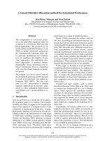
Detection of E3-independent polyubiquitination of AtUBC22 by luminescent analysisFigure 1
Detection of E3-independent polyubiquitination of
AtUBC22 by luminescent analysis. A, Polyubiquitin
chain on AtUBC22 but not on AtUBC35 was detected by
immunoblot analysis. In this assay, polyubiquitination reaction
was carried out with FLAG-tagged ubiquitin, and detected by
immunoblot analysis using anti-FLAG antibody. B, Schematic
diagram of detection of polyubiquitin chains by luminescent
analysis. Protein A-conjugated acceptor beads and streptavi-
din-coated donor beads are bound to anti-FLAG antibody
bound to FLAG-tagged ubiquitin and biotinylated E2, respec-
tively, and these two beads are in closed proximity when
polyubiquitin chain formed. Upon excitation 680 nm, a singlet
oxygen is generated from the donor beads, and then trans-
ferred to the acceptor beads within 200 nm, and the singlet
oxygen reacts the acceptor beads which in turn emits light at
520–620 nm. This light is measured by AlphaScreen kit and
change to signal value. C, Polyubiquitin chain on purified
recombinant E2 was detected by luminescent analysis in the
presence (E1 +) or absence (E1 -) of exogenous E1. Error
bars represent standard deviations from three independent
experiments.
BMC Plant Biology 2009, 9:39 />Page 4 of 11
(page number not for citation purposes)
and biotinylated ubiquitin were used. When biotinylated
ubiquitin is conjugated to the UPL-FLAG, a high lumines-
cent signal is obtained (Fig. 2B). As a result of the analysis,
ubiquitin-conjugation of UPL5 was observed (Fig. 2C). In
addition, polyubiquitin chains formed by UPLs were
detected with the luminescence assay using His-tagged
and biotinylated ubiquitin. To subtract polyubiquitin
chain formation from endogenous E2 and E3 in wheat
cell-free system, the assay was performed without recom-
binant UPL and only low signal was detected (Fig. 2D,
"UPL-" lane). As expected, luminescent signal was
observed in recombinant UPL5 and UPL7 (Fig. 2D).
Although the luminescent signal of UPL7 was lower than
that of UPL5, the signal was still two-fold higher than the
endogenous background signal. These results were con-
firmed by immunoblot analysis that showed distinct
mobility shifts of UPL5 (Fig. 2E) when detecting FLAG-
tagged UPLs, and polyubiquitin chain formation of UPL5
monitoring Alexa488-conjugated streptavidin (Fig. 2F).
Comparing the amount of polyubiquitin chain formation
in absence of UPLs (Fig. 2F, "UPL-" lane), UPL7 formed
weak but distinct polyubiquitin chains in presence of
AtUBC8. These luminescent signals were consistent with
immunoblot data. Interestingly, polyubiquitin chains
were formed by UPL5 without supplementing exogenous
E2 protein (Fig. 2D and 2F, "AtUBC8-" lane), suggesting
that wheat germ extract has endogenous E2 activity as well
as endogenous E1 activity. These data indicate that the
wheat cell-free production system is able to produce high
molecular weight proteins in functional forms and that
our luminescence method can detect activity of HECT-
type E3 ligases without purification. This is the first data
showing that full length recombinant HECT-type E3s have
ubiquitin-conjugating and polyubiquitination activity.
Taken together, the luminescent method based on the
wheat cell-free system could be useful for biochemical
analysis of HECT-type E3 ligases.
Detection of Polyubiquitin Chains by RING-Type CIP8 E3
Ligase
It is reported that at least 469 predicted RING-type E3
ligases are encoded in the Arabidopsis genome [25]. Like
the HECT-type E3, we attempted to express and carry out
the functional analysis of the RING-type E3 ligases. In this
study, we selected CIP8 as a model RING-type E3 ligase,
which is reported to possess a RING finger motif and have
typical features of an E3 ligase [26]. At first, polyubiquiti-
nation activity of purified CIP8 in presence or absence of
exogenous E1 and purified E2 (AtUBC8) was investigated
by luminescence. As shown in Fig 3A, luminescence anal-
ysis using His-tagged and biotinylated ubiquitin showed
the polyubiquitination of purified CIP8 only when exog-
enous E1 and purified E2 were added to the reaction mix-
ture. The CIP8-dependent polyubiquitination was
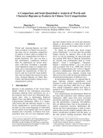
Analysis of recombinant Arabidopsis HECT-type E3 ligases (UPL7 and UPL5)Figure 2
Analysis of recombinant Arabidopsis HECT-type E3
ligases (UPL7 and UPL5). A, Production of FLAG-tagged
recombinant UPL proteins was detected by immunoblot
analysis. For analysis, 5 μl of crude recombinant UPL proteins
were loaded, and detected by immunoblot analysis using anti-
FLAG antibody. B, Schematic diagram of detection of ubiqui-
tin-conjugation of UPLs by luminescent analysis. Protein A-
conjugated acceptor beads and streptavidin-coated donor
beads were bound to anti-FLAG antibody bound to FLAG-
tagged recombinant UPLs and biotinylated ubiquitin, respec-
tively, and detected by same principle and procedure
described in Figure 1B. C, The ubiquitination of crude
recombinant UPL7 and UPL5 was detected by luminescent
analysis described in B. Bio-Ub means biotinylated ubiquitin.
D, polyubiquitination of crude recombinant UPL7 and UPL5
was detected by luminescent analysis with anti-His antibody.
Mix-Ub indicated the mixture of His-tagged and biotinylated
ubiquitin. E and F, Mobility shift of UPLs (E) and formation of
polyubiquitin chains (F) were detected by immunoblot using
anti-FLAG antibody and Alexa488-conjugated streptavidin,
respectively. The polyubiquitination reaction was done with
FLAG-tagged recombinant UPLs in presence or absence of
crude AtUBC8, and then recombinant UPLs were purified by
anti-FLAG antibody-conjugated agarose. Error bars repre-
sent standard deviations from three independent experi-
ments.
BMC Plant Biology 2009, 9:39 />Page 5 of 11
(page number not for citation purposes)
confirmed by immunoblot analyses detecting both FLAG-
CIP8 and His-tagged ubiquitin (Fig. 3B). On the other
hand, luminescent analysis with crude CIP8 protein
showed high polyubiquitination activity both in the pres-
ence or absence of purified E2 (Fig. 3C), and was con-
firmed by immunoblot analysis with crude protein (Fig.
3D). These data indicated that, like recombinant UPL5,
crude CIP8 also utilized endogenous wheat extract E1 and
E2 proteins, and therefore we could carry out the simple
polyubiquitination analysis of E3 without addition of
exogenous E1 and E2 proteins. Furthermore, immunoblot
analysis detecting purified CIP8 (Fig. 3B) showed a mobil-
ity shift of FLAG-tagged CIP8 to higher molecular weights
due to ubiquitination, whereas the mobility of the E2 was
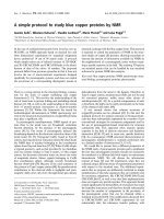
Detection of polyubiquitination and self-ubiquitination of CIP8Figure 3
Detection of polyubiquitination and self-ubiquitination of CIP8. A to D, The polyubiquitination assay was carried out
with purified (A and B) or crude recombinant CIP8 (C and D) and detected by luminescent analysis with anti-FLAG antibody (A
and C) and immunoblot analysis (B and D). His-Ub or Mix-Ub indicate His-tagged ubiquitin or the mixture of FLAG-tagged and
biotinylated ubiquitin, respectively. The polyubiquitination assay using luminescent analysis was carried out with recombinant
CIP8 without tag in the presence or absence of ubiquitin related components indicated below each graph. E, Ubiquitination of
crude recombinant CIP8 was observed by luminescent analysis with anti-FLAG antibody. The assay was carried out with or
without biotinylated ubiquitin and crude AtUBC8 recombinant protein. Bio-Ub means biotinylated ubiquitin. Error bars repre-
sent standard deviations from three independent experiments.
BMC Plant Biology 2009, 9:39 />Page 6 of 11
(page number not for citation purposes)
not altered (data not shown). This result indicates that the
CIP8-dependent polyubiquitin chains might be elongated
on CIP8 itself. This data is consistent with a recent report
showing that TRIM5a, a typical RING-type E3 ligase in
human, also undergoes self-ubiquitination, forming poly-
ubiquitin chains on itself [27]. To clarify whether the
mobility shift of CIP8 was concomitant with polyubiqui-
tin chain formation resulting from self-ubiquitination, we
tried to detect ubiquitination of CIP8 by the luminescent
method using crude FLAG-CIP8 protein and biotinylated
ubiquitin. The luminescent method clearly detected the
binding of biotinylated ubiquitin to FLAG-tagged CIP8
both in the presence and absence of exogenous E2 (Fig.
3E). Similar to polyubiquitin formation, the ubiquitina-
tion of CIP8 also occurred without the addition of exoge-
nous E2 protein (Fig. 3E, "AtUBC8-" lane). Taken
together, these data demonstrate that the luminescent
method could detect formation of RING-type CIP8-
dependent polyubiquitin chains and self-ubiquitination
of crude CIP8.
Screening of RING-Type E3 Ligases Having
Polyubiquitination Activity
Recent papers have reported that the polyubiquitin chain
is an important biological regulator. Identification of
activity and features of E3 ligases offers important infor-
mation about the ubiquitin-dependent regulation system.
Our luminescent method based on the wheat cell-free sys-
tem produced a simple and high-sensitivity detection of
CIP8-dependent polyubiquitin chains without any purifi-
cation (Fig. 3C). Using these tools, we screened new E3
ligases for the ability to form polyubiquitin chains like
CIP8.
The RING-type E3 ligases in Arabidopsis were divided into
30 subgroups based on domain structure, and CIP8 is cat-
egorized into subgroup 6 as it contains a coiled-coil
domain [25]. Eight other RING-type E3s from subgroup 6
were selected for screening, and the simple polyubiquiti-
nation assay was carried out with FLAG-tagged and bioti-
nylated ubiquitins, and the crude recombinant RING-type
E3s without addition of exogenous E1 and E2. The screen-
ing result showed significant polyubiquitination activity
of At1g55530, whereas other RING-E3 proteins were not
active (Fig. 4A). Immunoblot analysis of purified recom-
binant At1g55530 confirmed the polyubiquitination
activity and indicated that At1g55530 was self-ubiquiti-
nated (Fig. 4B). The polyubiquitination activity of
At1g55530 suggests that it may have a biological role for
proteasome-mediated degradation like CIP8 [26]. These
results show that the wheat cell-free protein expression
system and the luminescent ubiquitination detection
method could support functional high-throughput
screening of E3 proteins.
Analysis of the Wheat Cell-free Based Ubiquitination in
the Presence of Proteasome Inhibitor
It is known that some cell extracts, such as rabbit reticulo-
cyte or HeLa S-100 fraction, have 26S proteasome-
dependent proteolytic activity [28,29]. Based on the pres-
ence of endogenous E1 and E2 ubiquitination and polyu-
biquitination in the wheat cell-free system, it is expected
that the 26S proteasome activity will be very low (Fig. 2, 3
and 4). It was previously reported that the wheat germ
extract had little 26S proteasome-dependent protein deg-
radation activity [30]. Thus, we determined whether the
wheat cell-free system contains active 26S proteasome.
Using the crude recombinant proteins that formed polyu-
biquitin chains in this study, the polyubiquitination reac-
tion was carried out in presence or absence of MG132,
and accrual of the polyubiquitinated recombinant pro-
Screening of RING-type E3 ligases having polyubiquitination activityFigure 4
Screening of RING-type E3 ligases having polyubiqui-
tination activity. A, Polyubiquitination reaction of crude
recombinant E3 proteins was carried out with mixture of
FLAG-tagged and biotinylated ubiquitins, and investigated by
luminescent analysis with anti-FLAG antibody. B, Polyubiqui-
tination activity of At1g55530 was confirmed by immunoblot
analysis. The assay was carried out using purified recom-
binant AtUBC8 and At1g55530, and mobility shift of FLAG-
tagged At1g55530 and polymer of His-ubiquitin were
detected by immunoblot analysis using anti-FLAG and anti-
His antibodies, respectively. Error bars represent standard
deviations from three independent experiments.
BMC Plant Biology 2009, 9:39 />Page 7 of 11
(page number not for citation purposes)
teins and its polyubiquitin chain was estimated. As shown
in Fig 5, the amounts of UBC22, UPL5, UPL7 and
At1g55530 (Fig. 5A) and of its polyubiquitin chains (Fig.
5B) were hardly altered by MG132 treatment. This result
indicates that the proteolytic activity of the 26S proteas-
ome in the wheat cell-free system was below the detection
level. Thus, the wheat cell-free system could be suitable for
ubiquitination analysis.
Discussion
The ubiquitin signal is an important protein modification
in eukaryotes. Binding of a single ubiquitin to a target pro-
tein, mono-ubiquitination, is essential for membrane traf-
ficking, protein functions and protein-protein interaction
[7]. As for polyubiquitination, both Lys-48- and Lys-63-
linked polyubiquitin chains have been well characterized
in mammals and yeast. Lys-48 linked chains cause prote-
olysis of target proteins [6], and Lys-63 linked chains reg-
Effect of proteasome inhibitor on stability of polyubiquitinated proteinsFigure 5
Effect of proteasome inhibitor on stability of polyubiquitinated proteins. Polyubiquitination assays of crude FLAG-
tagged E2s and E3s were carried out in the presence or absence of biotinylated ubiquitin and 20 μM MG132. A, FLAG-tagged
recombinant proteins were detected by immunoblot analysis using anti-FLAG antibody. B, Polyubiquitination chain formed by
each recombinant protein was detected by Alexa488-conjugated streptavidin.
BMC Plant Biology 2009, 9:39 />Page 8 of 11
(page number not for citation purposes)
ulate signal transduction such as cellular localization of
protein or protein-protein interactions [7]. In mammals,
the multi-functional activities of NF-κB are regulated by
the Lys-63 linked chain [31]. In plants, the function of the
Lys-63 linked chain is still obscure. However, Arabidopsis
E2 and its variants promote formation of the Lys-63
linked chain [32], suggesting that the Lys-63 linked chain
in plant cells might also function similar to animal cells.
Hence, comprehensive analysis of the ubiquitin-related
plant proteins would open a door for elucidation of the
plant ubiquitin pathway. In this study, we developed a
simple and highly sensitive ubiquitination assay method
by combination of the wheat cell-free protein synthesis
system and luminescent detection. In general, in vivo pro-
tein production requires many time-consuming steps
such as vector construction, cell culture and purification
to obtain the recombinant protein. In contrast, this cell-
free based luminescence method could analyze a large
amount of ubiquitin reactions without these steps.
Using this method, we conveniently detected polyubiqui-
tin chain formation of E2 and E3s by using two tagged
ubiquitins (Fig. 1, 2, 3 and 4). The result of polyubiquiti-
nation analysis of the E2s obtained from luminescent-
based detection method was verified by immunoblot
analysis (Fig. 1). Our analysis also produced recombinant
protein of HECT-type E3 ligases without truncation and
detected their ubiquitin-conjugation and polyubiquitina-
tion activity by luminescent analysis (Fig. 2C and 2D).
The ubiquitin-conjugation of UPL5 was not observed
when a reductant was added to the reaction (data not
shown), suggesting that UPL5 formed a thioester bond
with ubiquitin. In addition, the model RING-type E3
CIP8 possessed high polyubiquitin formation activity
without substrate, consistent with what was reported pre-
viously [26]. Crude recombinant CIP8 formed polyubiq-
uitin chains in the absence of exogenous E1 and E2 (Fig.
3C and 3D), suggesting that the wheat cell-free system
might include enough endogenous E1 and E2 activity. It
was reported that wheat germ extracts have only a partial
ubiquitin pathway [30]. Although the process to isolate
wheat germ extract is different from the conventional
methods [33], this report strongly supports the existence
of endogenous ubiquitin pathway in our wheat cell-free
system. Indeed, luminescent analysis using crude recom-
binant protein showed slight polyubiquitin chain forma-
tion even in absence of recombinant E3 (Fig. 2D, Fig. 3C
and Fig. 4A, "E3-" lane), indicating that wheat cell-free
system might include not only E1 and E2, but E3s or other
factors that accelerates the polyubiquitin chain formation.
Further, quantitative immunoblot analysis using anti-
ubiquitin antibody showed that free ubiquitin was also
present in wheat germ extract at a concentration of at least
10 nM (data not shown). This is similar to the ubiquitin
concentration supplied in the in vitro assay. Although we
developed a convenient screening method to detect E3
activity in this study, removal of the endogenous ubiqui-
tin and ubiquitin related components such as E1, E2 and
E3, would yield a more sensitive assay. However, wheat
cell-free system does not have 26S proteasome proteolytic
activity (Fig. 5), indicating that using crude recombinant
protein is sufficient for in vitro ubiquitination assays.
By using this method, we found that a previously unchar-
acterized RING type E3, At1g55530, possessed high poly-
ubiquitination activity without exogenous E1 and E2
proteins (Fig. 4). This result suggested that the method
developed here is expected to find the activity of other
unknown E3 ligases such as At1g55530. Despite having
only 32% sequence similarity, the E3s CIP8 and
At1g55530 showed similar biochemical functions. Polyu-
biquitin chains formed by CIP8 and At1g55530 elongated
on themselves, while another report showed that polyu-
biquitin chains were formed on E2 before transferring
them to substrates [34]. This reflects that the pattern of
polyubiquitin chain formation differs between individual
E3s and that the detailed mechanisms are still unknown.
These studies suggest the importance of functional analy-
sis using active recombinant proteins. Although we devel-
oped a simple screen using crude recombinant E3s in
absence of exogenous E1 and E2 (Fig. 4), this method
could not detect the activity of some E3 ligases that were
unable to utilize endogenous ubiquitination components
in wheat cell-free system. The polyubiquitination activity
of At5g20910 recombinant protein, expressed in E. coli in
the presence of AtUBC8 [25], was not active in our in vitro
system (Fig. 4A), suggesting that in some cases exogenous
E2 and/or other components are necessary additions.
Such modifications to the ubiquitination assays detailed
here would help elucidate the biochemical features of E3s
(e.g., addition of recombinant E2s to reaction mixture
could give us further information about the E2–E3 specif-
icity, and of other E3 components would lead to the elu-
cidation of structure of complex type E3 ligase such as
SCF).
Conclusion
In this study, we found that the wheat cell-free system was
an excellent expression system to produce recombinant
protein efficiently and to carry out in vitro ubiquitination
assays without the interference of proteolytic activity.
Coupled with luminescent analysis, detection of these
ubiquitin reactions in the crude translation reaction mix-
ture was possible. Thus, this method should be helpful for
solving the complicated ubiquitin pathway in plant.
Methods
Construction of DNA Templates for Transcription
We used RAFL as templates. DNA templates of E2s and
E3s for transcription were constructed by "Split-Primer"
BMC Plant Biology 2009, 9:39 />Page 9 of 11
(page number not for citation purposes)
PCR as described previously [17]. Primers used in this
study are summarized in Additional file 1. The first round
of PCR was performed on each cDNA template using 10
nM of each of the following primers: a target protein spe-
cific primer (5'-CCACCCACCACCACCAatgnnnnnnnn
nnnnnnnn-3'; lowercase indicates the 5'-coding region of
the target gene) and the AODA2306 primer. Then, a sec-
ond round of PCR was carried out to construct the tem-
plates for protein synthesis using a portion (5 μl) of the
first PCR mix, 100 nM SPu primer, 100 nM AODA2303
primer and 1 nM deSP6E02 primer. GST tags were used
according to the methods we described previously [17].
The transcription templates of two HECT-type E3 ligases,
UPL7 and UPL5, were generated as C-terminal FLAG-
tagged proteins using the Gateway System
®
(Invitrogen,
Carlsbad, CA, USA). Briefly, the ORF sequences of UPL7
and UPL5 were amplified by PCR with sense and anti-
sense primers containing attB1 and FLAG-attB2
sequences, respectively. According to the manufacturer's
instructions (Invitrogen), these DNA fragments were sub-
cloned into pDONR221 vector by BP reaction and then
inserted into the Gateway-based pEU vector (pEU-E01-
GW) by LR reaction. Using these recombinant vectors as
templates, PCR was carried out with 100 nM SPu primer
and 100 nM AODA2303 primer and used as transcription
templates.
Cell-free Protein Synthesis
In vitro transcription and cell-free protein synthesis were
performed as described [18]. Transcript was made from
each of the DNA templates mentioned above using the
SP6 RNA polymerase. The synthetic mRNAs were then
precipitated with ethanol and collected by centrifugation
using a Hitachi R10H rotor. Each mRNA (usually 30–35
μg) was washed and transferred into a translation mixture.
The translation reaction was performed in the bilayer
mode [35] with slight modifications. The translation mix-
ture that formed the bottom layer consisted of 60 A260
units of the wheat germ extract (CellFree Sciences, Yoko-
hama, Japan) and 2 μg creatine kinase (Roche Diagnostics
K. K., Tokyo, Japan) in 25 μl of SUB-AMIX
®
(CellFree Sci-
ences). The SUB-AMIX
®
contained (final concentrations)
30 mM Hepes/KOH at pH 8.0, 1.2 mM ATP, 0.25 mM
GTP, 16 mM creatine phosphate, 4 mM DTT, 0.4 mM
spermidine, 0.3 mM each of the 20 amino acids, 2.7 mM
magnesium acetate, and 100 mM potassium acetate. SUB-
AMIX
®
(125 μl) was placed on the top of the translation
mixture, forming the upper layer. After incubation at
16°C for 15 h, the synthesized proteins were confirmed
by SDS-PAGE. For biotin labeling, 1 μl of crude biotin
ligase (BirA) produced by the wheat cell-free expression
system was added to the bottom layer, and 0.5 μM (final
concentration) of D-biotin (Nacalai Tesque, Inc., Kyoto,
Japan) was added to both upper and bottom layers, as
described previously [22].
Purification of E2 and E3 Proteins
Purification of GST-tagged protein was carried out accord-
ing to the procedure described previously [36] with slight
modification. Crude GST-tagged recombinant protein
(450 μl) produced by the cell-free reaction was precipi-
tated with glutathione sepharose™ 4B (GE Healthcare,
Buckinghamshire, UK). The recombinant proteins were
eluted with PBS buffer containing 0.1 U of AcTEV protease
(Invitrogen) in order to cleave the GST tag from the pro-
tein.
Detection of Polyubiquitination by the Luminescent
Method
In vitro polyubiquitination assays were carried out in a
total volume of 15 μl consisting of 20 mM Tris-HCl pH
7.5, 0.2 mM DTT, 5 mM MgCl
2
, (10 μM zinc acetate in the
assays for RING-type E3s only), 3 mM ATP, 1 mg/ml BSA,
25 nM biotinylated ubiquitin, 25 nM FLAG-tagged ubiq-
uitin, 1 μl of recombinant E2 (purified or crude) and 1 μl
of recombinant E3 (purified or crude) in the presence or
absence of 0.05 μM rabbit E1 (Boston Biochem, Cam-
bridge, MA, USA) at 30°C for 1 hr in a 384-well Optiplate
(PerkinElmer, Boston, MA, USA). In accordance with the
AlphaScreen IgG (ProteinA) detection kit (Perkin Elmer)
instruction manual, 10 μl of detection mixture containing
20 mM Tris-HCl pH 7.5, 0.2 mM DTT, 5 mM MgCl
2
, 5 μg/
ml Anti-FLAG antibody (Sigma-Aldrich, St. Louis, MO,
USA), 1 mg/ml BSA, 0.1 μl streptavidin-coated donor
beads and 0.1 μl anti-IgG acceptor beads were added to
each well of the 384 Optiplate followed by incubation at
23°C for 1 hr. Luminescence was analyzed by the AlphaS-
creen detection program.
Detection of Ubiquitinated E2 by Immunoblot Analysis
Crude biotinylated recombinant E2 proteins (40 μl) were
used for the ubiquitin-conjugating assay in a total reaction
volume of 50 μl containing 20 mM Tris-HCl pH 7.5, 0.2
mM DTT, 5 mM MgCl
2
, 3 mM ATP and 4 μM FLAG-tagged
ubiquitin (Sigma) for 3 hr at 30°C. The reaction products
were purified by Streptavidin Magnesphere Paramagnetics
particles (Promega, Madison, WI, USA). After washing the
beads with PBS buffer, recombinant E2s were boiled in 15
μl of SDS sample buffer containing 50 mM Tris-HCl pH
6.8, 2% SDS, 10% glycerol and 0.2% bromophenol blue,
and then separated from the magnet beads. The proteins
were separated by SDS-PAGE and transferred to PVDF
membrane (Millipore Bedford, MA, USA) according to
standard procedures. The blots were detected by the ECL
plus detection system (GE Healthcare) with anti-FLAG
antibody (Sigma) according to the manufacturer's proce-
dure.
Detection of Polyubiquitination by the Immunoblot
Analysis
For polyubiquitination of HECT-type E3 ligases, crude
FLAG-tagged UPL recombinant protein (20 μl) was ubiq-
BMC Plant Biology 2009, 9:39 />Page 10 of 11
(page number not for citation purposes)
uitinated in a total reaction volume of 50 μl consisting of
20 mM Tris-HCl pH 7.5, 0.2 mM DTT, 5 mM MgCl
2
, 3
mM ATP, 4 μM biotinylated ubiquitin and 20 μl of crude
recombinant AtUBC8 for 3 hr at 30°C. Then, recom-
binant UPL protein was gathered by anti-FLAG M2 agar-
ose (Sigma). After washing the agarose with PBS buffer,
the recombinant UPL protein was boiled in 15 μl of SDS
sample buffer and then separated from beads by centrifu-
gation. For polyubiquitination of RING-type E3 ligases,
the assay was carried out in 10 μl of reaction mixture con-
taining 20 mM Tris-HCl pH 7.5, 0.2 mM DTT, 5 mM
MgCl
2
, 10 μM zinc acetate, 3 mM ATP, 1 mg/ml BSA, 4 μM
FLAG- or His-tagged ubiquitin, 1 μl of purified or crude
recombinant E2 and 1 μl of purified or crude recombinant
E3 at 30°C for 3 hr. Then, 5 μl of three-fold concentrated
SDS sample buffer was added to the reaction mixture and
boiled for 5 min. Proteins were separated by SDS-PAGE
and transferred to Hybond-LFP PVDF membrane (GE
Healthcare) according to standard procedures. Immunob-
lot analysis was carried out with anti-FLAG antibody
(Sigma) or anti-His antibody (GE Healthcare) according
to the procedure described above. When detecting bioti-
nylated ubiquitin, blots were treated with 5 μg/ml
Alexa488-conjugated streptavidin (Invitrogen) in PBS
buffer. After washing with PBS containing 0.1% Tween-
20, the blot was analyzed by a Typhoon Imager (GE
Healthcare) using the 532 nm laser and 526 emission fil-
ters.
Polyubiquitination Assay with 26S Proteasome Inhibitor
Polyubiquitination reaction was carried out as same pro-
cedure described above except addition of MG132 (Calbi-
ochem, San Diego, CA, USA) at a final concentration of 20
μM to reaction mixture. Then, the protein on blot was
detected by immunoblot analysis with anti-FLAG anti-
body or Alexa488-conjugated streptavidin.
Authors' contributions
HT conceived the study and performed the experiments,
and contributed to writing the manuscript. MS and KS
provided RAFL cDNA clones. AN conceived the study. YE
conceived the study and supervised the work. TS con-
ceived and designed the study, supervised the work and
contributed to writing the manuscript.
Additional material
Acknowledgements
This work was partially supported by the Special Coordination Funds for
Promoting Science and Technology by the Ministry of Education, Culture,
Sports, Science and Technology, Japan (T. S. and Y. E.). We thank Michael
Andy Goren for proofreading this manuscript.
References
1. Bai C, Sen P, Hofmann K, Ma L, Goebl M, Harper JW, Elledge SJ:
SKP1 Connects Cell Cycle Regulators to the Ubiquitin Pro-
teolysis Machinery through a Novel Motif, the F-Box. Cell
1996, 86(2):263-274.
2. Chen Z, Hagler J, Palombella VJ, Melandri F, Scherer D, Ballard D,
Maniatis T: Signal-induced site-specific phosphorylation tar-
gets IκBα to the ubiquitin-proteasome pathway. Genes Dev
1995, 9(13):1586-1597.
3. Pickart CM: Mechanisms underlying ubiquitination. Annu Rev
Biochem 2001, 70:503-533.
4. Smalle J, Vierstra RD: The ubiquitin 26S proteasome proteo-
lytic pathway. Annu Rev Plant Biol 2004, 55:555-590.
5. Borden KL: RING domains: master builders of molecular scaf-
folds? J Mol Biol 2000, 295(5):1103-1112.
6. Glickman MH, Ciechanover A: The ubiquitin-proteasome prote-
olytic pathway: destruction for the sake of construction. Phys-
iol Rev 2002, 82(2):373-428.
7. Schnell JD, Hicke L: Non-traditional functions of ubiquitin and
ubiquitin-binding proteins. J Biol Chem 2003,
278(38):35857-35860.
8. Hofmann RM, Pickart CM: Noncanonical MMS2-encoded ubiq-
uitin-conjugating enzyme functions in assembly of novel
polyubiquitin chains for DNA repair. Cell 1999, 96(5):645-653.
9. Yin XJ, Volk S, Ljung K, Mehlmer N, Dolezal K, Ditengou F, Hanano
S, Davis SJ, Schmelzer E, Sandberg G, Teige M, Palme K, Pickart C,
Bachmair A: Ubiquitin lysine 63 chain forming ligases regulate
apical dominance in Arabidopsis. Plant Cell 2007,
19(6):1898-1911.
10. Hicke L: A new ticket for entry into budding vesicles – ubiqui-
tin. Cell 2001, 106(5):527-530.
11. Vierstra RD: The ubiquitin/26S proteasome pathway, the
complex last chapter in the life of many plant proteins.
Trends
Plant Sci 2003, 8(3):135-142.
12. Nelson DC, Lasswell J, Rogg LE, Cohen MA, Bartel B: FKF1, a
Clock-Controlled Gene that Regulates the Transition to
Flowering in Arabidopsis. Cell 2000, 101(3):331-340.
13. Osterlund MT, Hardtke CS, Wei N, Deng XW: Targeted destabi-
lization of HY5 during light-regulated development of Arabi-
dopsis. Nature 2000, 405(6785):462-466.
14. Stone SL, Williams LA, Farmer LM, Vierstra RD, Callis J: KEEP ON
GOING, a RING E3 ligase essential for Arabidopsis growth
and development, is involved in abscisic acid signaling. Plant
Cell 2006, 18(12):3415-3428.
15. Rosebrock TR, Zeng L, Brady JJ, Abramovitch RB, Xiao F, Martin GB:
A bacterial E3 ubiquitin ligase targets a host protein kinase
to disrupt plant immunity. Nature 2007, 448(7151):370-374.
16. Kraft E, Stone SL, Ma L, Su N, Gao Y, Lau OS, Deng XW, Callis J:
Genome analysis and functional characterization of the E2
and RING-type E3 ligase ubiquitination enzymes of Arabi-
dopsis. Plant Physiol 2005, 139(4):1597-1611.
17. Sawasaki T, Ogasawara T, Morishita R, Endo Y: A cell-free protein
synthesis system for high-throughput proteomics. Proc Natl
Acad Sci USA 2002, 99(23):14652-14657.
18. Sawasaki T, Gouda MD, Kawasaki T, Tsuboi T, Tozawa Y, Takai K,
Endo Y: The wheat germ cell-free expression system: meth-
ods for high-throughput materialization of genetic informa-
tion. Methods Mol Biol 2005, 310:131-144.
19. Kobayashi T, Kodani Y, Nozawa A, Endo Y, Sawasaki T: DNA-bind-
ing profiling of human hormone nuclear receptors via fluo-
rescence correlation spectroscopy in a cell-free system. FEBS
Lett 2008, 582(18):2737-2744.
20. Seki M, Narusaka M, Kamiya A, Ishida J, Satou M, Sakurai T, Nakajima
M, Enju A, Akiyama K, Oono Y, Muramatsu M, Hayashizaki Y, Kawai
J, Carninci P, Itoh M, Ishii Y, Arakawa T, Shibata K, Shinagawa A, Shi-
nozaki K: Functional annotation of a full-length Arabidopsis
cDNA collection.
Science 2002, 296(5565):141-145.
21. Kus B, Gajadhar A, Stanger K, Cho R, Sun W, Rouleau N, Lee T, Chan
D, Wolting C, Edwards A, Bosse R, Rotin D: A high throughput
Additional file 1
AGI code of Arabidopsis genes and primer sequences used in this
study.
AGI code of Arabidopsis genes and primer sequences used in this study.
Click here for file
[ />2229-9-39-S1.xls]
Publish with BioMed Central and every
scientist can read your work free of charge
"BioMed Central will be the most significant development for
disseminating the results of biomedical research in our lifetime."
Sir Paul Nurse, Cancer Research UK
Your research papers will be:
available free of charge to the entire biomedical community
peer reviewed and published immediately upon acceptance
cited in PubMed and archived on PubMed Central
yours — you keep the copyright
Submit your manuscript here:
/>BioMedcentral
BMC Plant Biology 2009, 9:39 />Page 11 of 11
(page number not for citation purposes)
screen to identify substrates for the ubiquitin ligase Rsp5. J
Biol Chem 2005, 280(33):29470-29478.
22. Sawasaki T, Kamura N, Matsunaga S, Saeki M, Tsuchimochi M, Moris-
hita R, Endo Y: Arabidopsis HY5 protein functions as a DNA-
binding tag for purification and functional immobilization of
proteins on agarose/DNA microplate. FEBS Lett 2008,
582(2):221-228.
23. Bates PW, Vierstra RD: UPL1 and 2, two 405 kDa ubiquitin-pro-
tein ligases from Arabidopsis thaliana related to the HECT-
domain protein family. Plant J 1999, 20(2):183-195.
24. Downes BP, Stupar RM, Gingerich DJ, Vierstra RD: The HECT
ubiquitin-protein ligase (UPL) family in Arabidopsis: UPL3
has a specific role in trichome development. Plant J 2003,
35(6):729-742.
25. Stone SL, Hauksdóttir H, Troy A, Herschleb J, Kraft E, Callis J: Func-
tional analysis of the RING-type ubiquitin ligase family of
Arabidopsis. Plant Physiol 2005, 137(1):13-30.
26. Hardtke CS, Okamoto H, Deng XW: Biochemical evidence for
ubiquitin ligase activity of the Arabidopsis COP1 interacting
protein 8 (CIP8). Plant J 2002, 30(4):385-394.
27. Yamauchi K, Wada K, Tanji K, Tanaka M, Kamitani T: Ubiquitina-
tion of E3 ubiquitin ligase TRIMa and its potential role. FEBS
J 2008, 275(7):1540-1555.
28. Waxman L, Fagan JM, Goldberg AL: Demonstration of two dis-
tinct high molecular weight proteases in rabbit reticulo-
cytes, one of which degrades ubiquitin conjugates. J Biol Chem
1987, 262(6):2451-2457.
29. Chen ZJ, Parent L, Maniatis T: Site-Specific Phosphorylation of
IκBα by a Novel Ubiquitination-Dependent Protein Kinase
Activity. Cell 1996, 84(6):853-862.
30. Hatfield PM, Vierstra RD: Ubiquitin-dependent proteolytic
pathway in wheatgerm: Isolation of multiple forms of ubiqui-
tin-activating enzyme, E1. Biochemistry 1989, 28:735-742.
31. Wu CJ, Conze DB, Li T, Srinivasula SM, Ashwell JD: Sensing of Lys
63-linked polyubiquitination by NEMO is a key event in NF-
κB activation. Nature Cell Biol 2006, 8(4):398-406.
32. Yin XJ, Volk S, Ljung K, Mehlmer N, Dolezal K, Ditengou F, Hanano
S, Davis SJ, Schmelzer E, Sandberg G, Teige M, Palme K, Pickart C,
Bachmaira A: Ubiquitin lysine 63 chain-forming ligases regu-
late apical dominance in Arabidopsis. Plant Cell 2007,
19(6):1898-1911.
33. Madin K, Sawasaki T, Ogasawara T, Endo Y: A highly efficient and
robust cell-free protein synthesis system prepared from
wheat embryos: Plants apparently contain a suicide system
directed at ribosomes. Proc Natl Acad Sci USA 2000,
97(2):559-564.
34. Li W, Tu D, Brunger AT, Ye Y: A ubiquitin ligase transfers pre-
formed polyubiquitin chains from a conjugating enzyme to a
substrate. Nature 2007, 446(7133):333-337.
35. Sawasaki T, Hasegawa Y, Tsuchimochi M, Kamura N, Ogasawara T,
Kuroita T, Endo Y: A bilayer cell-free protein synthesis system
for high-throughput screening of gene products. FEBS Lett
2002, 514(1):102-105.
36. Masaoka T, Nishi M, Ryo A, Endo Y, Sawasaki T: The wheat germ
cell-free based screening of protein substrates of calcium/
calmodulin-dependent protein kinase II delta. FEBS Lett 2008,
582(13):1795-1801.