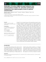
báo cáo khoa học: " Integration of tomato reproductive developmental landmarks and expression profiles, and the effect of SUN on fruit shape" pot
Bạn đang xem bản rút gọn của tài liệu. Xem và tải ngay bản đầy đủ của tài liệu tại đây (5.08 MB, 21 trang )
BioMed Central
Page 1 of 21
(page number not for citation purposes)
BMC Plant Biology
Open Access
Research article
Integration of tomato reproductive developmental landmarks and
expression profiles, and the effect of SUN on fruit shape
Han Xiao
†1
, Cheryll Radovich
†1
, Nicholas Welty
†1
, Jason Hsu
3
, Dongmei Li
3
,
Tea Meulia
2
and Esther van der Knaap*
1
Address:
1
Horticulture and Crop Science, The Ohio State University/OARDC, Wooster, OH 44691, USA,
2
Molecular and Cellular Imaging Center,
The Ohio State University/OARDC, Wooster, OH 44691, USA and
3
Department of Statistics, The Ohio State University, Columbus, OH 43210,
USA
Email: Han Xiao - ; Cheryll Radovich - ; Nicholas Welty - ;
Jason Hsu - ; Dongmei Li - ; Tea Meulia - ; Esther van der
Knaap* -
* Corresponding author †Equal contributors
Abstract
Background: Universally accepted landmark stages are necessary to highlight key events in plant
reproductive development and to facilitate comparisons among species. Domestication and
selection of tomato resulted in many varieties that differ in fruit shape and size. This diversity is
useful to unravel underlying molecular and developmental mechanisms that control organ
morphology and patterning. The tomato fruit shape gene SUN controls fruit elongation. The most
dramatic effect of SUN on fruit shape occurs after pollination and fertilization although a detailed
investigation into the timing of the fruit shape change as well as gene expression profiles during
critical developmental stages has not been conducted.
Results: We provide a description of floral and fruit development in a red-fruited closely related
wild relative of tomato, Solanum pimpinellifolium accession LA1589. We use established and propose
new floral and fruit landmarks to present a framework for tomato developmental studies. In
addition, gene expression profiles of three key stages in floral and fruit development are presented,
namely floral buds 10 days before anthesis (floral landmark 7), anthesis-stage flowers (floral
landmark 10 and fruit landmark 1), and 5 days post anthesis fruit (fruit landmark 3). To demonstrate
the utility of the landmarks, we characterize the tomato shape gene SUN in fruit development. SUN
controls fruit shape predominantly after fertilization and its effect reaches a maximum at 8 days
post-anthesis coinciding with fruit landmark 4 representing the globular embryo stage of seed
development. The expression profiles of the NILs that differ at sun show that only 34 genes were
differentially expressed and most of them at a less than 2-fold difference.
Conclusion: The landmarks for flower and fruit development in tomato were outlined and
integrated with the effect of SUN on fruit shape. Although we did not identify many genes
differentially expressed in the NILs that differ at the sun locus, higher or lower transcript levels for
many genes involved in phytohormone biosynthesis or signaling as well as organ identity and
patterning of tomato fruit were found between developmental time points.
Published: 7 May 2009
BMC Plant Biology 2009, 9:49 doi:10.1186/1471-2229-9-49
Received: 30 December 2008
Accepted: 7 May 2009
This article is available from: />© 2009 Xiao et al; licensee BioMed Central Ltd.
This is an Open Access article distributed under the terms of the Creative Commons Attribution License ( />),
which permits unrestricted use, distribution, and reproduction in any medium, provided the original work is properly cited.
BMC Plant Biology 2009, 9:49 />Page 2 of 21
(page number not for citation purposes)
Background
Plants display a diverse array of shapes, sizes and catego-
ries of fruit. Within the Solanaceae family fruit categories
range from capsules, drupes, pyrenes, berries, to several
other types of non-capsular dehiscent fruits [1]. Within
one species such as tomato (Solanum lycopersicum L.), fruit
morphology varies dramatically among cultivated acces-
sions. The dramatic diversity in tomato fruit shape and
size is due to domestication and continued selection for
its fruit characters [2,3]. Fruit formation starts with the
development of the floral meristem. Within the floral
meristem, the expression of organ identity genes gives rise
to the four whorls namely the sepals, petals, stamen and
gynoecium. The coordinate spatial and temporal expres-
sion of several classes of homeotic genes specifies the
identity of floral organs [4-7]. A class genes control sepal
identity, A and B class genes specify the identity of petals,
B and C genes define stamen identity, and C genes control
carpel identity. The E class genes act redundantly in spec-
ifying the identity of floral whorls in combinations with
the A, B and C genes [5-7].
After organ specification within the floral meristem, a
complex growth patterning is observed in the fourth floral
whorl comprising the gynoecium, which will become the
fruit after fertilization of the ovules. Along the apical-basal
axis, the developing tissue types of the gynoecium are the
stigma, style, ovary and gynophore, whereas along the
mediolateral axis of the ovary the valves or pericarp, sep-
tum or columella, placenta and ovules are formed. In fruit
such as that of Arabidopsis, the gynoecium also includes
two dehiscence-related tissues, replum and valve margin
[8,9]. Combined with the organ and tissue identity genes,
patterning is controlled by the expression of genes deter-
mining organ polarity [10]. A critical stage of fruit pattern-
ing occurs at fertilization which, when successful, results
in seed formation. Fruit of most species will abort if there
is none or limited fertilization and seed set. Phytohor-
mones, particularly auxin and gibberellins (GA), play crit-
ical roles in fruit set and early growth triggered by
pollination and fertilization. Auxin and GA can also
induce parthenocarpic fruits by triggering pollination-
independent fruit growth in several species including
tomato [11-15].
Descriptions of flower and fruit developmental stages
have been established for several species. The stages have
been used to interpret gene function, and to determine the
spatial and temporal expression of genes involved in
organ identity and patterning. In addition, detailed
descriptions of developmental stages are needed for com-
parative analyses to unravel genetic and molecular mech-
anisms that give rise to floral and fruit diversity. Ideally,
these stages should describe key developmental events
that are shared among flowering plant species, so that the
landmarks could be compared and queried across data-
bases using key morphological developmental features.
Buzgo et al (2004) compared three distant angiosperm
species and proposed ten floral landmark stages. These
landmarks comprise "inflorescence formation and flower
initiation", "sepal initiation", "petal initiation", "stamen
initiation", "carpel initiation", "microsporangia forma-
tion", "ovule initiation", "male meiosis", "female meio-
sis", and "anthesis" [16], which have been adopted in
studies of several other species [17,18]. However, key fruit
landmark stages that are applicable across species have
not been described to date. For example, whereas Arabi-
dopsis fruit development is described in eight stages,
tomato fruit development is described in four [19,20].
Phase I of tomato fruit development comprises ovary
development ending with fertilization. Phase II describes
early fruit growth following fertilization and spans cell
division and early embryo development. Phase III spans
cell expansion and embryo maturation. The final phase IV
is the ripening phase [19]. Both cell division and elonga-
tion occur concomitantly in the different parts of the
tomato fruit, thus these two phases are not well separated
during growth of the organ [21,22]. More importantly, the
stages described for Arabidopsis and tomato detail spe-
cies-specific events that are not applicable across species.
Therefore, the establishment of universally applied fruit
developmental landmarks would allow comparative anal-
ysis of data obtained from different species.
Tomato, classified as a berry fruit, represents an excellent
model for floral and fruit development and is used exten-
sively in comparative studies within the Solanaceae family
[2,3,19,23]. Whereas some information is known about
the regulation of organ identity and specification [24-29],
information about fruit patterning in Solanaceous species
is rather limited. Varieties that differ in fruit morphology
offer an important resource to further our understanding
on its patterning. Fruit size and shape of tomato are con-
trolled by major and minor QTL loci [2,3,30]. For some of
these major QTL, the underlying genes are known. SUN
and OVATE control fruit elongation and therefore affect
patterning along the apical-basal axis [31,32]. FW2.2 and
FAS control fruit mass via increases of the placenta area
and locule number, respectively, and thus affect pattern-
ing along the medio-lateral axis [33,34]. SUN encodes a
member of the IQD protein family [32]. The founding
member of the IQD protein family AtIQD1 is localized in
the nucleus and its overexpression leads to increases in
glucosinolate production in Arabidopsis [35]. The high
expression of SUN in tomato leads to elongated fruit,
whichis hypothesized to control increases in secondary
metabolites and/or hormone levels. In the near-isogenic
lines (NILs) that differ at SUN, the most significant fruit
shape changes occur after anthesis during fruit set [32].
However a detailed developmental time-course describing
BMC Plant Biology 2009, 9:49 />Page 3 of 21
(page number not for citation purposes)
fruit shape changes that would aid in understanding the
mechanism by which SUN acts has not been described.
Moreover, an evaluation of flower and fruit expression
profiles in the S. pimpinellifolium LA1589 background has
not been performed to date.
In this study, we adopt the floral landmarks established
previously [16], and also propose new landmarks of fruit
development that are applicable across angiosperm plant
species. These landmarks are superimposed onto the fruit
shape changes controlled by SUN and combined with
gene expression profiles of floral buds 10 days prior to
anthesis, anthesis-stage flowers and fruit 5 days post pol-
lination.
Results
We used S. pimpinellifolium accession LA1589 for the
tomato flower and fruit developmental studies due to its
indeterminate growth habit and the abundant number of
flowers and inflorescences throughout its life cycle. For
example, LA1589 carries on average 20 flowers per inflo-
rescence (Fig. 1A and 1B), whereas a typical cultivated
variety carries only 3 to 7 flowers per inflorescence [36]. In
addition, flower development is highly regular in the wild
relative LA1589 compared to most cultivated types [36].
To time the developmental stages of consecutive buds and
then fruits on an inflorescence, we recorded the time of
anthesis for each flower in a total of 83 inflorescences
investigated over four independent experiments. As
shown in Figure 1C, the second flower opened 70% of the
time one day after the first flower, 29% of the time on the
same day as the first flower, and 1% of the time two days
after the first flower and so on. In general, consecutive
flower opening occurred at one-day intervals 75% of the
time, until the 16th flower on a given inflorescence (Fig.
1C). Flower buds developed after the 16
th
on a given inflo-
rescence tended to open more irregularly and often at an
interval of 2-days or more. By inference, this result
implied that the first 16 floral meristems arose 75% of the
time in one-day interval from one another. Therefore, we
concluded that until the 16
th
flower on a given inflores-
cence, the developing flower and fruit respectively, are
staged at close to one-day intervals from one another.
Initiation of floral organ primordia
The first landmark represented inflorescence formation
and flower initiation (Table 1). The transition to flower-
ing and inflorescence formation in LA1589 has been
described previously [36]. Briefly, transition to flowering
commenced with the termination of the vegetative meris-
tem into an inflorescence meristem. Floral initiation
occurred through the apparent bifurcation of the inflores-
cence meristem resulting in bud number 1 (Fig. 2A and
2B). The flatter inflorescence meristem continued its inde-
terminate growth pattern, while the more domed meris-
tem developed into a flower (Fig. 2A and 2B). Following
flower initiation, the emergence of the sepal primordia
around the perimeter of the floral apex of bud number 2
marked the second landmark (Fig. 2A). The five tomato
sepals initiated in a helical pattern of 144° (Fig. 2C). The
sepals continued to grow and covered the floral meristem
approximately 4 days after floral initiation (Fig. 2D and
2E). At the time of sepal enclosure, petal primordia started
to arise, representing landmark 3. Following petal primor-
dia emergence, stamen primordia emerged in alternate
positions to the petals (Fig. 2F and 2G), at approximately
5 days after floral initiation, representing landmark 4.
Sepals and petals continued to elongate while carpel pri-
Characterization of the S. pimpinellifolium accession LA1589 inflorescenceFigure 1
Characterization of the S. pimpinellifolium accession
LA1589 inflorescence. (A) Series of consecutive floral
buds 7 (right) to 19 (left) days since floral bud initiation. (B)
Series of consecutive developing fruits on a given inflores-
cence. Note that two days after anthesis, the flower has
senesced. (C) Timing of consecutive flower opening in
LA1589 starting with the second oldest flower (2). The black
bar indicates the percentage of flowers that opened at one-
day time intervals at the position on the inflorescence listed
on the X-axis. The white bar indicates the percentage of
flowers that opened at two-day time intervals and the grey
bar indicates the percentage of flowers that opened within
the same day. Size bar represents 1 mm.
BMC Plant Biology 2009, 9:49 />Page 4 of 21
(page number not for citation purposes)
Table 1: Flower developmental landmarks.
Flower Development
Landmarks; Buzgo et
al. (2004)
Days after flower
initiation in tomato
Perianth organs Reproductive organs
Ovary and ovule
development
Stamen and pollen
development
(1) Inflorescence formation
and flower initiation
1 Flattened inflorescence
apex becomes dome-
shaped.
(2) Initiation of outermost
perianth organs
2 Emergence of sepal
primordia in a helical
pattern.
(3) Initiation of inner
perianth organs.
4 Simultaneous emergence of
petal primordia in
alternating positions to the
sepals. Sepals overlay the
floral meristem
(4) Stamen initiation 5 Sepals and petals elongate. Simultaneous initiation of
stamen primordia.
(5) Carpel initiation 6 Petals start curling over the
stamens
Carpel primordia arise.
7 Central column that will
form the locular cavities
arise.
Stamen filament start
developing and two anther
lobes become visible.
(6) Microsporangia
initiation
8 Central column continues
to elongate. Carpels fuse at
the apex of the ovary. Style
initiation. Initiation of
placental development.
Primary pariety cells
develop into endothecium,
middle layers and tapetum.
Sporogenous layers visible.
(7) Ovule initiation 9 Ovule primordia begin to
emergence from the
placenta.
The two lobes of the
anther and the locule are
distinguishable,
microsporocyte and tapetal
cells are distinguishable.
Binucleate tapetal cells.
(8) Male meiosis 10 Microsporogenesis.
Microsporocytes or
microspore mother cells
undergo meiosis I and II
and forming tetrads.
(9) Female meiosis 11 Megasporogenesis.
Megaspore mother cell
(meiocyte or
megasporocyte) is visible.
Meiosis I. The nucellus is
small resulting in a tenui-
nucellate ovule.
12 Petals grow to the top of
sepals
The single integument
begins to grow over the
nucellus resulting in
unitegmic ovules.
Callose wall surrounding
the tetrads degrades
releasing the microspores.
Tapetum starts
degenerating.
BMC Plant Biology 2009, 9:49 />Page 5 of 21
(page number not for citation purposes)
mordia began to emerge in the floral center (Fig. 2G),
marking landmark 5, which occurred approximately 6
days after floral bud initiation. The carpel walls or valves
continued to enlarge, while the central part comprising
the septum and the central column formed congenitally
with the carpel walls, revealing the formation of the two
locular cavities of wild type tomato ovary (Fig. 2H). The
carpel walls elongated slightly faster than the central col-
umn revealing the locular cavity prior to ovary enclosure
and initiation of the style, which occurred 8 days post bud
initiation (Fig. 2I and 2J).
Reproductive organ formation
Male reproductive development initiated with microspor-
angia development, which represented landmark 6, and
occurred approximately eight days after floral bud initia-
tion (Table 1 and Fig. 3A). The primary sporogenous lay-
ers were visible at this stage (Fig. 3A). Nine days after
floral bud formation, the tapetal cells were binucleate,
and the developing microsporocytes were also visible (Fig.
3B and 3C). At 10 days after floral bud initiation, micro-
sporocytes or pollen mother cells were undergoing meio-
sis (Fig. 3D), marking landmark 8. A callose wall
surrounded the four haploid nuclei of the tetrads (Fig.
3E). One day later, the callose walls began to degrade and
the microspores were being released (Fig. 3F). At 13 days
after floral bud initiation, the tapetum was degenerating;
and the microspores were single and encapsulated in a
thick wall (Fig. 3G and 3H). One day later, the micro-
spores became vacuolated (Fig. 3I) and underwent one
asymmetric mitosis. Fifteen days after floral bud initia-
tion, the microspores were bi-cellular (Fig. 3J) and a day
later, the generative and vegetative cells were clearly dis-
tinguishable within the developing pollen (Fig. 3K). At
day 17 after floral bud initiation, the generative cell dis-
played the characteristic crescent shaped nucleus (Fig. 3L
and 3M). The second mitosis of the generative cell did not
occur until after pollination.
Female reproductive development initiated with the
development of the ovules and represented landmark 7
(Fig. 4A). Approximately 9 days after floral bud initiation,
the style and the ovary were nearly equal in length, and
ovule primordia were emerging on the placental tissues
(Fig. 4A). Ovules were clearly visible one day later (Fig.
4B). Two days after ovule primordia initiation and 11
days after floral bud initiation, a single integument started
to envelope the single cell layered nucellus and the devel-
oping megasporocyte, resulting in a unitegmic tenui-
nucleate ovule representing landmark 9 (Fig. 4C). Appar-
ently the megasporocyte underwent the first meiotic divi-
sion at this stage (Fig. 4D). A day later, the single
integument at the base of the nucellus was clearly visible,
while the megasporocyte is undergoing the second mei-
otic division, representing the first stage of megagame-
togenesis (Fig. 4E). Fourteen days after floral bud
initiation, the integument enveloped the nucellus com-
pletely and the micropyle was well defined. The embryo
sac development was taking place as evidenced by concen-
trated dark staining at the micropyle end. The presence of
the megaspore at the chalaza end of the ovule indicated
the development of the egg apparatus (Fig. 4F).
Fertilization and fruit set
Anthesis or flower opening was the final floral landmark
as well as the first fruit landmark (Table 1 and 2). At the
time of anthesis, the anther lobes dehisced to release the
pollen, which after landing on the receptive stigma, ger-
minated. Pollen tubes had grown close to the base of the
style 6 hours after pollination, and reached the ovules
approximately 2 hours later (Fig. 5A and 5B). Ten to 12
hours after pollination, the pollen tubes had released their
content resulting in fertilization of the ovules (Fig. 5C)
and representing fruit development landmark 2 (Table 2).
Senescence of floral organs, namely petal, stamens and
style is associated with successful fertilization and was vis-
13 Petals emerge from the
sepals.
Micropyle development. Free microspores are being
incased in a thick
polysaccharide wall;
tapetum degenerated.
14 Onset of sepal opening Megagametogenesis and
development of the
embryo sac.
Microspores come
vacuolated, and begins
asymmetric mitosis
15 Bi-cellular pollen grain.
16 Ovule development nears
completion.
The vegetative cell and
generative cell are well
distinguishable
(10) Anthesis 19 Petal opening
The timing of the landmarks described by Buzgo et al (2004) in S. pimpinellifolium accession LA1589 floral development.
Table 1: Flower developmental landmarks. (Continued)
BMC Plant Biology 2009, 9:49 />Page 6 of 21
(page number not for citation purposes)
ible approximately two days after anthesis as shown in
Fig. 1B.
Development of the pericarp after pollination
Following fertilization, tomato fruit growth consists of
cell division and cell expansion [19]. We analyzed the
growth of the pericarp following pollination to establish
the timing of cell division and cell elongation in the devel-
oping LA1589 pericarp. Pericarp width doubled from
anthesis to 2 days post anthesis (dpa), and then further
doubled at 5 and 10 dpa, respectively (Fig. 6). Cell
number across the pericarp increased from 10 at anthesis
to 17 at 2 dpa, and reached the final number of 19–21 at
5 dpa (Fig. 6F), implying that cell division ended at or
before that time. Mesocarp cell expansion started as early
as 2 dpa (Fig. 6B). These results indicated that cell division
and expansion occurred concurrently in the pericarp of
the early developing fruit. Note the presence of the cuticle
layer and starch granules in the epicarp and mesocarp
respectively, of 10 dpa fruit (Fig. 6D).
Seed development
As indicated above, cell division overlapped with cell
elongation during the early stages of fruit development.
Moreover, the cell division stage was short, ending before
5 dpa in LA1589, whereas the cell elongation stage
spanned fruit development from 2 dpa until mature green
stage. Thus, these two fruit developmental stages, which
correspond to tomato development phases II and III, pro-
vided limited guides for referencing. To develop addi-
tional landmarks for the developmental stages of tomato
fruit growth, we analyzed morphological changes in
embryo development, which occur concomitantly with
fruit growth in most angiosperm plant species.
We propose the third fruit developmental landmark as the
stage of 4–16 celled embryo, which occurred approxi-
mately 4 dpa (Fig. 7A and 7B). The fourth landmark was
represented by the globular embryo stage at 6 to 10 dpa
(Fig. 7C and 7D). Heart shape embryo was the fifth land-
mark and occurred between 10 and 12 dpa (Fig. 7E and
7F) highlighting the beginning of cotyledon growth. The
13–16 dpa embryo was torpedo shape, marking the sixth
landmark (Fig. 7G and 7H). After the sixth landmark, the
cotyledons grew into a coil and reached the seventh land-
mark approximately at 20 dpa. At this stage, the embryo
approached its final size, but the seed was not yet viable
for germination (Fig. 7I and 7J). The eighth fruit develop-
mental landmark was reached when the seeds harvested
from the maturing fruit were viable for germination. Seed
were collected from maturing fruit starting at 26 dpa until
33 dpa. Up until 29 dpa, there was little or no seed germi-
nation (Fig. 8). However, at 30 dpa, the germination rate
Early flower developmental landmarksFigure 2
Early flower developmental landmarks. (A) Scanning
electron microscopy image of a young inflorescence with the
shoot meristem terminating into the inflorescence meristem,
and the sympodial shoot meristem initiating the youngest leaf
axil on the flank of the inflorescence, the youngest floral bud
1, and the second youngest bud 2 had also emerged from the
inflorescence meristem. (B) Light microscopy image of a sec-
tion from a young inflorescence showing the floral meristem,
the youngest bud 1 and the third youngest bud 3. (C) Scan-
ning electron microscopy images of a floral bud three days
after flower initiation with sepal primordia, and (D) four days
after floral initiation, with sepals enclosing over the floral
meristem. (E) Light microscopy images of a section across
two consecutive floral buds, three and four days after initia-
tion, and (F) a floral bud six days after floral initiation, with
petals and stamens emerging under the sepals. (G) Scanning
electron microscopy images of floral buds at six days after
floral initiation, with carpel primordia starting to emerge.
The sepals were removed to visualize the developing petal,
stamen and carpel. (H) Six days after floral initiation, with the
central column rising and displaying the formation of the two
locular cavities. (I) Seven days after floral initiation the carpel
walls continue to elongate with the central column lagging
behind. (J) Eight days after floral initiation, the ovary is closed
and style has initiated. 1, youngest bud; 2, second youngest
bud; 3, third youngest bud; 4, fourth youngest bud; IF, inflo-
rescence meristem; SU, sympodial unit. Size bar in all images
measure 50 μm.
BMC Plant Biology 2009, 9:49 />Page 7 of 21
(page number not for citation purposes)
increased dramatically thus reaching landmark eight. At
32 dpa, nearly 100% of the seeds germinated.
Fruit ripening
Tomato fruit ripening stages consist of mature green,
breaker and red ripe [19,23]. At the mature green stage,
ethylene treatment will result in a rapid reddening of the
fruit [23,37-39]. We measured ethylene sensitivity in half
of the harvested fruits while determining the germination
ability of the seed in the other half that were collected at
selected times (see above). Ethylene sensitivity was
achieved over a short period of up to two days, and coin-
cided with the time when the seed became viable for ger-
mination (Fig. 8). Forty percent of fruit had responded to
ethylene at 30 dpa when 43% of the seeds were viable for
germination. Fruit younger than 29 dpa did not respond
to ethylene treatment (Fig. 8). The ninth landmark is the
onset of fruit ripening, coinciding with the breaker stage
when color began to change at approximately 32 dpa. This
stage is followed by the tenth and final landmark of ripe
fruit.
Gene expression profiles of floral and fruit development
To obtain a global overview of gene expression in flower
and fruit, we compared the profiles between three critical
developmental time points. The first stage was young
flower buds at floral landmark 7, representing ovule initi-
ation (10 days pre-anthesis). The second stage was the
anthesis-stage, representing flower landmark 10 and fruit
landmark 1. The third and last stage was 5 dpa fruits, rep-
resenting the 4–16 cell embryo stage and fruit landmark
3. Differentially expressed genes were identified using the
resampling-based multiple testing method [40]. Without
the cutoff of fold-change applied, 2495 genes with
adjusted p < 0.01 were differentially expressed in at least
one of the three stages (see Additional file 1). Among
them, 1232 genes showed higher expression at anthesis,
Flower landmarks representing male reproductive develop-mentFigure 3
Flower landmarks representing male reproductive
development. (A) Eight days after initiation, the primary
sporogenous layers (arrowheads) have formed. (B) Nine days
after floral initiation, the microsporocytes (MS) were visible
in the sporogenous tissue as well as the tapetal cells (T). (C)
Tapetal cells are binucleated (arrowhead). (D) Microsporo-
cytes 10 days after floral initiation are undergoing meiotic
divisions marking landmark 8. (E) Tetrads are enclosed by
callose walls (arrowhead). (F) Release of microspores. (G –
H) The tapetum is degenerating and the microspores are
released 13 days after floral initiation. (I) The microspores
become vacuolated 14 days after floral initiation. (J) Bi-cellu-
lar microspores. (K) Generative and vegetative cells are visi-
ble in microspores. (L) Seventeen days after floral initiation,
the microspores show a crescent generative nucleus. (M)
Pollen at anthesis. Scale bar, 50 μm (A-I), 20 μm (J-M).
Floral landmarks representing female reproductive develop-mentFigure 4
Floral landmarks representing female reproductive
development. (A) Landmark 7 occurs nine days after floral
initiation. (B) Ten days after floral initiation, the developing
ovules become visible. (C) Eleven days after floral initiation,
the megaspore mother cell forms, marking female meiosis
and floral landmark 9. (D) Landmark 9 megaspore mother
cell showing the nuclei (orange color) and the tubulin (green
color). (E) Twelve days after floral initiation, the single integ-
ument has nearly covered the developing embryo sac. (F)
The developing ovule with a clear micropyle is visible 14 days
after floral initiation. Scale bar, 50 μm (A, B, F), 10 μm (C, D),
20 μm (E).
BMC Plant Biology 2009, 9:49 />Page 8 of 21
(page number not for citation purposes)
whereas 527 and 736 genes showed higher expression in
young flower buds and 5 dpa fruits, respectively (Table 3).
Functional classification of the differentially expressed
genes showed a distinct distribution of genes involved in
various biological processes for the three stages investi-
gated. For example, more genes involved in developmen-
tal processes were found in flower buds during ovule
initiation and anthesis-stage flowers than in 5 dpa fruit.
On the other hand, phytohormone-related genes were
predominantly found in anthesis-stage flowers and 5 dpa
fruits compared to flower buds (Table 3).
Expression of organ identity and patterning genes
Of the genes representing the developmental processes,
key floral and fruit patterning genes were examined for
their expression profiles during reproductive develop-
ment (see Additional file 2, Fig. 9). Genes orthologous or
homologous to the Arabidopsis ABCE genes required for
floral organ identity have been identified in tomato
[41,42]. On our array, the tomato floral organ identity
genes differentially expressed at the three stages include B
class genes TAP3 (TC116723) [26], TPI (TC117703) and
SlMBP1/LePI-B (TC119919) [42], C class gene TAG1
(TC124766) [43], and E class gene TM29 [44]. The tomato
ortholog TC121763 of Arabidopsis NAP that is directly
activated by B class gene APETALA3 and PISTILLATA in
Arabidopsis [45] was also differentially expressed. All the
above-mentioned genes showed higher expression in flo-
ral buds and/or anthesis-stage flowers (see Additional file
2), in agreement with their previously identified expres-
sion patterns. Another tomato B class gene TM6
(TC117238) was not differentially expressed, likely due to
its more ubiquitous expression in floral organs [26].
While there is no clear ortholog of Arabidopsis A class
genes in tomato [42], the closest related AP1 gene, MADS-
MC (TC118643) [46], showed no expression changes in
Table 2: Fruit developmental landmarks.
Fruit Development Landmarks Days post anthesis Descriptions of fruit development in tomato
Fruit growth (Gillaspy et al 1993) Embryo/seed development
(1) Anthesis 0 Mature ovary, phase I. Mature gametes. Pollen is shed, which will
land on the stigma and germinate. Pollen
tubes growth through the style.
(2) Fertilization 1–2 End of phase I, beginning of phase II. Fusion of sperm and egg nuclei.
(3) 4–16 Cell Stage Embryo 3–6 Phase II and III, cell division and
elongation stage.
First embryo divisions.
(4) Globular Stage Embryo 6–10 Phase III, cell expansion stage. Globular embryo.
(5) Heart Stage Embryo 10–12 Phase III, cell expansion stage. Heart Stage embryo lasts approximately
one day and occurs 10–12 dpa.
(6) Torpedo Stage Embryo 13–16 Phase III, continued fruit enlargement. Torpedo Stage embryo lasts
approximately one day and occurs 13–16
dpa.
(7) Coiled Stage Embryo 20 Phase III, continued fruit enlargement. Cotyledon expansion and curl as they
elongate. Embryo appears physically
mature, but the seed is not yet viable.
20–28 Seed maturation period
(8) Seed germination 29–31 The fruit has reached the mature green
stage. Fruit becomes sensitive to
ethylene.
Seeds are becoming viable for
germination.
(9) Fruit ripening 33–40 Ripening starts at the onset of the
breaker stage. Changes in pigmentation
are visible.
After ripening of seed.
(10) Ripe Fruit 40 Red ripe stage of tomato.
Timing of the fruit landmarks in S. pimpinellifolium LA1589.
BMC Plant Biology 2009, 9:49 />Page 9 of 21
(page number not for citation purposes)
the three developmental stages. Many of the organ iden-
tity genes encode MADS box proteins of MIKC-type, and
in vitro interaction analysis of twenty-two tomato MADS
box proteins show modified as well as novel interaction
patterns that had evolved for the family members in this
species [47].
In addition to these organ identity genes, other genes play
key roles in patterning of the fruit. In Arabidopsis, these
include the apical-basal patterning genes: ETTIN (ETT)
[48], LEUNIG (LUG) [49], TOUSLED (TSL) [50], STYLISH
(STY1 and STY2) [51], SPATULA (SPT) [52], NO TRANS-
MITTING TRACT (NTT) [53], and HECATE (HEC1, HEC2
and HEC3) [54], involved in basal valve growth, carpel
and septum fusion, elongation of apical tissues, and style
and transmitting tract formation, respectively. There are
also genes patterning valve and valve margin of the fruit
along the medio-lateral axis, including SHATTERPROOF
(SHP) [55], ALCATRAZ (ALC) [56], INDEHISCENCE
(IND) [57], REPLUMLESS (RPL) [58], and FRUITFULL
(FUL) [59]. The Arabidopsis gene SEEDSTICK (STK) is
required for ovule identity and patterning as well as seed
disposal [60], and ERECTA (ER) regulates fruit shape by
controlling cell expansion and cell division [61]. JAGGED
(JAG) acts redundantly with the polarity genes FILAMOUS
FLOWER (FIL) and YABBY3 (YAB3) to activate FUL and
SHP [10]. Additional polarity genes required for proper
patterning and establishment of organ boundaries are
CRABS CLAW (CRC) [62], KANADI (KAN1 and KAN2)
[63],
GYMNOS (GYM) [64], PHAVOLUTA (PHV) and
PHABULOSA (PHB) [65]. Tomato genes homologous to
Arabidopsis patterning genes FIL (TC126122), FUL
(TC125305 and TC126125), CRC (TC125410), ER
(TC121018, TC122809 and TC123029), PHB
(TC130887), and SPT (TC126307) were more abundantly
expressed in tomato flower buds compared to the other
tissues. The tomato SHP homolog TC118705 showed
higher expression in anthesis-stage flowers and fruits at 5
dpa than in floral buds. The STK homolog in tomato
TAGL11 (TC119398), which is expressed in the inner
integument of the ovules and the endothelium in devel-
oping seeds [41], was expressed higher in fruits at 5 dpa
compared to other time points (see Additional file 2), sug-
gesting that it may also play a role in tomato fruit devel-
opment. Tomato genes with high similarity to
Arabidopsis fruit patterning genes ETT, GYM, KAN2, LUG,
PHV, RPL, HEC1, STY1 and TSL were not differentially
expressed between the three stages, whereas no tomato
homologs for JAG, NTT, ALC, IND, YAB3, STY2 were
included on our array. Further, the hierarchical clustering
of all the 122 differentially expressed developmental proc-
esses genes revealed that flower bud and 5 dpa fruit shared
expression profiles of the same developmental genes,
whereas anthesis-stage flower showed a distinctive profile
(Fig. 9, see Additional file 2), which is in agreement with
results from other gene profiling studies in Arabidopsis
[66-68].
Expression of phytohormone-related genes
Phytohormones play essential roles in many aspects of
plant development. Among the three developmental time
points, 79 phytohormone-related genes were differen-
tially expressed (see Additional file 3). Of these genes, 30
FertilizationFigure 5
Fertilization. (A) Style at 6 hours after pollination. (B) Style
at 10 hours after pollination. (C) Detail from an ovary at 10
hours after pollination. Several pollen tubes are penetrating
the ovules. Scale bar, 400 μm (A and B), 100 μm (C). Styles
were stained with aniline blue. VB, vascular bundles that fluo-
resce in a slightly different color blue compared to pollen
tubes.
BMC Plant Biology 2009, 9:49 />Page 10 of 21
(page number not for citation purposes)
were involved in auxin conjugation, transport or signal-
ing. Most of the auxin-related genes (22 of 30) were either
up- or down-regulated in 5 dpa fruit (Fig. 10, see Addi-
tional file 3). Moreover, most of the genes with similarity
to GH3 involved in IAA conjugation were repressed after
pollination, whereas three auxin response factor genes
TC118569 (ARF4), TC122720 (ARF8), and TC122700
(ARF9), were expressed at the lowest level in anthesis stage
flowers. Further, transcripts of three auxin transporter
genes, TC127164, TC123055 and TC120936, homolo-
gous to AUX1, PIN4 and an auxin efflux carrier family pro-
tein, respectively, were less abundant in 5 dpa fruit (Fig.
10, see Additional file 3). Several genes involved in bio-
synthesis of tryptophan (TC119571, TC121695,
TC125473, TC127841, TC129375, and TC130235), a pre-
cursor of IAA, were not developmentally regulated in this
study, neither was the ortholog of Arabidopsis auxin
receptor TRANSPORT INHIBITOR RESPONSE1 (TIR1,
TC121284) [69]. The ortholog of ALDEHYDE OXIDASE 1
(AAO1, TC117167) involved in auxin biosynthesis [70],
was expressed at higher level in anthesis flower. This may
imply that many components in auxin pathway are chan-
neled to the increasing demand for auxin-dependent pro-
grams to fulfill rapid fruit growth after pollination.
Some GA-related genes were also differentially expressed
in the three developmental stages. Transcript levels of the
tomato ortholog TC124105 of AtKAO2 that catalyzes the
conversion of ent-kaurenoic acid to GA
12
in gibberellin
biosynthesis pathway [71], was more abundant in 5 dpa
fruit compared to other stages. In contrast, the expression
of SlGA2ox2 (TC127124), involved in catabolism of GA
[72], was lower in the developing fruits than in flower
buds at 10 days preanthesis and anthesis-stage flowers.
Interestingly, transcripts of three tomato homologs
TC118018, TC121133 and TC124715 of Arabidopsis GA
receptors GA INSENTIVE DWARF1B and C (GID1B and
GID1C) [73], were less abundant in 5 dpa fruit. This sug-
gests that although GA levels may increase in 5 dpa fruit
as a result of increased biosynthesis and reduced catabo-
lism, the sensitivity to the hormone may decrease as a
result of reduced expression of the receptor. GA biosyn-
thesis genes of the GA 20-oxidase and GA 3-oxidase fami-
lies were either not differentially expressed (SlGA20ox-3,
SlGA3ox-2) or not included on the array (SlGA20ox-1, -2
and SlGA3ox-1, -3). Most of the seven GA responsive genes
were not differentially expressed following pollination
with the exception of tomato gene TC126562 encoding
GASA/GAST/Snakin family protein that was upregulated
after anthesis (Fig. 10, see Additional file 3).
Transcripts of all the eight brassinosteroid-related genes
were more abundant in 5 dpa fruit, whereas the majority
of jasmonate- and ethylene-related genes were less abun-
dant in 5 dpa fruit (see Additional file 3). Expression of
genes involved in ABA biosynthesis and response like
were also lower in 5 dpa fruits. The putative ortholog of
Arabidopsis gene CYP707A3 (TC129465), encoding the
major ABA 8'-hydroxylase involved in ABA catabolism
[74], is expressed at higher level in 5 dpa fruit compared
to the other stages, suggesting that the ABA levels are
reduced during the early fruit growth.
Fruit shape changes in LA1589 NILs differing at sun
We used the floral and fruit developmental landmarks
described above to determine when SUN affects tomato
fruit shape. SUN controls fruit elongation and its high
expression results in oval shaped fruit [32]. We analyzed
the changes in fruit shape from anthesis onward in NILs
in LA1589 background differing at the sun locus because
at anthesis the ovary shape is only marginally different
Pericarp growth following anthesisFigure 6
Pericarp growth following anthesis. (A) Pericarp at 0
dpa. (B) Pericarp at 2 dpa. (C) Pericarp at 5 dpa. (D) Pericarp
at 10 dpa. (E) Thickness of the pericarp as a function of dpa.
(F) Cell number across the pericarp as a function of dpa. (G)
Cell size measured in the epicarp, mescocarp and endocarp
was calculated from measured length (L) and width (W) using
the following formula V = L*W*((L+W)/2). The log (volume)
is plotted as a function of dpa. Epi, epicarp; meso, mesocarp;
endo, endocarp. Size bar, 50 μm.
BMC Plant Biology 2009, 9:49 />Page 11 of 21
(page number not for citation purposes)
(Fig. 11). The LA1589pp has round fruit and carries the
wild type allele, while LA1589ee carries the Sun1642
allele of sun resulting in an elongated fruit (Fig. 11A). The
difference in fruit shape between the two NILs became
apparent immediately following fertilization and was
most pronounced between 6 and 10 dpa coinciding with
the globular embryo stage of fruit landmark 4. At the end
of the sixth fruit landmark, representing the seed torpedo
stage, the fruit shape index of the LA1589ee NIL started to
decrease. After the landmark of seed germination corre-
sponding to mature green stage, the fruit shape index
remained constant. LA1589pp fruit showed a decrease in
fruit shape index from > 1 at anthesis to < 1 at 5 dpa (Fig.
11A). We examined SUN expression in the developing
fruits of the LA1589 NILs starting from anthesis-stage ova-
ries until ripe fruit. In LA1589ee, SUN was expressed at a
high level until fruit landmark 7 coinciding with coiled
embryo and seed maturation stage at 20 dpa (Fig. 11B). A
detailed investigation of its expression immediately
before and after anthesis showed that SUN transcript lev-
els increased from 2 days prior to anthesis to 2 dpa and
thus showed a similar kinetics to that of the changes in
fruit shape (Fig. 11B).
Gene expression profiles associated with SUN
To further investigate the effect of SUN on tomato fruit
shape and to identify genes that may interact with SUN in
regulating morphology, we compared transcriptional pro-
files of three floral and fruit developmental stages in the
NILs in LA1589 background that differ at sun. The stages
selected represented the three important events in flower
and early fruit development when SUN exhibited the
greatest differential gene expression (Fig. 11B), namely 10
days pre-anthesis, anthesis and 5 days post-anthesis fruit.
In total, we found 34 differentially expressed genes
between the NIL pairs (p < 0.05 and fold change FC > 1.4)
(see Additional file 4, Table 4). One of the genes, DEFL2
encoding a member of plant defensins, was differentially
expressed at all three time points. Another gene encoding
maternal effect embryo arrest 59 (MEE59, TC125885) was
upregulated in oval shaped fruit at two time points.
Twenty four genes were differentially expressed only in
anthesis-stage flowers and eight genes were differentially
expressed only in 5 dpa fruit. The differences in the tran-
script levels of the 34 genes were less than two-fold with
the exception of DEFL2. The latter gene is located very
close to SUN on chromosome 7. Therefore, decreased
DEFL2 expression in the NIL carrying elongated fruit was
likely due to the mutation at the locus and not a conse-
quence of increased expression of SUN (see sequence
annotation EF094940). The remaining differentially
expressed genes did not fall into known developmental
pathways. Note that SUN and DEFL1, which are differen-
tially expressed in these NILs [32] were not on the array.
SUN has been hypothesized to affect fruit shape by alter-
ing hormone levels such as auxin [32]. However, several
auxin biosynthesis genes, including ALDEHYDE OXI-
DASE 1 (AAO1) and most genes encoding tryptophan
biosynthesis enzymes that were present on the array, were
not changed in the NILs. Gibberellins (GA) also play
important roles in cell division and elongation [75,76].
Similarly, none of the GA biosynthesis genes on the array
were differentially expressed. We also performed North-
ern blots on GA biosynthesis genes that were not on the
array and found that none were differentially expressed in
the NILs either (data not shown). This implied that SUN
is not directly involved in regulating auxin and GA levels.
Discussion
The formation of the flower and fruit can be described by
a series of landmarks that coincide with key development
events. Floral landmarks described by Buzgo et al. (2004)
and fruit landmarks proposed herein provide the frame-
work for comparative analyses of floral and fruit develop-
ment among angiosperm species. Moreover,
understanding the common mechanisms of reproductive
development also provides the basis from which to dissect
the differences observed among species and the evolution
of fruit form [77].
For tomato, S. pimpinellifolium accession LA1589 is an
excellent model for flower and fruit development because
of its predictable growth pattern, large numbers of flowers
per inflorescence and inflorescences per plant. Previous
studies in cherry tomato (S. lycopersicum var. cerasiforme)
described flower development in 20 stages from sepal ini-
tiation to anthesis and established the correlation
between major cellular events in reproductive organs with
perianth markers [78]. The main floral developmental
Fruit landmarks described by stages of seed developmentFigure 7
Fruit landmarks described by stages of seed develop-
ment. (A-B) Landmark 3 corresponding to a 4 dpa fruit. (C-
D) Landmark 4 corresponding to an 8 dpa fruit. (E-F) Land-
mark 5 corresponding to 10 dpa fruit. (G-H) Landmark 6
corresponding to 14 dpa fruit. (I-J) Landmark 7 correspond-
ing to 20 dpa fruit. A, C, E, G, I are light microscopy sections
stained with safranin O and astra blue. B, D, F, H, J show
scanned fresh developing fruit images. Size bars are 50 μm
for A, C, E, G, and I. Size bars are 2 mm for B, D, F, H, and J.
BMC Plant Biology 2009, 9:49 />Page 12 of 21
(page number not for citation purposes)
events we described for LA1589 are in agreement with
those observations in cherry tomato, although we started
floral development with inflorescence formation and flo-
ral initiation rather than sepal initiation. Inflorescence
formation and floral initiation is a major event in floral
development, and the critical transformation from vegeta-
tive meristem to floral meristem is tightly regulated by flo-
ral meristem identity genes, such as LEAFY and APETALA1
[79,80]. Therefore, floral landmark 1 will be of great inter-
est in dissecting functions and expression patterns of flo-
ral meristem identity genes in tomato as well as genes that
play a role in fruit size and shape. Previous fruit develop-
ment of cultivated tomato has been divided into phases
based on cell division activities [19]. We observed a very
short duration of cell division in the pericarp of LA1589
fruit (less than 5 dpa), in contrast to ~7 to 10 dpa in cul-
tivated tomato [19]. Embryogenesis and seed formation
in many flowering plants occur concomitantly with fruit
development, therefore we described the ontogeny of the
fruit following key events in embryogenesis and seed for-
mation. Thus, herein we provide a complete set of consen-
sus landmarks for flower and fruit stages starting from
floral initiation until fruit ripening. These landmarks
highlight major events in reproductive development and
serve as a guide in floral and fruit developmental research.
The use of common terminology will make data and
information from different species queryable, while also
facilitates comparative analysis across species.
Recently, a genome-wide analysis of the transcriptional
changes induced by pollination and GA application of
ovaries was performed [81]. A comparison between ours
and the previously published study showed that some
phytohormone related genes were shared in the two stud-
ies. Four auxin-related genes, encoding GH3.3
(TC118161), auxin responsive family protein
(TC130798), amino acid permease (TC122973) and
auxin efflux carrier family protein (TC120936), shared the
same expression patterns between the two experiments.
However, none of the GA-related genes were shared in the
two studies. Abscisic acid (ABA) and ethylene may also
play roles in fruit set and fruit growth post pollination as
genes involved in biosynthesis and signaling of these phy-
tohormones were differentially expressed after pollina-
tion [81]. Similar to the Vriezen et al study (2008), several
ACC synthase genes were differentially expressed and all
the ethylene biosynthesis genes were less abundant in 5
dpa fruits, suggesting reduced levels of this hormone after
pollination. The expression of ABA biosynthesis genes,
such as neoxantin synthase (NSY) and 9-cis-epoxycarote-
noid dioxygenase (LeNCED), is reduced in fruits post pol-
lination [81]. Similarly, in our study zeaxanthin
epoxidase (ZEP/ABA1) was less abundant in 5 dpa fruit
compared to flower. In both studies, an ABA 8'-hydroxylase
gene (cytochrome P450 family member) involved in ABA
catabolism [74], was more abundant in fruits post polli-
nation. This suggests that ABA, like ethylene, is in low
demand during fruit set and early growth. Recently, Gal-
paz et al (2008) determined that tomato high-pigment 3
(hp3) mutant with a mutation in the ZEP gene produces a
higher level of fruit lycopene linked to increased plastid
number as a result of ABA deficiency [82]. Because the hp3
mutant makes smaller fruit [82], certain amounts of ABA
may be required for fruit growth after anthesis.
Transcriptional profiles of other classes of genes were also
similar between the previously published study [81] and
ours. More than half (13 of 22) of cell cycle-related genes
and half (13) of the cell wall-related genes were shared
between the two studies (see Additional file 5) [81]. Two
cyclin genes TC120949 and TC128804, showing highest
similarities to Arabidopsis CYCLIN D3;1 (CYCD3;1) and
CYCLIN B1;4 (CYCB1;4), were induced by pollination,
but not by GA treatment based on previous observations
[81]. However, their higher expression before and after
anthesis in our experiments suggests that the two genes
are not only inducible by pollination but also involved in
pre-anthesis activation of cell division possibly in
response to other hormone cues such as cytokinin. In Ara-
bidopsis, CYCD3;1 responds to cytokinin to activate cell
division at the G1-S cell cycle phase [83].
After establishing the morphological landmarks for flower
and fruit development in tomato, we superimposed the
effect of SUN on fruit formation. SUN controls fruit shape
after anthesis [32]. From the landmark fertilization to the
landmark globular embryo stage, the fruit shape index
Ethylene sensitivity of fruit and the corresponding seed viabil-ityFigure 8
Ethylene sensitivity of fruit and the corresponding
seed viability. The ethylene response and seed germination
rate is plotted as a function of days post anthesis. Seed were
extracted from half of the fruit prior to ethylene treatment
of the remaining half.
BMC Plant Biology 2009, 9:49 />Page 13 of 21
(page number not for citation purposes)
dramatically increased in the accession that expresses SUN
to a high level (Fig. 11). The coincidence between the
dynamics of fruit shape index mediated by SUN and fruit
growth suggests that SUN mainly acts in fast growing tis-
sues, which is further supported by high expression of
SUN in the oval shaped fruits during early fruit growth.
Although we hypothesized that SUN may indirectly affect
hormone or secondary metabolite levels and as such alter-
ing organ shape [32], the identified differentially
expressed genes did not support that notion. Moreover,
the very low number of differentially expressed genes was
surprising considering that the expression of SUN was
quite high in the lines carrying oval-shaped fruit at the
time points sampled.
Conclusion
Following the universal landmarks proposed by Buzgo et
al (2004), we outlined flower and fruit developmental
landmarks in tomato. Transcriptional profiles of flower
and developing fruit at three main stages have been inte-
grated with their corresponding landmarks, which will be
useful for identifying important regulatory components
responsible for key developmental processes. We identi-
fied genes encoding patterning, phytohormone and cell
cycle-related proteins modulated during flower and early
fruit development, which will provide basis for further
studies on tomato fruit growth. The usefulness of the
landmarks was demonstrated by examining the fruit
shape changes mediated by SUN.
Methods
Plant materials
Seeds of S. pimpinellifolium accession LA1589 were
obtained from the C.M. Rick Tomato Genetics Resource
Center, Davis, California, USA. Nearly Isogenic Lines
(NILs) that differ at sun locus were resulted from the high-
resolution recombinant screens conducted to fine map
the locus [84]. After multiple backcrosses and molecular
marker analysis, we estimated that the introgression of the
Sun1642 allele in the LA1589 background is less then 100
kb with very few, if any, other regions of the genome har-
boring the Sun1642 allele. Plants were grown under
standard conditions with supplemental lighting in the
greenhouse.
Timing of flower opening on individual inflorescences
Eighty three inflorescences from four independent experi-
ments were tagged before flower opening. Anthesis was
recorded each day at the same time, and two flowers that
opened on the same day were recorded as 0 days between
flowerings.
Seed viability determination
Seeds were extracted from the fruit harvested on tagged
inflorescences that were hand pollinated to ensure uni-
form fruit set. The dates of pollination were recorded and
the fruits were harvested based on days after anthesis.
Seeds were extracted and incubated for 20 min in 25%
HCl to remove the gelatinous layer surrounding the seed,
rinsed with distilled water and germinated for one week in
the dark at 30°C on moist Whatman paper.
Ethylene sensitivity of developing fruit
Tagged flowers were hand pollinated and the dates were
recorded. Fifteen to 20 fruit from mature green to breaker
(26–33 dpa) were treated for 16 hours in a sealed cham-
ber with 10 μl/L ethylene and the color changes were
monitored two days later. Color for each fruit was
recorded into different categories (green, color turning,
orange, yellow and red) before and after ethylene treat-
ment, and ethylene sensitivity was expressed by fruits with
changed colors in total fruits assayed.
Table 3: Functional classification of differentially expressed genes during flower and early fruit development
10 days preanthesis Anthesis 5 DPA fruit
Category number percentage number percentage number percentage
Cell cycle and Cell wall 14 2.66 20 1.62 14 1.90
Defense related 14 2.66 14 1.14 12 1.63
Developmental processes 32 6.07 74 6.01 16 2.17
Electron transport or energy pathway 11 2.09 27 2.19 13 1.77
Phytohormone related 10 1.90 48 3.90 21 2.85
Metabolism and other cellular processes 174 33.02 398 32.31 287 38.99
Regulation of transcription 17 3.23 43 3.49 21 2.85
Response to stimuli 25 4.74 63 5.11 26 3.53
Signal transduction 9 1.71 18 1.46 11 1.49
Structural 51 9.68 26 2.11 122 16.58
Transport 26 4.93 106 8.60 39 5.30
Unclassified 144 27.32 395 32.06 154 20.92
Total 527 100 1232 100 736 100
BMC Plant Biology 2009, 9:49 />Page 14 of 21
(page number not for citation purposes)
Hierarchical clustering of expression for the differentially expressed genes involved in developmental processesFigure 9
Hierarchical clustering of expression for the differentially expressed genes involved in developmental proc-
esses. Differentially expressed genes putatively involved in developmental process were selected by multtest package in R.
Shown is the heat map representation for averaged expression intensities. All data are presented as log
2
(RMA expression
value).
BMC Plant Biology 2009, 9:49 />Page 15 of 21
(page number not for citation purposes)
Timing of fertilization
Flowers were emasculated one day prior to anthesis and
hand-pollinated the next day. Pistils were collected at 6, 8,
10, 12 and 24 hours after pollination. Dissected pistils
were fixed in 3:1 95% ethanol:glacial acetic acid overnight
at room temperature. Samples were subsequently sof-
tened for 24 hours in 10 N NaOH, rinsed five times in
ddH
2
O and stained using 0.1% aniline blue (aniline blue
fluorochrome, Biosupplies Australia) in 0.1 M potassium
phosphate buffer pH8.0 for 4 hours in the dark. Samples
were mounted in 30% glycerol and viewed on a Leica DM
IRB epifluorescence microscope using the UV filter set
(Chroma filter A, BP340-380, LP425).
Fruit shape changes during development
Data were collected from five individual NIL plants per
genotype homozygous for sun. For ovary and developing
fruits from anthesis to 34 dpa, developing fruit were cut in
half longitudinally and images were obtained using cam-
era connected to dissection microscope (0–7 dpa) or
using scanner (fruit older than 7 dpa). Shape index
(length divided by width) were obtained with ImageJ
Hierarchical clustering of expression for the differentially expressed genes involved in plant hormone biosynthesis and signalingFigure 10
Hierarchical clustering of expression for the differentially expressed genes involved in plant hormone biosyn-
thesis and signaling. Differentially expressed genes related to hormone were selected by multtest package in R. Shown are
heat map representations for averaged expression intensities. All data are presented as log
2
(RMA expression value).
BMC Plant Biology 2009, 9:49 />Page 16 of 21
(page number not for citation purposes)
Fruit developmental landmarks superimposed with shape changes controlled by SUNFigure 11
Fruit developmental landmarks superimposed with shape changes controlled by SUN. (A) Fruit landmark stages
are color coded and indicated above the graph. Fruit shape index (length/width ratio, Y-axis) is shown as a function of dpa (X-
axis). The kinetics of fruit shape change is overlaid on the fruit developmental landmarks. The largest difference in fruit shape
indices is achieved at fruit landmark 3 and 4, coinciding with the landmarks 4–16 cell and globular stage of the embryo. Data
shown are mean (± se) from three inflorescences per plant and from five plants per genotype. (B) SUN expression in the devel-
oping fruit and flowers of LA1589 NILs as determined by Northern blot analysis. Tissues were harvested in days post anthesis
(DPA) as indicated above the lanes. LA1589ee carries oval shaped fruit and the sun allele of Sun1642. LA1589pp carries round
fruit and the sun allele of LA1589 which is wildtype.
BMC Plant Biology 2009, 9:49 />Page 17 of 21
(page number not for citation purposes)
on images taken. Each time
point has at least three ovaries or fruits from each individ-
ual plant.
Scanning electron microscopy of floral development
Flowers were processed in its entirety or partially dissected
under the dissecting microscope prior to fixation. Samples
were infiltrated and fixed with 3% gluteraldehyde, 2%
paraformaldehyde in 0.1 M potassium phosphate buffer
pH7.4 for two hours at room temperature and then over
night at 4°C. After 3 washes with ddH
2
O samples were
post fixed with 1% osmium tetroxide, washed 3 times
with ddH
2
O and dehydrated following a graded ethanol
series (once for 25%, 50%, 70%, 90%, twice 100%). Crit-
ically point dried (Samdri-790, Tousimis Research Corpo-
ration) samples were mounted on aluminum stubs, and
sputter-coated with platinum (Polaron). When necessary,
flower buds were further dissected after platinum coating.
Samples were viewed and images recorded with a Hitachi
3500N scanning electron microscope under high vacuum.
Light microscopy
Flower and fruit samples were infiltrated and fixed in 3%
glutaraldehyde, 4% paraformaldehyde, 0.05% Triton X-
100 in 0.1 M potassium phosphate buffer at pH 7.2 for
two hours at room temperature and then over night at
Table 4: Differentially expressed genes in LA1589 sun NILs
Gene ID* Fold Changes Gene annotation Category
Flower bud
TC119205 -2.0 Defensin, DEFL2 Defense response
Flower
TC119205 -3.4 Defensin, DEFL2 Defense response
TC123918 -1.6 Pyridoxal 5'-phosphate-dependant histidine decarboxylase Metabolism
TC120795 -1.5 Harpin-induced protein 1 (Hin1) (AT5G11890). Unknown
TC130586 -1.5 Putative GPI protein (At5g53870) Energy pathways
TC118655 -1.5 Unknown Unknown
TC129091 -1.5 Weakly similar to potato resistance gene cluster AF265664. Defense response
TC119275 -1.5 Auxin-responsive family protein (AT3G25290) Developmental processes
TC128245 -1.5 Hypothetical protein Unknown
TC131486 -1.5 Hypothetical protein Unknown
TC121636 -1.5 Unknown Unknown
TC130702 -1.4 Plant thionin family protein (AT1G12663) Unknown
TC122761 -1.4 Unknown Unknown
TC116706 -1.4 Unknown Unknown
TC123023 -1.4 Plastocyanin-like domain-containing protein (AT5G53870) Energy pathways
TC126072 1.4 DNAJ-LIKE 20 (At4g13830) Metabolism
TC120357 1.4 Universal stress protein (USP) family protein (At3g62550) Stress response
TC127119 1.4 Thiamine biosynthesis family protein/thiC family protein (AT2G29630) Biosynthetic process
TC124373 1.4 Unknown protein (AT4G32480) Unknown
TC131247 1.4 alternative oxidase 2 (AT5G64210) Energy pathways
TC116590 1.4 60S ribosomal protein L6 (RPL6A) (AT1G18540) Biosynthetic process
TC125885 1.5 MEE59 (maternal effect embryo arrest 59) (AT4g37300) Developmental processes
TC127729 1.5 ALPHA-CRYSTALLIN DOMAIN 31.2 (At1g06460 mRNA) Stress response
TC116513 1.5 Single-stranded DNA binding protein precursor (AT2G37220) Stress response
TC123370 1.6 HEPTAHELICAL TRANSMEMBRANE PROTEIN1 (AT5g20270) Developmental processes
TC124422 1.7 Phi-1. Arabidopsis thaliana phosphate-responsive protein (EXO) Developmental processes
TC116452 1.8 Pectin methylesterase inhibitor isoform (AT5G62360) Metabolism
Fruit
TC119205 -2.9 Defensin, DEFL2 Defense response
TC130680 -1.5 unknown Unknown
TC116444 -1.4 Auxin/aluminum-responsive protein (AT4G27450) Unknown
TC122863 -1.4 Sulfate transporter (AT3G51895) Transport
TC122115 1.4 proteinase inhibitor isoform Stress response
TC126601 1.4 Gty37 protein; putative cell wall protein (AT2G20870) Unknown
TC124142 1.4 2OG-Fe(II) oxygenase family (AT2G36690) Biosynthetic process
TC123957 1.4 THI1 protein (AT5G54770) Biosynthetic process
TC123969 1.4 Late embryogenesis abundant protein Developmental processes
TC125885 1.6 MEE59 (maternal effect embryo arrest 59) (AT4g37300) Developmental processes
*SUN and DEFL1 were not on the Nimblegen Array; genes with RMA expression value smaller than 5 were considered to bee too low expressed
and removed from the analysis.
BMC Plant Biology 2009, 9:49 />Page 18 of 21
(page number not for citation purposes)
4°C. After three washes with potassium phosphate buffer,
samples were processed for embedding into London
Resin White (LRW) (EMS) or paraffin (PolyFin, Poly-
science).
For LRW embedding, samples were dehydrated in a
graded ethanol series (25%, 50%, 70%, twice 90%), infil-
trated with a graded resin and 90% ethanol series (1:3,
1:1, 3:1, twice 100% resin), embedded in airtight gelatine
capsules (EMS) and polymerized overnight at 60°C. Five
μm thick sections were collected on glass slides and
stained with 0.1% sodium bicarbonate, 0.5% toluidine
blue, in 25% EtOH before light microscopy observation.
For paraffin embedding, samples were dehydrated in a
graded ethanol series (50%, 80%, 90% twice 100%), and
subsequently infiltrated, first in a graded ethanol/tertiary
butyl alcohol (TBA) series at room temperature (2:1, 1:1,
1:2, twice 100% TBA), and then in a graded TBA/paraffin
series (1:3, 1:1, 3:1, twice 100% paraffin) at 56°C and
embedded in paraffin. 6–10 μm sections were collected
on silane treated glass slides (Polyscience). Deparaffin-
ized sections were stained 10 minutes with 10 mg/ml
safranin O in 50% ethanol, and 10 seconds with 5 mg/ml
astra blue containing 20 mg/ml tartaric acid following
Jensen procedure [85]. Sections were observed on the
Leica DM IRB light microscope (Leica Microsystems, Wet-
zlar Germany) and images were captured using the Mag-
naFire model S99802 digital camera (Optronics, Goleta,
CA).
For fluorescent microscopy, sections were deparaffinized,
blocked with 10 mM potassium phosphate buffer
(pH7.4), 150 mM NaCl (PBS) containing 10 mM
NaAzide, 0.2%BSA, 1% normal goat serum for 30 min-
utes. Tubulin was detected using a 1/500 dilution of the
mouse anti-tubulin monoclonal IgG1 (Molecular Probes)
as primary antibody, and AlexaFluor488 sheep anti-
mouse secondary antibody (Invitrogen, USA). Antibody
incubations were performed in incubation buffer (PBS
containing 10 mM NaAzide, 0.2%BSA) at room tempera-
ture for 4 hours for the primary antibody, and 2 hours for
the secondary antibody. After each incubation, the sec-
tions were washed five times with PBS. Cell nuclei were
counterstained for 8 minutes with 0.25 mM SytoxOrange
(Invitrogen, USA). Sections were then mounted with Gel-
Mount (Biomedia) and observed on a Leica TCS-NT con-
focal microscope.
Additional developing embryos were visualized using dif-
ferential interference contrast microscopy. Samples were
fixed in 10:3:1 ethanol, glacial acetic acid, chloroform
mixture. Tissue was rinsed in 90% ethanol twice, and then
cleared in modified Hoyer's solution consisting of 60 ml
of distilled water, 7.5 g arabic gum, 100 g chloral hydrate,
5 ml of glycerin. Samples were mounted in 70% glycerol,
smashed using the cover slip and viewed with a Nomarski
objective or phase contrast using the Leica DM IRB light
microscope.
Pericarp cell number and size measurements
Fruits were harvested at 0, 2, 5, and 10 dpa. Prior to fixa-
tion, fruit of 5 and 10 dpa were cut longitudinally and per-
pendicular to the septum, while fruit of 0 and 2 dpa were
fixed as a whole. The fixed tissues were embedded into
London Resin White as described above. Sections were
collected from 6 and 20 samples per time point. Pericarp
consists of epicarp (the single outermost cell layer), endo-
carp (the single innermost cell layer) and mesocarp com-
prising of cells in-between epicarp and endocarp. Cell
lengths of epicarp and endocarp were determined by aver-
aged lengths of 5–10 cells along. The length of the meso-
carp was measured in the middle region of the mesocarp
sampling 5–10 cells. Cell volume was calculated based on
formula V = L*W*((L+W)/2), where V represents cell vol-
ume, L = cell length, W = cell width.
Microarray analysis
The tomato microarray was custom-designed oligoarray
manufactured by Nimblegen (Nimblegen Inc. USA) based
on TIGR tomato Tentative Contigs sequences (release 9,
/>gimain.pl?gudb=tomato). It consists of 15270 TCs corre-
sponding to 7600 different clusters (transcripts) and each
TC was represented by 12 pairs of perfect and mismatch
probes of 24-mers.
Total RNA for microarray analysis were extracted from 10-
day preanthesis flower bud and anthesis flower and fruits
at 5 dpa using Trizol reagent (Invitrogen Inc. USA). Before
RNA extraction, tissues harvested at 7-day interval from
five plants were pooled for each genotype. Three biologi-
cal replicates were conducted with three sets of LA1589
sun NILs growing during different time periods resulting
in 3 time points × 2 genotypes × 3 replicates = 18 array
hybridizations. Microarray hybridizations, image scan-
ning and data extracting were performed by Nimblegen
Inc. Background correction and data normalization were
performed by Robust Microarray Analysis (RMA, [86]) in
Bioconductor. Differentially expressed genes (DEs)
among the three stages were selected by multiple testing
package multtest [40] of R
using
the RMA expression values. To update the gene descrip-
tion and annotation, sequences of the differentially
expressed genes were BLASTed against Arabidopsis pro-
tein database (version 7 released on July 24, 2007 by
TAIR,
.) using blastx. Descrip-
tion of proteins encoded by some differentially expressed
genes with low homology (p < 1e-10) to Arabidopsis pro-
teins was assigned with the annotation of the newest TC
BMC Plant Biology 2009, 9:49 />Page 19 of 21
(page number not for citation purposes)
(release version 11 by TIGR) or those with best hit in
NCBI database
. The data
discussed in this publication have been deposited in
NCBI's Gene Expression Omnibus [87] and are accessible
through GEO Series accession number GSE15453 http://
www.ncbi.nlm.nih.gov/geo/query/
acc.cgi?acc=GSE15453.
Northern blot
RNA was isolated from the whole fruit or flower using Tri-
zol reagent (Invitrogen Inc. USA) (for ovary and fruits of
20 dpa or younger), or the hot borate method (for fruits
of 25, 30, and 34 dpa old) [88]. For Northern blot, 10
μ
g
of the total RNA of each sample was separated in 1.2%
Agarose gel in 1XMOPS buffer with formaldehyde, trans-
ferred onto Hybond N membrane (Amersham Bio-
sciences) and hybridized at 42°C in formamide-
containing hybridization buffer with radiolabeled gene-
specific probes sequentially after previous probes were
stripped.
Authors' contributions
HX and NW conducted the experiments on ethylene and
seed germination, and fruit shape mediated by SUN. HX
conducted the Northern blots and together with JH and
DL the transcript profiling analysis. CR and TM conducted
the floral landmark study. NW and TM conducted the fruit
landmark study. EvdK supervised the project and con-
ducted the pollination experiment. HX, TM, NW and
EvdK wrote the paper with editorial comments from the
other authors.
Additional material
Acknowledgements
The authors thank J. Moyseenko and L. Duncan for plant care and the
Molecular and Cellular Imaging Center staff for technical help. We thank
Drs. S. Hogenhout and S. Kamoun for collaborations on the Nimblegen
microarray experiments. We also thank Dr. M. Buzgo for advice, and Drs
JC Jang and M. Jones for critical reading of this manuscript. This work was
funded by National Science Foundation grants DBI 0227541 to EVDK.
References
1. Knapp S: Tobacco to tomatoes: a phylogenetic perspective on
fruit diversity in the Solanaceae. J Exp Bot 2002, 53:2001-2022.
2. Paran I, Knaap E van der: Genetic and molecular regulation of
fruit and plant domestication traits in tomato and pepper. J
Exp Bot 2007, 58:3841-3852.
3. Tanksley SD: The genetic, developmental, and molecular
bases of fruit size and shape variation in tomato. Plant Cell
2004, 16(Suppl):S181-189.
4. Coen ES, Meyerowitz EM: The war of the whorls: genetic inter-
actions controlling flower development. Nature 1991,
353:31-37.
5. Ditta G, Pinyopich A, Robles P, Pelaz S, Yanofsky MF: The SEP4
gene of Arabidopsis thaliana functions in floral organ and
meristem identity. Curr Biol 2004, 14:1935-1940.
6. Pelaz S, Ditta GS, Baumann E, Wisman E, Yanofsky MF: B and C flo-
ral organ identity functions require SEPALLATA MADS-box
genes. Nature 2000, 405:200-203.
7. Theissen G, Saedler H: Plant biology. Floral quartets. Nature
2001, 409:469-471.
8. Balanza V, Navarrete M, Trigueros M, Ferrandiz C: Patterning the
female side of Arabidopsis: the importance of hormones. J
Exp Bot 2006, 57:3457-3469.
9. Dinneny JR, Yanofsky MF: Drawing lines and borders: how the
dehiscent fruit of Arabidopsis is patterned. Bioessays 2005,
27(1):42-49.
10. Dinneny JR, Weigel D, Yanofsky MF: A genetic framework for
fruit patterning in Arabidopsis thaliana. Development 2005,
132:4687-4696.
11. Fos M, Proano K, Nuez F, Garcia-Martinez JL:
Role of gibberellins
in parthenocarpic fruit development induced by the genetic
system pat-3/pat-4 in tomato. Physiol Plant 2001, 111:545-550.
12. Gorguet B, van Heusden AW, Lindhout P: Parthenocarpic fruit
development in tomato. Plant Biol (Stuttg) 2005, 7:131-139.
Additional file 1
Differentially expressed genes at 10 day pre-anthesis flower, anthesis
flower and 5 dpa fruit. The spreadsheet contains all the transcripts that
were differentially expressed between any of the three stages: 10 days pre-
anthesis flower buds (pre), anthesis flowers (ant) and 5 dpa fruit (post).
Click here for file
[ />2229-9-49-S1.xls]
Additional file 2
Diffentially expressed genes involved in developmental processes. The
spreadsheet contains the differentially expressed transcripts that encode
proteins involved in developmental processes.
Click here for file
[ />2229-9-49-S2.xls]
Additional file 3
Differentially expressed genes related to phytohormones. The spread-
sheet contains the differentially expressed transcripts that encode proteins
involved in biosynthesis, signal perception or transport of phytohormones.
Click here for file
[ />2229-9-49-S3.xls]
Additional file 4
Plot of RMA intensities between LA1589 NILs that differ at sun. Back-
ground collection and normalization of hybridization signals were per-
formed on the 18 arrays using the Robust Multichip Average algorithm
(RMA). (A) Plot of RMA values for flower buds at 10 days preanthesis
(Bud). (B) Plot of RMA values for anthesis-stage flower (Flower). (C)
Plot of RMA values for 5 dpa fruit (Fruit). Data shown in the graphs are
averaged log
2
(RMA intensities) of the three biological replicates. The
number in each plot is the Pearson's coefficient (R) between the two gen-
otypes, LA1589ee, homozygous LA1589 NILs with SUN, and LA1589pp,
homozygous for the wild-type allele.
Click here for file
[ />2229-9-49-S4.doc]
Additional file 5
Differentially expressed genes involved in the cell cycle and cell wall
modification. The spreadsheet contains the differentially expressed tran-
scripts that encode proteins involved in regulation of cell cycle and cell wall
modification.
Click here for file
[ />2229-9-49-S5.xls]
BMC Plant Biology 2009, 9:49 />Page 20 of 21
(page number not for citation purposes)
13. Olimpieri I, Siligato F, Caccia R, Mariotti L, Ceccarelli N, Soressi GP,
Mazzucato A: Tomato fruit set driven by pollination or by the
parthenocarpic fruit allele are mediated by transcriptionally
regulated gibberellin biosynthesis. Planta 2007, 226:877-888.
14. Serrani JC, Ruiz-Rivero O, Fos M, García-Martínez JL: Auxin-
induced fruit-set in tomato is mediated in part by gibberel-
lins. Plant J 2008, 56:922-934.
15. Vivian-Smith A, Koltunow AM: Genetic analysis of growth-regu-
lator-induced parthenocarpy in Arabidopsis. Plant Physiol 1999,
121:437-451.
16. Buzgo M, Soltis DE, Soltis PS, Ma H: Towards a comprehensive
integration of morphological and genetic studies of floral
development. Trends Plant Sci 2004, 9:164-173.
17. Becker A, Gleissberg S, Smyth DR: Floral and vegetative mor-
phogenesis in California Poppy (Eschscholzia californica
Cham.). Int J Plant Sci 2005, 166:537-555.
18. Buzgo M, Chanderbali AS, Kim S, Zheng Z, G Oppenheimer D, Soltis
PS, Soltis DE: Floral developmental morphology of Persea
americana (Avocado, Lauraceae): The oddities of male organ
identity. Int J Plant Sci 2007, 168:261-284.
19. Gillaspy G, Ben-David H, Gruissem W: Fruits: a developmental
perspective. Plant Cell 1993, 5:1439-1451.
20. Smyth DR, Bowman JL, Meyerowitz EM: Early flower develop-
ment in Arabidopsis. Plant Cell 1990, 2:755-767.
21. Bertin N, Genard M, Fishman S: A model for an early stage of
tomato fruit development: cell multiplication and cessation
of the cell proliferative activity. Ann Bot (Lond) 2003, 92:65-72.
22. Joubes J, Phan T-H, Just D, Rothan C, Bergounioux C, Raymond P,
Chevalier C: Molecular and biochemical characterization of
the involvement of cyclin-dependent kinase A during the
early development of tomato fruit. Plant Physiol 1999,
121:857-869.
23. Giovannoni JJ: Genetic regulation of fruit development and rip-
ening. Plant Cell 2004, 16(Suppl):S170-180.
24. Angenent GC, Franken J, Busscher M, Colombo L, van Tunen AJ:
Petal and stamen formation in petunia is regulated by the
homeotic gene fbp1. Plant J 1993, 4:101-112.
25. Colombo L, Franken J, Koetje E, van Went J, Dons HJ, Angenent GC,
van Tunen AJ: The petunia MADS box gene FBP11 determines
ovule identity. Plant Cell 1995, 7:1859-1868.
26. de Martino G, Pan I, Emmanuel E, Levy A, Irish VF: Functional anal-
yses of two tomato APETALA3 genes demonstrate diversifi-
cation in their roles in regulating floral development. Plant
Cell 2006, 18:1833-1845.
27. Krol AR van der, Brunelle A, Tsuchimoto S, Chua NH: Functional
analysis of petunia floral homeotic MADS box gene pMADS1.
Genes Dev 1993, 7:1214-1228.
28. Vandenbussche M, Zethof J, Royaert S, Weterings K, Gerats T: The
duplicated B-class heterodimer model: whorl-specific effects
and complex genetic interactions in Petunia hybrida flower
development. Plant Cell 2004, 16:741-754.
29. Vandenbussche M, Zethof J, Souer E, Koes R, Tornielli GB, Pezzotti
M, Ferrario S, Angenent GC, Gerats T: Toward the analysis of the
petunia MADS box gene family by reverse and forward
transposon insertion mutagenesis approaches: B, C, and D
floral organ identity functions require SEPALLATA-like
MADS box genes in petunia. Plant Cell 2003, 15:2680-2693.
30. Grandillo S, Ku HM, Tanksley SD: Identifying the loci responsible
for natural variation in fruit size and shape in tomato. Theor
Appl Genet 1999, 99:978-987.
31. Liu J, Van Eck J, Cong B, Tanksley SD: A new class of regulatory
genes underlying the cause of pear-shaped tomato fruit. Proc
Natl Acad Sci USA 2002, 99:
13302-13306.
32. Xiao H, Jiang N, Schaffner E, Stockinger EJ, Knaap E van der: A ret-
rotransposon-mediated gene duplication underlies morpho-
logical variation of tomato fruit. Science 2008, 319:1527-1530.
33. Cong B, Barrero LS, Tanksley SD: Regulatory change in YABBY-
like transcription factor led to evolution of extreme fruit size
during tomato domestication. Nat Genet 2008, 40:800-804.
34. Frary A, Nesbitt TC, Frary A, Grandillo S, Knaap Evd, Cong B, Liu J,
Meller J, Elber R, Alpert KB, et al.: fw2.2: a quantitative trait locus
key to the evolution of tomato fruit size. Science 2000,
289:85-88.
35. Levy M, Wang Q, Kaspi R, Parrella MP, Abel S: Arabidopsis IQD1,
a novel calmodulin-binding nuclear protein, stimulates glu-
cosinolate accumulation and plant defense. Plant J 2005,
43:79-96.
36. Welty N, Radovich C, Meulia T, Knaap E van der: Inflorescence
development in two tomato species. Can J Bot 2007,
85:111-118.
37. Deikman J, Kline R, Fischer RL: Organization of ripening and eth-
ylene regulatory regions in a fruit-specific promoter from
tomato (Lycopersicon esculentum). Plant Physiol 1992,
100:2013-2017.
38. Giovannoni J: Molecular biology of fruit maturation and ripen-
ing. Annu Rev Plant Physiol Plant Mol Biol 2001, 52:725-749.
39. Klee HJ: Ethylene signal transduction. Moving beyond Arabi-
dopsis. Plant Physiol 2004, 135:660-667.
40. Ge Y, Dudoit S, Speed T: Resampling-based multiple testing for
microarray data analysis. Test 2003, 12:1-44.
41. Busi MV, Bustamante C, D'Angelo C, Hidalgo-Cuevas M, Boggio SB,
Valle EM, Zabaleta E: MADS-box genes expressed during
tomato seed and fruit development. Plant Mol Biol 2003,
52:801-815.
42. Hileman LC, Sundstrom JF, Litt A, Chen M, Shumba T, Irish VF:
Molecular and phylogenetic analyses of the MADS-Box gene
family in tomato.
Mol Biol Evol 2006, 23:2245-2258.
43. Pnueli L, Hareven D, Rounsley SD, Yanofsky MF, Lifschitz E: Isolation
of the tomato AGAMOUS gene TAG1 and analysis of its
homeotic role in transgenic plants. Plant Cell 1994, 6:163-173.
44. Ampomah-Dwamena C, Morris BA, Sutherland P, Veit B, Yao JL:
Down-regulation of TM29, a tomato SEPALLATA homolog,
causes parthenocarpic fruit development and floral rever-
sion. Plant Physiol 2002, 130:605-617.
45. Sablowski RW, Meyerowitz EM: A homolog of NO APICAL MER-
ISTEM is an immediate target of the floral homeotic genes
APETALA3/PISTILLATA. Cell 1998, 92:93-103.
46. Vrebalov J, Ruezinsky D, Padmanabhan V, White R, Medrano D,
Drake R, Schuch W, Giovannoni J: A MADS-box gene necessary
for fruit ripening at the tomato ripening-inhibitor (rin) locus.
Science 2002, 296:343-346.
47. Leseberg CH, Eissler CL, Wang X, Johns MA, Duvall MR, Mao L:
Interaction study of MADS-domain proteins in tomato. J Exp
Bot 2008, 59:2253-2265.
48. Sessions A, Nemhauser JL, McColl A, Roe JL, Feldmann KA, Zam-
bryski PC: ETTIN patterns the Arabidopsis floral meristem
and reproductive organs. Development 1997, 124:4481-4491.
49. Liu Z, Franks RG, Klink VP: Regulation of gynoecium marginal
tissue formation by LEUNIG and AINTEGUMENTA. Plant Cell
2000, 12:1879-1892.
50. Roe JL, Nemhauser JL, Zambryski PC: TOUSLED participates in
apical tissue formation during gynoecium development in
Arabidopsis. Plant Cell 1997, 9:
335-353.
51. Kuusk S, Sohlberg JJ, Long JA, Fridborg I, Sundberg E: STY1 and STY2
promote the formation of apical tissues during Arabidopsis
gynoecium development. Development 2002, 129:4707-4717.
52. Alvarez J, Smyth DR: CRABS CLAW and SPATULA, two Arabi-
dopsis genes that control carpel development in parallel
with AGAMOUS. Development 1999, 126:2377-2386.
53. Crawford BCW, Ditta G, Yanofsky MF: The NTT gene is required
for transmitting-tract development in carpels of Arabidopsis
thaliana. Curr Biol 2007, 17:1101-1108.
54. Gremski K, Ditta G, Yanofsky MF: The HECATE genes regulate
female reproductive tract development in Arabidopsis thal-
iana. Development 2007, 134:3593-3601.
55. Liljegren SJ, Ditta GS, Eshed Y, Savidge B, Bowman JL, Yanofsky MF:
SHATTERPROOF MADS-box genes control seed dispersal in
Arabidopsis. Nature 2000, 404:766-770.
56. Rajani S, Sundaresan V: The Arabidopsis myc/bHLH gene ALC-
ATRAZ enables cell separation in fruit dehiscence. Curr Biol
2001, 11:1914-1922.
57. Liljegren SJ, Roeder AHK, Kempin SA, Gremski K, Østergaard L,
Guimil S, Reyes DK, Yanofsky MF: Control of fruit patterning in
Arabidopsis by INDEHISCENT. Cell 2004, 116:843-853.
58. Roeder AHK, Ferrándiz C, Yanofsky MF: The role of the REPLUM-
LESS homeodomain protein in patterning the Arabidopsis
fruit. Curr Biol 2003, 13:1630-1635.
59. Ferrandiz C, Liljegren SJ, Yanofsky MF: Negative regulation of the
SHATTERPROOF
genes by FRUITFULL during Arabidopsis
fruit development. Science 2000, 289:436-438.
Publish with BioMed Central and every
scientist can read your work free of charge
"BioMed Central will be the most significant development for
disseminating the results of biomedical research in our lifetime."
Sir Paul Nurse, Cancer Research UK
Your research papers will be:
available free of charge to the entire biomedical community
peer reviewed and published immediately upon acceptance
cited in PubMed and archived on PubMed Central
yours — you keep the copyright
Submit your manuscript here:
/>BioMedcentral
BMC Plant Biology 2009, 9:49 />Page 21 of 21
(page number not for citation purposes)
60. Pinyopich A, Ditta GS, Savidge B, Liljegren SJ, Baumann E, Wisman E,
Yanofsky MF: Assessing the redundancy of MADS-box genes
during carpel and ovule development. Nature 2003, 424:85-88.
61. Torii KU, Mitsukawa N, Oosumi T, Matsuura Y, Yokoyama R, Whit-
tier RF, Komeda Y: The Arabidopsis ERECTA gene encodes a
putative receptor protein kinase with extracellular leucine-
rich repeats. Plant Cell 1996, 8:735-746.
62. Bowman JL, Smyth DR: CRABS CLAW, a gene that regulates car-
pel and nectary development in Arabidopsis, encodes a
novel protein with zinc finger and helix-loop-helix domains.
Development 1999, 126:2387-2396.
63. Kerstetter RA, Bollman K, Taylor RA, Bomblies K, Poethig RS:
KANADI regulates organ polarity in Arabidopsis. Nature 2001,
411:706-709.
64. Eshed Y, Baum SF, Bowman JL: Distinct mechanisms promote
polarity establishment in carpels of Arabidopsis. Cell 1999,
99:199-209.
65. McConnell JR, Emery J, Eshed Y, Bao N, Bowman J, Barton MK: Role
of PHABULOSA and PHAVOLUTA in determining radial pat-
terning in shoots. Nature 2001, 411:709-713.
66. Alves-Ferreira M, Wellmer F, Banhara A, Kumar V, Riechmann JL,
Meyerowitz EM: Global expression profiling applied to the
analysis of Arabidopsis stamen development. Plant Physiol
2007, 145:747-762.
67. Gomez-Mena C, de Folter S, Costa MM, Angenent GC, Sablowski R:
Transcriptional program controlled by the floral homeotic
gene AGAMOUS during early organogenesis. Development
2005, 132:429-438.
68. Wellmer F, Riechmann JL, Alves-Ferreira M, Meyerowitz EM:
Genome-wide analysis of spatial gene expression in Arabi-
dopsis flowers. Plant Cell 2004, 16:1314-1326.
69. Dharmasiri N, Dharmasiri S, Jones AM, Estelle M: Auxin action in a
cell-free system. Curr Biol 2003, 13:
1418-1422.
70. Woodward AW, Bartel B: Auxin: regulation, action, and inter-
action. Ann Bot 2005, 95:707-735.
71. Helliwell CA, Sullivan JA, Mould RM, Gray JC, Peacock WJ, Dennis ES:
A plastid envelope location of Arabidopsis ent-kaurene oxi-
dase links the plastid and endoplasmic reticulum steps of the
gibberellin biosynthesis pathway. Plant J 2001, 28:201-208.
72. Serrani JC, Sanjuan R, Ruiz-Rivero O, Fos M, Garcia-Martinez JL: Gib-
berellin regulation of fruit set and growth in tomato. Plant
Physiol 2007, 145:246-257.
73. Ueguchi-Tanaka M, Ashikari M, Nakajima M, Itoh H, Katoh E, Koba-
yashi M, Chow TY, Hsing YI, Kitano H, Yamaguchi I, et al.: GIB-
BERELLIN INSENSITIVE DWARF1 encodes a soluble receptor
for gibberellin. Nature 2005, 437:693-698.
74. Umezawa T, Okamoto M, Kushiro T, Nambara E, Oono Y, Seki M,
Kobayashi M, Koshiba T, Kamiya Y, Shinozaki K: CYP707A3, a
major ABA 8'-hydroxylase involved in dehydration and rehy-
dration response in Arabidopsis thaliana. Plant J 2006,
46:171-182.
75. Foster R, Mattsson O, Mundy J: Plants flex their skeletons. Trends
Plant Sci 2003, 8:202-204.
76. Teale WD, Paponov IA, Palme K: Auxin in action: signalling,
transport and the control of plant growth and development.
Nat Rev Mol Cell Biol 2006, 7:847-859.
77. Zahn LM, Kong H, Leebens-Mack JH, Kim S, Soltis PS, Landherr LL,
Soltis DE, dePamphilis CW, Ma H: The evolution of the SEPAL-
LATA subfamily of MADS-box genes: a preangiosperm origin
with multiple duplications throughout angiosperm history.
Genetics 2005, 169:2209-2223.
78. Brukhin V, Hernould M, Gonzalez N, Chevalier C, Mouras A: Flower
development schedule in tomato Lycopersicon esculentum cv.
Sweet cherry. Sex Plant Reprod 2003, 15:311-320.
79. Mandel MA, Gustafson-Brown C, Savidge B, Yanofsky MF: Molecular
characterization of the Arabidopsis floral homeotic gene
APETALA1. Nature 1992, 360:273-277.
80. Weigel D, Alvarez J, Smyth DR, Yanofsky MF, Meyerowitz EM: LEAFY
controls floral meristem identity in Arabidopsis. Cell 1992,
69:843-859.
81. Vriezen WH, Feron R, Maretto F, Keijman J, Mariani C: Changes in
tomato ovary transcriptome demonstrate complex hormo-
nal regulation of fruit set. New Phytol 2008, 177:60-76.
82. Galpaz N, Wang Q, Menda N, Zamir D, Hirschberg J: Abscisic acid
deficiency in the tomato mutant high-pigment 3 leading to
increased plastid number and higher fruit lycopene content.
Plant J 2008, 53:717-730.
83. Riou-Khamlichi C, Huntley R, Jacqmard A, Murray JAH: Cytokinin
activation of Arabidopsis cell division through a D-type cyc-
lin. Science 1999, 283:1541-1544.
84. Knaap E van der, Sanyal A, Jackson SA, Tanksley SD: High-resolu-
tion fine mapping and fluorescence in situ hybridization anal-
ysis of sun, a locus controlling tomato fruit shape, reveals a
region of the tomato genome prone to DNA rearrange-
ments. Genetics 2004, 168:2127-2140.
85. Jensen WA: Botanical Histochemistry: Principles and Prac-
tice. San Francisco: W H Freeman; 1962.
86. Irizarry RA, Bolstad BM, Collin F, Cope LM, Hobbs B, Speed TP:
Summaries of Affymetrix GeneChip probe level data. Nucl
Acids Res 2003, 31:e15.
87. Edgar R, Domrachev M, Lash AE: Gene expression omnibus:
NCBI gene expression and hybridization array data reposi-
tory. Nucleic Acids Res 2002, 30:207-210.
88. Wan CY, Wilkins TA: A modified hot borate method signifi-
cantly enhances the yield of high-quality RNA from cotton
(Gossypium hirsutum L.). Anal Biochem 1994, 223:7-12.