báo cáo khoa học: " A membrane-bound matrix-metalloproteinase from Nicotiana tabacum cv. BY-2 is induced by bacterial pathogens" pdf
Bạn đang xem bản rút gọn của tài liệu. Xem và tải ngay bản đầy đủ của tài liệu tại đây (1.7 MB, 12 trang )
BioMed Central
Page 1 of 12
(page number not for citation purposes)
BMC Plant Biology
Open Access
Research article
A membrane-bound matrix-metalloproteinase from Nicotiana
tabacum cv. BY-2 is induced by bacterial pathogens
Andreas Schiermeyer*
1
, Hanna Hartenstein
2
, Manoj K Mandal
2
,
Burkhard Otte
2
, Verena Wahner
3
and Stefan Schillberg
1
Address:
1
Fraunhofer Institute for Molecular Biology and Applied Ecology (IME), Department Plant Biotechnology, Forckenbeckstrasse 6, 52074
Aachen, Germany,
2
RWTH Aachen University, Institute for Molecular Biotechnology, Worringerweg 1, 52074 Aachen, Germany and
3
Aachen
University for Applied Sciences, Campus Juelich, Ginsterweg 1, 52428 Juelich, Germany
Email: Andreas Schiermeyer* - ; Hanna Hartenstein - ;
Manoj K Mandal - ; Burkhard Otte - ;
Verena Wahner - ; Stefan Schillberg -
* Corresponding author
Abstract
Background: Plant matrix metalloproteinases (MMP) are conserved proteolytic enzymes found
in a wide range of monocotyledonous and dicotyledonous plant species. Acting on the plant
extracellular matrix, they play crucial roles in many aspects of plant physiology including growth,
development and the response to stresses such as pathogen attack.
Results: We have identified the first tobacco MMP, designated NtMMP1, and have isolated the
corresponding cDNA sequence from the tobacco suspension cell line BY-2. The overall domain
structure of NtMMP1 is similar to known MMP sequences, although certain features suggest it may
be constitutively active rather than dependent on proteolytic processing. The protein appears to
be expressed in two forms with different molecular masses, both of which are enzymatically active
as determined by casein zymography. Exchanging the catalytic domain of NtMMP1 with green
fluorescent protein (GFP) facilitated subcellular localization by confocal laser scanning microscopy,
showing the protein is normally inserted into the plasma membrane. The NtMMP1 gene is
expressed constitutively at a low level but can be induced by exposure to bacterial pathogens.
Conclusion: Our biochemical analysis of NtMMP1 together with bioinformatic data on the
primary sequence indicate that NtMMP1 is a constitutively-active protease. Given its induction in
response to bacterial pathogens and its localization in the plasma membrane, we propose a role in
pathogen defense at the cell periphery.
Background
Matrix metalloproteinases (MMPs) are protein-digesting
enzymes that are widely distributed in the plant kingdom.
Genes encoding MMPs have been cloned from several
plant species including soybean, cucumber and the model
legume Medicago trunculata, and have also been identified
in sugarcane [1-6]. In Arabidopsis thaliana, a family of five
very similar intronless MMP genes has been identified [7]
encoding proteins with the same characteristic domain
structure as animal MMPs [8]. This comprises an N-termi-
nal signal peptide, a propeptide including a cysteine
switch motif, and a zinc-binding region with the con-
Published: 29 June 2009
BMC Plant Biology 2009, 9:83 doi:10.1186/1471-2229-9-83
Received: 11 February 2009
Accepted: 29 June 2009
This article is available from: />© 2009 Schiermeyer et al; licensee BioMed Central Ltd.
This is an Open Access article distributed under the terms of the Creative Commons Attribution License ( />),
which permits unrestricted use, distribution, and reproduction in any medium, provided the original work is properly cited.
BMC Plant Biology 2009, 9:83 />Page 2 of 12
(page number not for citation purposes)
served sequence HEXGHXXGXXH followed by a methio-
nine turn motif. Four of the Arabidopsis MMPs are
predicted to integrate into the plasma membrane via a C-
terminal hydrophobic helix, while the presence of an
uncleavable signal peptide suggests the remaining family
member resides in the ER lumen.
Although the natural substrates of plant MMPs are
unknown, they play important roles in a variety of physi-
ological processes including senescence [3], pathogen
defense [1] and growth and development [9]. Very
recently an MMP-like protein from M. trunculata
(MtMMPL1) has been shown to be involved in the estab-
lishment of symbiotic interactions with Sinorhizobium
meliloti [4]. In this case the protein's function might not
depend on proteolytic activity since it has an amino acid
substitution in a normally conserved position within the
catalytic domain.
MMPs are usually expressed at low levels in a variety of tis-
sues but are strongly induced under certain conditions.
The levels of soybean SMEP1 and Arabidopsis At2-MMP
mRNA in leaf tissue increase in line with the age of the
plant [2,9] and Cs1-MMP mRNA levels in cucumber
increase sharply after the onset of senescence in cotyle-
dons and leaves [3]. GmMMP2 mRNA in soybean is
induced by certain types of stress, including wounding,
dehydration and infection with the oomycete pathogen
Phytophtora sojae or the bacterial pathogen Pseudomonas
syringae pv. glycinea [1]. At3-MMP mRNA in Arabidopsis is
induced > 30-fold 30 minutes after exposure of seedlings
to the P. syringae derived flg22 peptide [10].
Here we describe the cloning of a tobacco MMP gene from
tobacco BY-2 suspension cells and functional analysis of
the encoded product, NtMMP1 using zymographic assays
on artificial substrates. We determined the subcellular
localization of NtMMP1 using a fluorescent reporter pro-
tein, and analyzed the expression profile during normal
fermentation and after challenge with bacterial patho-
gens. Structural and functional differences between
NtMMP1 and the well-characterized vertebrate MMPs are
discussed.
Results
Cloning the NtMMP1 cDNA
Degenerate MMP primers were designed by reverse trans-
lation of the conserved zinc-binding motif in the collec-
tion of plant MMP sequences in the GenBank
®
database.
These were used to amplify MMP cDNA sequences from
BY-2 cell total RNA in combination with an oligo(dT)
primer. A putative partial MMP sequence was identified
by sequencing several of the cloned PCR products and
completed by amplification of the 5'-end of the cDNA
using specific primers. The complete cDNA was 1270 bp
in length and contained an open reading frame of 1098
bp encoding a 365-amino-acid MMP named NtMMP1
(Figure 1).
The NtMMP1 protein sequence contained all the compo-
nents found in other MMPs, including a signal peptide (aa
1–20), a potential propeptide (aa 21–145) containing a
cysteine switch motif (aa 116–123), a putative peptidog-
lycan binding motif (aa 55–117), two zinc-binding sites
(structural and catalytic), a methionine turn motif (aa
292–296), a potential transmembrane domain, and seven
potential N-glycosylation sites. According to the MEROPS
classification of proteases [11], NtMMP1 belongs to the
M10A subfamily of plant matrixins. NtMMP1 is closely
related to At2-MMP, At3-MMP and At5-MMP from A. thal-
iana with 65.6%, 65.3% and 63.8% identity at the amino
acid sequence level, respectively. Figure 2 shows NtMMP1
aligned with other plant MMP sequences described in the
literature.
Subcellular localization of NtMMP1
In silico analysis using InterProScan [12] and PSORT [13]
predicted that NtMMP1 is targeted to the secretory path-
way and integrated into the plasma membrane via a C-ter-
minal 17-amino-acid hydrophobic domain. To test this
prediction, the catalytic domain of NtMMP1 was
exchanged with the sequence for Emerald GFP (EmGFP),
a variant of the green fluorescent protein [14]. Tobacco
BY-2 cells were stably transformed with this construct and
the localization of NtMMP1-GFP was analyzed by laser
scanning confocal microscopy.
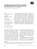
By subculture day 6, confocal analysis revealed clear labe-
ling of the plasma membranes but no significant staining
in other cell compartments (Figure 3A). Additional stain-
ing of the ER was observed prior to day 6 (data not
shown) indicating transit of the protein through the secre-
tory pathway. To exclude the possibility that NtMMP1-
GFP is secreted to the apoplast and not associated with the
plasma membrane, cells were rinsed with 0.5 M KNO
3
to
induce plasmolysis. Under these conditions GFP staining
was clearly associated with the protoplasts, whereas no
GFP was detected in the surrounding cell walls, confirm-
ing membrane integration (Figure 3B).
Transient expression of recombinant NtMMP1 and
analysis of proteolytic activity
To facilitate analysis of NtMMP1 enzymatic activity, two
recombinant NtMMP1 versions designated NtMMP1-apo
and NtMMP1-KDEL were produced. In both variants the
C-terminal hydrophobic domain was omitted to facilitate
protein extraction. NtMMP1-apo contained a C-terminal
His
6
tag for purification, NtMMP1-KDEL contained the
His
6
tag followed by the ER retention sequence. The corre-
sponding NtMMP1-apo and NtMMP1-KDEL cDNAs were
BMC Plant Biology 2009, 9:83 />Page 3 of 12
(page number not for citation purposes)
Nucleotide and amino acid sequences of NtMMP1Figure 1
Nucleotide and amino acid sequences of NtMMP1. The signal peptide sequence (aa 1–20) is shown in bold. The seven
potential N-glycosylation sites are shown in bold italics. The so-called cysteine switch motif is underlined, the zinc binding
region within the catalytic domain is double underlined and the predicted hydrophobic transmembrane helix is underlined in
bold.
BMC Plant Biology 2009, 9:83 />Page 4 of 12
(page number not for citation purposes)
Multiple sequence alignment of ten plant MMPs described in the literatureFigure 2
Multiple sequence alignment of ten plant MMPs described in the literature. The protein sequences were retrieved
from GenBank with the following accession numbers: At1-MMP [GenBank: AAO42162 />28393482], At2-MMP [GenBank: NP_177174 At3-MMP [GenBank:
NP_173824 />], At4-MMP [GenBank: NP_182030 />protein/15225398], At5-MMP [GenBank: NP_176205 SMEP1 GenBank:
P29136 />], GmMMP2 [GenBank:AAL27029 />16901508], Cs1-MMP [GenBank: CAB76364 NtMMPL1 [GenBank: CAA77093
/>]. Amino acid residues that are identical in all ten sequences are shown with a
dark grey background, blocks of similar amino acids are shown with a light grey background.
1 60
At5-MMP (1) MRTLLLTILIFFFTVNPISAKFYTNVSSIPPL QFLNATQNAWET
At2-MMP (1) MRFCVFGFLSLFLIVSPASAWFFPNSTAVPP SLRNTTRVFWDA
At3-MMP (1) MVRICVFMVFLLFFAPSPVSAGFYTNSSAIPPQ LLRNATGNPWNS
NtMMP1 (1) MRIPLFIAIVLVLSLSPASAHFSPNISSIPP SLLKPNNTAWDA
SMEP1 (1) MTLRNHQELLVALATLYFLATSLPSV SAHGPYAWDGEA
At1-MMP (1) MSRNLIYRRNRALCFVLILFCFPYRFGARITPEAEQST AKATQIIHVSNSTWHD
At4-MMP (1) MHHHHHPCNRKPFTTIFSFFLLY LN LHNQQIIEARNPSQFT
Cs1-MMP (1) MASPKALQIIFPFTLLFLSLFPNPNTSSPIILKHS SQNMNSSNSLMF
GmMMP2 (1) MMKSSSHLSAIFLLFFLLTALSPSDGVSFSSFLKQLKQKLEKSPTLKDFLKPTTIGDIYY
MtMMPL1 (1) MNMMKLYQFELLLSLLFIIVN TTLSGYIP
61 120
At5-MMP (45) FSKLAGCHIGEN INGLSKLKQYFRRFGYITTT G-N CTDDFDDVLQSAINTYQK
At2-MMP (44) FSNFTGCHHGQN VDGLYRIKKYFQRFGYIPET-FSGN FTDDFDDILKAAVELYQT
At3-MMP (46) FLNFTGCHAGKK YDGLYMLKQYFQHFGYITETNLSGN FTDDFDDILKNAVEMYQR
NtMMP1 (44) FHKLLGCHAGQK VDGLAKIKKYFYNFGYIPSL S-N FTDDFDDALESALKTYQQ
SMEP1 (39) TYKFTTYHPGQN YKGLSNVKNYFHHLGYIPNAP H FDDNFDDTLVSAIKTYQK
At1-MMP (55) FSRLVDVQIGSH VSGVSELKRYLHRFGYVNDGS EI FSDVFDGPLESAISLYQE
At4-MMP (42) TNPSPDVSIP EIKRHLQQYGYLPQN KESDDVSFEQALVRYQK
Cs1-MMP (48) LKNLQGCHLGDT KQGIHQIKKYLQRFGYITTNIQKHSNPIFDDTFDHILESALKTYQT
GmMMP2 (61) TLNFTEIFSSEERSAPPVSLIKDYLSNYGYIESSG P LSNSMDQETIISAIKTYQQ
MtMMPL1 (30) QLSPSLGKQTEE IQGLSKIKQHLYHFKYLQGLYLVG FDDYLDNKTISAIKAYQQ
121 180
At5-MMP (97) NFNLKVTGKLDSSTLRQIVKPRCGNPDLIDGVSEMNGGK ILR TTEKY
At2-MMP (98) NFNLNVTGELDALTIQHIVIPRCGNPDVVNGTSLMHGGRRKTFEVNFSR THLHAVKRY
At3-MMP (101) NFQLNVTGVLDELTLKHVVIPRCGNPDVVNGTSTMHSGR-KTFEVSFAGRGQRFHAVKHY
NtMMP1 (96) NFNLNTTGVLDAPTIQHLIRPRCGNADVVNGTSTMNSGK PPAG-SQNMHTVAHF
SMEP1 (91) NYNLNVTGKFDINTLKQIMTPRCGVPDIIINTNKTTSFG MIS DY
At1-MMP (108) NLGLPITGRLDTSTVTLMSLPRCGVSDTHMTINNDFLHT TAH Y
At4-MMP (84) NLGLPITGKPDSDTLSQILLPRCGFPD-DVEPKTAPFHT GKK Y
Cs1-MMP (106) NHNLAPSGILDSNTIAQIAMPRCGVQDVIKNKKTKKRNQ N FTNNGHTHFHKVSHF
GmMMP2 (116) YYCLQPTGKLNNETLQQMSFLRCGVPDINIDYNFTDDNMS
MtMMPL1 (84) FFNLQVTGHLDTETLQQIMLPRCGVPDINPDINPDFGFAR
181 240
At5-MMP (144) SFFPGKPRWPKRKR-DLTYAFAPQ NNLTDEVKRVFSRAFTRWAEVT-PLNFTRSES
At2-MMP (156) TLFPGEPRWPRNRR-DLTYAFDPK NPLTEEVKSVFSRAFGRWSDVT-ALNFTLSES
At3-MMP (160) SFFPGEPRWPRNRR-DLTYAFDPR NALTEEVKSVFSRAFTRWEEVT-PLTFTRVER
NtMMP1 (149) SFFPGRPRWPDSKT-DLTYAFLPQ NGLTDNIKSVFSRAFDRWSEVT-PLSFTETAS
SMEP1 (135) TFFKDMPRWQAGTT-QLTYAFSPE PRLDDTFKSAIARAFSKWTPVV-NIAFQETTS
At1-MMP (151) TYFNGKPKWNRDT LTYAISKTHKLDYLTSEDVKTVFRRAFSQWSSVI-PVSFEEVDD
At4-MMP (126) VYFPGRPRWTRDVPLKLTYAFSQENLTPYLAPTDIRRVFRRAFGKWASVI-PVSFIETED
Cs1-MMP (161) TFFEGNLKWPSSK-LHLSYGFLPN YPIDAIKPVSRAFSKWSLNT-HFKFSHVAD
GmMMP2 (156) -YPKAGHRWFPHTN LTYGFLPE NQIPANMTKVFRDSFARWAQASGVLNLTETT-
MtMMPL1 (124) AQGNKWFPKGTKELTYGFLPE SKISIDKVNVFRNAFTRWSQTTRVLKFSEATS
241 300
At5-MMP (198) ILRADIVIGFFSGEHG DGEPFDGAMGTLAHASSPPTGMLHLDGDEDWLISNGE-ISRR
At2-MMP (210) FSTSDITIGFYTGDHG DGEPFDGVLGTLAHAFSPPSGKFHLDADENWVVSG DLDS
At3-MMP (214) FSTSDISIGFYSGEHG DGEPFDGPMRTLAHAFSPPTGHFHLDGEENWIVSGE GGDG
NtMMP1 (203) FQSADIKIGFFAGDHN DGEPFDGPMGTLAHAFSPPGGHFHLDGDENWVIDGVPIVEGN
SMEP1 (189) YETANIKILFASKNHG DPYPFDGPGGILGHAFAPTDGRCHFDADEYWVASG DVT
At1-MMP (207) FTTADLKIGFYAGDHG DGLPFDGVLGTLAHAFAPENGRLHLDAAETWIVDDDL
At4-MMP (185) YVIADIKIGFFNGDHG DGEPFDGVLGVLAHTFSPENGRLHLDKAETWAVDFDE
Cs1-MMP (213) YRKADIKISFERGEHG DNAPFDGVGGVLAHAYAPTDGRLHFDGDDAWSVGAIS
GmMMP2 (208) YDNADIQVGFYNFTYLGIDIEVYGGSLIFLQPDSTKKGVILLDGTNKLWALPSEN G-R
MtMMPL1 (177) YDDADIKIGFYNISYN SKEVIDVVVSDFFINLRS FTIRLEAS
301 360
At5-MMP (255) ILPVTTVVDLESVAVHEIGHLLGLGHSSVEDAIMFPAISGGD-RKVELAKDDIEGIQHLY
At2-MMP (265) FLSVTAAVDLESVAVHEIGHLLGLGHSSVEESIMYPTITTGK-RKVDLTNDDVEGIQYLY
At3-MMP (270) FISVSEAVDLESVAVHEIGHLLGLGHSSVEGSIMYPTIRTGR-RKVDLTTDDVEGVQYLY
NtMMP1 (261) FFSILSAVDLESVAVHEIGHLLGLGHSSVEDSIMFPSLAAGT-RRVELANDDIQGVQVLY
SMEP1 (243) KSPVTSAFDLESVAVHEIGHLLGLGHSSDLRAIMYPSIPPRT-RKVNLAQDDIDGIRKLY
At1-MMP (260) KGSSEVAVDLESVATHEIGHLLGLGHSSQESAVMYPSLRPRT-KKVDLTVDDVAGVLKLY
At4-MMP (238) EKSS-VAVDLESVAVHEIGHVLGLGHSSVKDAAMYPTLKPRS-KKVNLNMDDVVGVQSLY
Cs1-MMP (266) GYFDVETVALHEIGHILGLQHSTIEEAIMFPSIPEG VTKGLHGDDIAGIKALY
GmMMP2 (265) LSWEEGVLDLESAAMHEIGHLLGLDHSNKEDSVMYPCILPSHQRKVQLSKSDKTNVQHQF
MtMMPL1 (219) KVWDLETVAMHQIGHLLGLDHSSDVESIMYPTIVPLHQKKVQITVSDNQAIQQLY
361 418
At5-MMP (314) GGNPNGDGGGSKP SRESQSTGGDSVRRWRGWMISLSSIATCIFLISV
At2-MMP (324) GANPNFNGTTSPPSTTKHQRDTGGFSAAWRIDGSSRSTIVSLLLSTVGLVLWFLP
At3-MMP (329) GANPNFNGSRSPP-PSTQQRDTGDSGAPGRSDGS-RSVLTNLLQYYFWIIFGLFLYLV
NtMMP1 (320) GSNPNFTG PNTVLNPTQENDTNGAPKFGSLWVHVVFAFFLSFLHLI
SMEP1 (302) GINP
At1-MMP (319) GPNPKLRLD SLTQSEDSIKNGTVSHRFLSGNFIGYVLLVVGLILFL
At4-MMP (296) GTNPNFTLN SLLASETSTNLADGSRIRSQGMIYSTLSTVIALCFLNW
Cs1-MMP (319) RV
GmMMP2 (325) ANVEDSAG HVGRLGVSLITTLSLVFAYLLLLLY
MtMMPL1 (274) TKQTNQDRDELGFFDYSGDFFESSSGLLNSLSLGFAFVALMNLAF
BMC Plant Biology 2009, 9:83 />Page 5 of 12
(page number not for citation purposes)
inserted into the plant expression vector pTRAkt and the
proteins transiently expressed in tobacco leaves. Total sol-
uble proteins were extracted from tobacco leaves using
mild detergents and recombinant NtMMP1 was purified
via the C-terminal histidine tag.
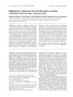
Immunoblot analysis revealed that the purified recom-
binant NtMMP1-apo exists in two forms with apparent
molecular masses of ~30 and 55 kDa (Figure 4A). The the-
oretical mass calculated from the amino acid sequence
lacking the signal peptide is 37.7 kDa. The difference
between the predicted and apparent values probably
reflects glycosylation at one or more of the seven potential
N-glycosylation sites. The microheterogeneity of the
upper band likely reflects differences in the glycosylation
pattern and represents the full-length NtMMP1 protein
including the propeptide. The lower molecular weight
form of NtMMP1 that appears as a double band likely rep-
resents differentially processed forms without the propep-
tide. Data for SMEP1 suggest that the protein could be
processed in the region of amino acid residue 150 [15],
which is consistent with the observed molecular mass of
~30 kDa for the low molecular weight forms of recom-
binant NtMMP1.
The zymography assay demonstrated that all forms of
NtMMP1-apo are enzymatically active and degrade co-
polymerized casein in a polyacrylamide gel, the same
being true for the KDEL-tagged version of the protein (Fig-
ure 4B). Preincubation of all recombinant forms with
APMA, a metallo-organic activator of metalloproteases
[16], did not enhance casein degradation, indicating that
recombinant NtMMP1 is already present in an active
form. In contrast, enzymatic activity was efficiently
blocked by the inclusion of 10 mM EDTA in the protease
buffer, showing that divalent cations are required as cofac-
tors for NtMMP1 activity (Figure 4C).
BY-2 confocal laser scanning microscopyFigure 3
BY-2 confocal laser scanning microscopy. Tobacco BY-2 cells stably transformed with NtMMP1-GFP were analyzed by
confocal laser scanning microscopy six days after sub-culturing. A: Untreated cells. B: Cells after treatment with 0.5 M KNO
3
to induce plasmolysis. In each case, white light transmission is shown on the left, green fluorescence in the middle, and the
overlaid images on the right. The scale bar indicates a distance of 50 μm.
BMC Plant Biology 2009, 9:83 />Page 6 of 12
(page number not for citation purposes)
Analysis of endogenous NtMMP1 expression in BY-2 cells
The expression of NtMMP1 mRNA and NtMMP1 protein
was monitored in wild type BY-2 cells between days 4 and
10 of a typical fermentation cycle. The mRNA could be
detected by Northern blot at all time points although a
slight increase was observed at day 10 (Figure 5A). How-
ever, the overall expression levels were quite low, perhaps
providing an explanation for the absence of NtMMP1
sequences in the BY-2 EST database [17]. In agreement
with the transient expression data, the NtMMP1 protein
was represented by two forms with molecular masses of >
55 kDa and > 35 kDa (Figure 5B). In contrast to the mRNA
data, the abundance of both proteins declined towards
the end of the cultivation. The mobility of the larger band
was slightly retarded compared to the recombinant form
of NtMMP1 reflecting the presence of the hydrophobic C-
terminus, which was removed from the recombinant pro-
tein.
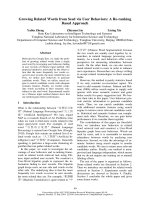
Induction of NtMMP1 by Pseudomonas syringae
To determine whether NtMMP1 can be induced by path-
ogens like other plant MMPs, BY-2 cells were incubated
with either Agrobacterium tumefaciens, Pseudomonas syrin-
gae pv tomato or xylanase from Trichoderma viridae [18].
Total RNA was isolated after 30 min and 1 h and Northern
blots were carried out using NtMMP1 as the probe (Figure
6). While NtMMP1 mRNA levels are induced after treat-
ment with P. syringae and A. tumefaciens, the xylanase
treatment had no effect on NtMMP1 mRNA levels indicat-
ing a lack of responsiveness toward fungal elicitors.
The induction level of NtMMP1 mRNA after one hour of
incubation with either P. syringae or A. tumefaciens were
calculated from three independent biological replicates
using the AIDA software. For the Agrobacterium treatment
the calculated induction factor is 2.4 (SD = 0.9) and for
the Pseudomonas treatment 5.1 (SD = 1.1).
Analysis of recombinant NtMMP1 produced transiently in tobacco leavesFigure 4
Analysis of recombinant NtMMP1 produced transiently in tobacco leaves. A: Immunoblot analysis of fractions from
immobilized metal affinity chromatography purification of NtMMP1-apo. Equal volumes of the different fractions were sepa-
rated by 12% (w/v) SDS PAGE, blotted onto nitrocellulose membranes and probed with a Penta-His antibody (Qiagen) diluted
1:5000, followed by detection with a goat anti-mouse AP-labeled Fc-specific antibody (Dianova) diluted 1:10.000 and develop-
ment with NBT/BCIP. Lane 1: protein extract from wild type plants; 2: flow through fraction; 3: wash fraction; 4–6: elution frac-
tions. B: Zymography of recombinant NtMMP1 (NtMMP1-apo and NtMMP1-KDEL). Equal amounts of NtMMP1-apo and
NtMMP1-KDEL were separated by 12% (w/v) SDS PAGE containing 0.1% (w/v) casein. Lane 1: NtMMP1-KDEL with APMA
treatment; 2: NtMMP1-KDEL without APMA treatment; 3: NtMMP1-apo with APMA treatment; 4: NtMMP1-apo without
APMA treatment. C: Zymography in the presence of 10 mM EDTA. Samples were applied as listed in B.
BMC Plant Biology 2009, 9:83 />Page 7 of 12
(page number not for citation purposes)
Discussion
We have cloned a cDNA encoding the matrix metallopro-
teinase NtMMP1 from tobacco BY-2 cells, which possess
all the expected features of a MMP including the cysteine
switch, and the zinc-binding region and methionine turn
motif in the catalytic domain. Although the overall struc-
ture is very similar to other MMPs, NtMMP1 also has
some novel features, including the substitution of alanine
for the second proline residue normally found within the
cysteine switch consensus sequence PRCXXPD [8]. Since
proline residues have a profound impact on protein struc-
ture, substitution with the non-polar amino acid alanine
may lead to the inactivation of the cysteine switch by pre-
venting the free cysteine residue coordinating the zinc ion
within the catalytic domain and maintaining the latency
of the proenzyme. The sensitivity of this motif towards
amino acid replacements has been shown for the human
MMP-26 where an arginine to histidine exchange within
this domain inactivates the cysteine switch [19]. This
amino acid substitution leads to structural changes within
the prodomain and hence to an alternative activation
mechanism that is independent of the cysteine switch
motif.
Another key feature is that NtMMP1 contains a second
cysteine residue (Cys 50) in the N-terminal portion of the
protein. According to the Scratch protein predictor server
[20] this residue is predicted to form a disulfide bridge
with Cys 118 in the cysteine switch motif. Therefore, it is
unlikely that NtMMP1 is regulated by the cysteine switch
mechanism that has been proposed for human MMP mol-
ecules [21]. The closest homologs to NtMMP1 are At2-
MMP, At3-MMP, and At5-MMP from A. thaliana which
also contain one (At2-MMP and At3-MMP) or two addi-
tional cysteine residues (At5-MMP). The additional
cysteine residues in these MMPs are also predicted to form
NtMMP1 expression in wild type BY-2 suspension cellsFigure 5
NtMMP1 expression in wild type BY-2 suspension
cells. A: Northern blot analysis of the endogenous NtMMP1
mRNA during BY-2 suspension cell cultivation. Total RNA
(12 μg) was loaded for each time point and the blot was
hybridized with a BglII/HindIII NtMMP1 probe. The ethidium
bromide bands confirm equal loading. Lane 1: day 4, 2: day 5;
3: day 6; 4: day 7; 5: day 8; 6: day 9; 7: day 10 after sub-cultur-
ing. B: Endogenous NtMMP1 protein was detected during
BY-2 suspension cell cultivation by immunoblot analysis.
Equal amounts of BY-2 cell extracts were separated by 12%
SDS PAGE and blotted onto a nitrocellulose membrane.
NtMMP1 was detected with anti-LeMMP antiserum diluted
1:2000 and a goat anti-rabbit HRP-labeled Fc-specific anti-
body diluted 1:5000 (Dianova) followed by the ECL proce-
dure. C: recombinant NtMMP1-apo transiently produced in
tobacco leaves as positive control. Lane 1: day 4; 2: day 5; 3:
day 6; 4: day 7; 5: day 8; 6: day 9; 7: day 10 after sub-culturing.
Induction of NtMMP1 in wild type BY-2 suspension cellsFigure 6
Induction of NtMMP1 in wild type BY-2 suspension
cells. BY-2 cells were treated with A. tumefaciens, P. syringae
pv tomato DC3000 or xylanase from T. viridae. The bacteria
were grown to OD
600
of 1.0 and diluted 1:100 in the BY-2
cell culture. Xylanase was used at a final concentration of 2
μg/ml. Total RNA was extracted at the indicated time points
and 12 μg were loaded per lane. NtMMP1 mRNA was
detected by probing with a radiolabeled BglII/HindIII NtMMP1
fragment. Signals were detected with a phosphorimager and
quantified using the AIDA software. The ethidium bromide
bands confirm equal loading. Lane 1: 30 min untreated cells;
lane 2: 30 min exposure to A. tumefaciens; lane 3: 30 min
exposure to P. syringae; lane 4: 30 min exposure to xylanase;
lane 5: 1 h untreated cells; lane 6: 1 h exposure to A. tumefa-
ciens; lane 7: 1 h exposure to P. syringae; lane 8: 1 h exposure
to xylanase.
BMC Plant Biology 2009, 9:83 />Page 8 of 12
(page number not for citation purposes)
disulfide bridges with the cysteine residue from the switch
motif, possibly representing constitutively active forms of
the enzyme. Like NtMMP1, they have a C-terminal hydro-
phobic domain and are believed to reside in the plasma
membrane [7].
The above data suggest that NtMMP1 does not require
proteolytic cleavage for activation, a hypothesis supported
by the finding that APMA treatment has no effect on
NtMMP1 activity. Although it is well established that
zymogens are activated stepwise during zymography [22],
APMA treatment is accompanied by a decrease in molecu-
lar mass due to autoproteolytic processing [23]. However,
we observed no shift to a lower molecular mass in the
NtMMP1 zymogram assay (Figure 4B). Furthermore both
recombinant forms NtMMP1-apo and NtMMP1-KDEL
show the same activity although they are expected to have
different subcellular localizations. While NtMMP1-KDEL
is expected to reside exclusively in the ER due to the C-ter-
minal KDEL sequence, NtMMP1apo can follow the entire
secretory pathway until it is finally secreted to the apo-
plast. Therefore NtMMP1 seems to gain enzymatic activity
immediately after synthesis in the ER. Since no endog-
enous MMP inhibitor proteins like the tissue inhibitors of
metalloproteases (TIMPs) in animals have been identified
thus far in plants, it is likely that NtMMP1 is constitutively
active.
NtMMP1 is expressed constitutively but at a low level dur-
ing BY-2 cell cultivation (Figure 5). The low expression
level is reflected by the absence of NtMMP1-related
sequences in an EST library of BY-2 cells containing more
than 9200 sequences [17]. NtMMP1 mRNA is induced
within 30 min after the treatment of BY-2 cells with P.
syringae and to a lesser extent by A. tumefaciens (Figure 6).
Other MMP genes induced by pathogenic bacteria include
soybean GmMMP2, which is induced after treatment with
compatible and incompatible P. syringae pathovars [1],
and Arabidopsis At3-MMP, which is rapidly induced after
treatment of Arabidopsis seedlings with a 22-amino-acid
peptide (flg22) derived from P. syringae flagellin [10]. The
normal substrates for NtMMP1 are unknown, so it may
act directly against invading bacteria or may help to gen-
erate signaling molecules that trigger further defense
responses of the plant cell. Given the constitutive expres-
sion and activity of NtMMP1, it might be an integral part
of the plant's surveillance system for pathogens or other
stress signals.
The N-terminal portion of NtMMP1 (aa 55–117) is pre-
dicted to form a peptidoglycan-binding motif comprising
three alpha helices, a structure initially described for the
Streptomyces albus Zn
2+
G peptidase [24]. According to the
Pfam protein families database [25] many matrixins con-
tain an N-terminal peptidoglycan-binding like motif
(PF01471). Whether this domain binds to bacterial path-
ogen-associated molecular patterns (PAMPs) such as pep-
tidoglycan [26] and flagellin [10] remains to be
determined. The plant cell usually recognizes specific pep-
tide fragments from PAMPs rather than the full length
proteins [27,28]. In the case of flagellin, a peptide frag-
ment from the DO domain is recognized by the corre-
sponding plant surface receptor [29]. Yet this domain, and
hence the flg22 peptide that binds to the plant FLS2 recep-
tor, is hidden inside the intact bacterial flagellum [30]. It
is therefore tempting to speculate that plasma membrane-
bound proteases such as NtMMP1 recognize PAMPs and
process them to generate specific peptides that subse-
quently bind to their corresponding transmembrane
receptors of the nucleotide-binding site/leucine-rich
repeat (NBS-LRR), receptor-like kinase (RLK) or receptor-
like protein (RLP) classes [31]. Although NtMMP1 did not
respond to the fungal elicitor xylanase (Figure 6) MMP
induction has been shown in soybean for GmMMP2
treated with the oomycete P. sojae and in tomato for
LeMMP1 treated with the fungal elicitor fusicoccin [32].
Therefore also certain PAMPs from fungal origin are able
to induce MMP expression. In future work we will aim to
determine the natural substrate(s) of NtMMP1 and its
potential role in PAMP recognition and processing. How-
ever, the induction of NtMMP1 by bacterial pathogens
indicates its involvement in pathogen recognition and
defense responses and therefore contributes to our under-
standing of pathogen-host interactions.
Conclusion
The matrix metalloproteinase NtMMP1 is localized in the
plasma membrane of tobacco BY-2 cells. Our biochemical
data indicate that the enzyme is constitutively active, and
this is supported by bioinformatic analysis of the primary
sequence. The low basal level of NtMMP1 expression
increases immediately after the exposure of tobacco BY-2
cells to bacterial pathogens. Given the low-level constitu-
tive activity of the protein, its induction in response to
bacterial pathogens and its localization at the cell surface,
we propose that NtMMP1 plays a role in pathogen recog-
nition and defense at the cell periphery.
Methods
Gene cloning
Degenerate primers were designed according to the
CODEHOP procedure [33] based on the known MMP
protein sequences from Arabidopsis thaliana, soybean, rice,
cucumber and Medicago trunculata [GenBank: NP_177174
/>, Gen-
Bank: NP_176205 />15218963, GenBank: NP_173824 http://
www.ncbi.nlm.nih.gov/protein/30688744, GenBank:
O65340 />75219926, GenBank: NP_182030 http://
BMC Plant Biology 2009, 9:83 />Page 9 of 12
(page number not for citation purposes)
www.ncbi.nlm.nih.gov/protein/15225398, GenBank:
AAM62476 />21553383, GenBank: O48680 http://
www.ncbi.nlm.nih.gov/protein/75219474, GenBank:
AAO42162 />28393482, GenBank: P29136 http://
www.ncbi.nlm.nih.gov/protein/2827777, GenBank:
1905425A />384337, GenBank: AAL27029 http://
www.ncbi.nlm.nih.gov/protein/16901508, GenBank:
AAK55464 />14165332, GenBank: AAK55462 http://
www.ncbi.nlm.nih.gov/protein/14165330, GenBank:
AAK55459 />14165327, GenBank: CAB76364 http://
www.ncbi.nlm.nih.gov/protein/7159629, GenBank:
CAA77093 />116874798]. Total RNA was prepared from logarithmi-
cally growing Nicotiana tabacum cv Bright Yellow 2 (BY-2)
cells using the RNeasy Plant Mini Kit (Qiagen, Hilden,
Germany) and a cDNA was synthesized using the MM3
primer (5'-CTC GAG GAT CCG CGG CCG C(T)
18
-3') and
the Superscript first strand cDNA synthesis system (Invit-
rogen, Karlsruhe, Germany). MMP-related sequences
from BY-2 cDNA were amplified with the primer pair Met-
allo-1 (5'-GAT CTG GAA TCT GTT GCT GTT CAY GAR
ATH GGN C-3') and MM3, in a 50-μl reaction volume
using the Expand High Fidelity PCR System (Roche, Man-
nheim, Germany). The program comprised 5 min at 95°C
followed by 35 cycles of denaturation at 95°C for 30 s,
annealing at 53°C for 30 s and extension at 72°C for 30
s. PCR products were gel purified and cloned in the
pCR2.1 vector using the TOPO Cloning Kit (Invitrogen).
Insert sequences were verified using the BigDye Sequenc-
ing Kit (Applied Biosystems, Darmstadt, Germany).
To clone the missing 5' portion of the MMP sequence, the
adapter ASLinker (5'-PO
4
-CTG CAG AAA GCT TGG TGG
ATC CTA-NH
2
-3') was ligated to single stranded cDNA as
described [34]. Using the complementary primer AS04
(5'-TAG GAT CCA CCA AGC TTT CTG CAG-3') and the
MMP-specific primer MMPRace1 (5'-GGG TTA GAC CCG
TAT AAC ACC TGG AC-3') the 5' end of the cDNA was
amplified using the PCR procedure described above and
the following program: 5 min at 95°C followed by 35
cycles of denaturation at 95°C for 1 min, annealing at 50–
70°C for 30 s and extension at 72°C for 1 min. PCR prod-
ucts were subcloned and sequenced as described above.
The final NtMMP1 full-length cDNA sequence was depos-
ited in GenBank
®
[GenBank: DQ508374].
Transient expression of recombinant NtMMP1
To produce recombinant NtMMP1 for functional analysis,
the 5' and 3' cDNA sequences were amplified, joined in-
frame by SOE-PCR [35] and inserted into the plant expres-
sion vector pTRAkt [36]. To facilitate extraction of the
recombinant protein, the hydrophobic C-terminal trans-
membrane domain was omitted. Two constructs were
generated, one with a C-terminal His
6
tag alone
(NtMMP1-apo) and another with a C-terminal His
6
tag
followed by a SEKDEL motif for ER retention (NtMMP1-
KDEL). The 5' portion of NtMMP1 was amplified using
primers NtMMP1-Nterm_for (5'-CCA TGG AAA TGA GGA
TTC CTT TAT TTA TCG CC-3') and NtMMP1-Nterm_rev
(5'-CCA CTC TTC GGG TAC CCG CTG C-3'). The 3' por-
tion was similarly amplified using primers NtMMP1-
Cterm_for (5'-AGC AGC GGG TAC CCG AAG AGT GGA
GC-3') and either NtMMP1-Cterm-apo_rev (5'-TCT AGA
CTA GTG ATG GTG ATG GTG ATG ACC AAA TTT CGG
GGC TCC ATT TGT GTC-3') or NtMMP1-Cterm-
KDEL_rev (5'-GCG GCC GCA CCA AAT TTC GGG GCT
CC-3'). Introduced restriction sites are shown in italic. The
amplified partial cDNAs were joined by SOE-PCR and
inserted into pTRAkt using the NcoI and XbaI sites for the
NtMMP1-apo construct or the NcoI and NotI sites for the
NtMMP-KDEL construct.
Both vectors were introduced into A. tumefaciens
GV3101::pMP90RK by electroporation [37]. The recom-
binant proteins were expressed transiently in detached
leaves of N. tabacum cv. Petite Havana SR1 by vacuum
infiltration [38] and partially purified via their His
6
tags
by immobilized metal-affinity chromatography (IMAC)
as described previously [39].
GFP fusions
To analyze the cellular localization of recombinant
NtMMP1 by fluorescence microscopy, the peptidase
domain was replaced with the cDNA encoding Emerald
GFP (EmGFP, Invitrogen). The 5' end of the NtMMP1
cDNA was amplified with the primer pair NtMMP1-
Nterm_for (5'-CCA TGG AAA TGA GGA TTC CTT TAT TTA
TCG CC-3') and NtMMP-Nterm+GFP_rev (5'-CTC GCC
CTT GCT CAC CAT ATT CTG AGA ACC TGC CGG CG-3'),
EmGFP was amplified with the primer pair GFP_for (5'-
CGC CGG CAG GTT CTC AGA ATA TGG TGA GCA AGG
GCG AG-3') and GFP_rev (5'-GGC CCA GTA AAA TTT
GGG TTA GAC TTG TAC AGC TCG TCC ATG CCG-3'),
and the 3' end of the NtMMP1 cDNA was amplified with
the primer pair NtMMP1-Cterm+GPF_for (5'-CGG CAT
GGA CGA GCT GTA CAA GTC TAA CCC AAA TTT TAC
TGG G-3') and NtMMP-Cterm_rev (5'-TCT AGA TTT AAA
TTA AAT GGA GAA ATG ATA AG-3'). Introduced restric-
tion sites are shown in italic. The three fragments were
joined by SOE-PCR, reamplified, and cloned in the plant
expression vector pTRAkt using the NcoI and XbaI restric-
tion sites.
BMC Plant Biology 2009, 9:83 />Page 10 of 12
(page number not for citation purposes)
Plant cell culture, transformation, and treatments
N. tabacum cv BY-2 cells [40] were maintained in MSMO
medium (Sigma, Taufkirchen, Germany) supplemented
with 0.15 μg/ml thiamin, 0.02 μg/ml KH
2
PO
4
and 3% (w/
v) sucrose (pH 5.6). The cells were passed each week into
fresh culture medium using a 2% (v/v) inoculum for wild
type and a 5% (v/v) inoculum for transgenic cells. The
cells were incubated in an orbital shaker (New Brunswick
Scientific, Edison, NJ, USA) at 180 rpm, 26°C in darkness.
Transgenic BY-2 cells were produced by co-cultivation
with A. tumefaciens as described [41]. The recombinant
pTRAkt vectors were transformed into A. tumefaciens
GV3101::pMP90RK [42] by electroporation using a mult-
iporator (Eppendorf, Hamburg, Germany).
A. tumefaciens was grown in YEB medium [43]. P. syringae
pv. tomato DC3000 was cultivated in KingsB medium
[44]. For the treatment of tobacco BY-2 cells, the bacteria
were grown to an OD
600
of 1.0 and diluted 1:100 with the
BY-2 culture. Xylanase from T. viridae (Sigma) was used at
a final concentration of 2 μg/ml.
Plant cell confocal imaging
Wild type and transgenic BY-2 cells were imaged using a
Leica TCS-SP spectral confocal microscope equipped with
an argon ion laser using a 40 × oil immersion Plan-Apo
objective (Leica, Wetzlar, Germany). EmGFP was excited
with the 488 nm wavelength argon laser line and confocal
images were taken at a 500–570 nm emission setting
using Leica TCS-SP software. Image overlays were gener-
ated using Adobe Photoshop CS2 software.
Northern blot
Total RNA was extracted from tobacco BY-2 suspension
cells using the RNeasy Plant Mini Kit (Qiagen), and 12 μg
were loaded onto denaturing formaldehyde agarose gels
followed by capillary blotting onto nylon membranes
(Hybond N
+
, GE Healthcare, Freiburg, Germany). The
membranes were probed with a 765-bp BglII/HindIII frag-
ment of the NtMMP1 cDNA radiolabeled with [α
32
]P-
dATP (GE Healthcare) using the DecaLabel DNA labeling
kit (Fermentas, St. Leon-Rot, Germany) according to the
manufacturer's instructions. After prehybridization (50%
(v/v) formamide, 10% (w/v) dextran sulfate, 1% (w/v)
SDS, 1 M NaCl) for three hours at 42°C, the denatured
probe was added to the prehybridization solution with
100 μg salmon sperm carrier DNA and hybridization was
carried out at 42°C overnight. The membranes were
washed twice for 30 min in 2× SSC containing 0.1% (w/
v) SDS at 65°C. The signals were visualized by exposing
the membranes on a phosphorimager plate overnight.
The plates were read with a phosphorimager (FLA-2000,
Fujifilm, Tokyo, Japan) and the images were processed
using AIDA software (Raytest, Straubenhardt, Germany).
Zymography
Protease activity was visualized by in-gel assays using
casein as a substrate [45]. The substrate was co-polymer-
ized with the acrylamide at a final concentration of 0.1%
(w/v). SDS-PAGE was carried out on a 12% (w/v) gel at a
constant current of 20 mA (MiniProteanII, Biorad,
Munich, Germany). The samples were neither reduced
nor boiled prior to loading and electrophoresis was car-
ried out in an ice bath. After electrophoresis the SDS was
removed by washing the gel twice for 15 min in 2.5% (v/
v) Triton X-100 followed by two further 15-min washes in
protease assay buffer (50 mM Tris, 5 mM CaCl
2
, 100 μM
ZnCl
2
, pH 7.6). The gels were incubated overnight in the
protease assay buffer then stained with Coomassie bril-
liant blue. Proteolytic activities were revealed after
destaining as clear bands on a blue background.
APMA treatment was done with a final concentration of
10 mM for 2 h at 37°C as described [46].
Immunoblot analysis
Protein samples from BY-2 cells were prepared as
described [39] and separated by SDS-PAGE. The proteins
were transferred onto nitrocellulose membrane by semi-
dry electroblotting using a Trans-blot SD device (Biorad)
and a standard transfer buffer (25 mM Tris, 192 mM, 20%
(v/v) methanol, as described [47] at a constant current of
2.5 mA/cm
2
for 40 min. Nonspecific binding sites were
blocked with 5% (w/v) skimmed milk in PBST at 4°C
overnight. The membrane was washed once with PBST
and NtMMP1 was detected with a rabbit anti-LeMMP
antiserum raised against LeMMP from tomato (Solanum
lycopersicum) at a dilution of 1:2000 in PBST for 1 h at
room temperature. The antiserum was kindly provided by
A. Schaller (University of Hohenheim, Germany). Mem-
branes were washed three times for 5 min in PBST and
incubated with a HRP-conjugated secondary goat-anti-
rabbit IgG Fc
γ
antibody (Dianova, Hamburg, Germany)
diluted 1:5000 in PBST. The membranes were washed
three times with PBST, once with PBS and then developed
with the ECL reagent (GE Healthcare). Images were
acquired using the LAS 3000 cooled CCD camera device
(Fujifilm).
Abbreviations
APMA: 4-aminophenylmercuric acid; ECL: enhanced
chemiluminescence; EST: expressed sequence tags; GFP:
green fluorescent protein; IMAC: immobilized metal
affinity chromatography; MMP: matrix metalloprotein-
ase; MSMO: Murahige & Skoog medium with minimal
organics; PAGE: polyacrylamide gel electrophoresis; SDS:
sodium dodecylsulfate; SOE-PCR: splicing by overlap
extension polymerase chain reaction.
BMC Plant Biology 2009, 9:83 />Page 11 of 12
(page number not for citation purposes)
Authors' contributions
AS conceived of the study, cloned the NtMMP1 cDNA
from BY-2 cells and participated in drafting the manu-
script. HH and MKM cloned constructs for transient
expression and characterized the recombinant enzyme.
BO analyzed the expression of native NtMMP1 in BY-2
cells. VW cloned GFP constructs and analyzed the subcel-
luar localization together with BO. SS participated in the
experiment design, interpretation of the data and drafting
of the manuscript. All authors have read and approved the
manuscript.
Acknowledgements
We are grateful to Prof. Dr. Andreas Schaller (University of Hohenheim)
for providing the antiserum against LeMMP and fruitful discussions. We
thank Dr. Flora Schuster (RWTH Aachen University) for expert assistance
in plant cell culture and transformation and Dr. Stefano Di Fiore (RWTH
Aachen University) for his advice on confocal microscopy. This work was
supported by Pharma-Planta (EU Integrated Project #503565 in FP 6) and
by a scholarship from RWTH Aachen University given to MKM.
References
1. Liu Y, Dammann C, Bhattacharyya MK: The matrix metalloprotei-
nase gene GmMMP2 is activated in response to pathogenic
infections in soybean. Plant Physiol 2001, 127(4):1788-1797.
2. Pak JH, Liu CY, Huangpu J, Graham JS: Construction and charac-
terization of the soybean leaf metalloproteinase cDNA. FEBS
Lett 1997, 404(2–3):283-288.
3. Delorme VG, McCabe PF, Kim DJ, Leaver CJ: A matrix metallo-
proteinase gene is expressed at the boundary of senescence
and programmed cell death in cucumber. Plant Physiol 2000,
123(3):917-927.
4. Combier JP, Vernie T, de Billy F, El Yahyaoui F, Mathis R, Gamas P:
The MtMMPL1 early nodulin is a novel member of the
matrix metalloendoproteinase family with a role in Medi-
cago truncatula infection by Sinorhizobium meliloti. Plant Physiol
2007, 144(2):703-716.
5. Ramos OHP, Selistre-de-Araujo HS: Identification of metallopro-
tease gene families in sugarcane. Genet Mol Biol 2001, 24(1–
4):285-290.
6. Flinn BS: Plant extracellular matrix metalloproteinases. Funct
Plant Biol 2008, 35(12):1183-1193.
7. Maidment JM, Moore D, Murphy GP, Murphy G, Clark IM: Matrix
metalloproteinase homologues from Arabidopsis thaliana –
Expression and activity. J Biol Chem 1999, 274(49):34706-34710.
8. Maskos K, Bode W: Structural basis of matrix metalloprotein-
ases and tissue inhibitors of metalloproteinases. Mol Biotechnol
2003, 25(3):241-266.
9. Golldack D, Popova OV, Dietz KJ: Mutation of the matrix metal-
loproteinase At2-MMP inhibits growth and causes late flow-
ering and early senescence in Arabidopsis. J Biol Chem 2002,
277(7):5541-5547.
10. Zipfel C, Robatzek S, Navarro L, Oakeley EJ, Jones JD, Felix G, Boller
T: Bacterial disease resistance in Arabidopsis through flagellin
perception. Nature
2004, 428(6984):764-767.
11. Rawlings ND, Morton FR, Kok CY, Kong J, Barrett AJ: MEROPS:
the peptidase database. Nucleic Acids Res 2008, 36:D320-D325.
12. Quevillon E, Silventoinen V, Pillai S, Harte N, Mulder N, Apweiler R,
Lopez R: InterProScan: protein domains identifier. Nucleic
Acids Res 2005, 33:W116-W120.
13. Nakai K, Horton P: PSORT: a program for detecting sorting
signals in proteins and predicting their subcellular localiza-
tion. Trends Biochem Sci 1999, 24(1):34-36.
14. Tsien RY: The green fluorescent protein. Annu Rev Biochem 1998,
67:509-544.
15. McGeehan G, Burkhart W, Anderegg R, Becherer JD, Gillikin JW,
Graham JS: Sequencing and characterization of the soybean
leaf metalloproteinase – Structural and functional similarity
to the matrix metalloproteinase family. Plant Physiol 1992,
99(3):1179-1183.
16. Sellers A, Cartwright E, Murphy G, Reynolds JJ: Evidence that
latent collagenases are enzyme-inhibitor complexes. Biochem
J 1977, 163(2):303-307.
17. Matsuoka K, Demura T, Fukuda H: Normalized EST microarray
of tobacco BY-2 cells and culture stage specific secretory
proteins. Plant Cell Physiol 2002, 43:S2-S2.
18. Yano A, Suzuki K, Shinshi H: A signaling pathway, independent
of the oxidative burst, that leads to hypersensitive cell death
in cultured tobacco cells includes a serine protease. Plant J
1999, 18(1):105-109.
19. Marchenko ND, Marchenko GN, Strongin AY: Unconventional
activation mechanisms of MMP-26, a human matrix metallo-
proteinase with a unique PHCGXXD cysteine-switch motif.
J Biol Chem 2002, 277(21):18967-18972.
20. Cheng J, Randall AZ, Sweredoski MJ, Baldi P: SCRATCH: a protein
structure and structural feature prediction server. Nucleic
Acids Res 2005, 33:
W72-W76.
21. Van Wart HE, Birkedal-Hansen H: The cysteine switch: a princi-
ple of regulation of metalloproteinase activity with potential
applicability to the entire matrix metalloproteinase gene
family. Proc Natl Acad Sci USA 1990, 87(14):5578-5582.
22. Kleiner DE, Stetler-Stevenson WG: Quantitative zymography:
detection of picogram quantities of gelatinases. Anal Biochem
1994, 218(2):325-329.
23. Cameron PM, Marcy AI, Rokosz LL, Hermes JD: Use of an active-
site inhibitor of stromelysin to elucidate the mechanism of
prostromelysin activation. Bioorg Chem 1995, 23(4):415-426.
24. Dideberg O, Charlier P, Dive G, Joris B, Frere JM, Ghuysen JM:
Structure of a Zn2+-containing D-alanyl-D-alanine-cleaving
carboxypeptidase at 2.5 resolution. Nature 1982,
299(5882):469-470.
25. Finn RD, Tate J, Mistry J, Coggill PC, Sammut SJ, Hotz HR, Ceric G,
Forslund K, Eddy SR, Sonnhammer EL, Bateman A: The Pfam pro-
tein families database. Nucleic Acids Res 2008, 36:D281-D288.
26. Gust AA, Biswas R, Lenz HD, Rauhut T, Ranf S, Kemmerling B, Gotz
F, Glawischnig E, Lee J, Felix G, Nurnberger T: Bacteria-derived
peptidoglycans constitute pathogen-associated molecular
patterns triggering innate immunity in Arabidopsis. J Biol Chem
2007, 282(44):32338-32348.
27. Kunze G, Zipfel C, Robatzek S, Niehaus K, Boller T, Felix G: The N
terminus of bacterial elongation factor Tu elicits innate
immunity in Arabidopsis plants. Plant Cell 2004,
16(12):3496-3507.
28. Brunner F, Rosahl S, Lee J, Rudd JJ, Geiler C, Kauppinen S, Rasmussen
G, Scheel D, Nurnberger T: Pep-13, a plant defense-inducing
pathogen-associated pattern from Phytophthora trans-
glutaminases. EMBO J 2002, 21(24):6681-6688.
29. Felix G, Duran JD, Volko S, Boller T: Plants have a sensitive per-
ception system for the most conserved domain of bacterial
flagellin. Plant J 1999, 18(3):265-276.
30. Yonekura K, Maki-Yonekura S, Namba K: Complete atomic
model of the bacterial flagellar filament by electron cryomi-
croscopy. Nature 2003, 424(6949):643-650.
31. Jones DA, Takemoto D: Plant innate immunity – direct and
indirect recognition of general and specific pathogen-associ-
ated molecules. Curr Opin Immunol 2004, 16(1):48-62.
32. Frick UB, Schaller A: cDNA microarray analysis of fusicoccin-
induced changes in gene expression in tomato plants. Planta
2002, 216(1):83-94.
33. Rose TM, Schultz ER, Henikoff JG, Pietrokovski S, McCallum CM,
Henikoff S: Consensus-degenerate hybrid oligonucleotide
primers for amplification of distantly related sequences.
Nucleic Acids Res 1998, 26(7):1628-1635.
34. Russinova E, Slater A, Atanassov AI, Elliott MC: Cloning novel
alfalfa cyclin sequences – a RACE-PCR approach. Cell Mol Biol
(Noisy-Le-Grand) 1995, 41(5):703-714.
35. Horton RM, Cai ZL, Ho SN, Pease LR: Gene splicing by overlap
extension: tailor-made genes using the polymerase chain
reaction. BioTechniques 1990, 8(5):528-535.
36. Sack M, Paetz A, Kunert R, Bomble M, Hesse F, Stiegler G, Fischer R,
Katinger H, Stoeger E, Rademacher T: Functional analysis of the
broadly neutralizing human anti-HIV-1 antibody 2F5 pro-
duced in transgenic BY-2 suspension cultures. FASEB J 2007,
21(8):1655-1664.
Publish with BioMed Central and every
scientist can read your work free of charge
"BioMed Central will be the most significant development for
disseminating the results of biomedical research in our lifetime."
Sir Paul Nurse, Cancer Research UK
Your research papers will be:
available free of charge to the entire biomedical community
peer reviewed and published immediately upon acceptance
cited in PubMed and archived on PubMed Central
yours — you keep the copyright
Submit your manuscript here:
/>BioMedcentral
BMC Plant Biology 2009, 9:83 />Page 12 of 12
(page number not for citation purposes)
37. Dower WJ, Miller JF, Ragsdale CW: High efficiency transforma-
tion of E. coli by high voltage electroporation. Nucleic Acids Res
1988, 16(13):6127-6145.
38. Kapila J, De Rycke R, Van Montagu M, Angenon G: An Agrobacte-
rium-mediated transient gene expression system for intact
leaves. Plant Sci 1997, 122(1):101-108.
39. Schiermeyer A, Schinkel H, Apel S, Fischer R, Schillberg S: Produc-
tion of Desmodus rotundus salivary plasminogen activator α1
(DSPAα1) in tobacco is hampered by proteolysis. Biotechnol
Bioeng 2005, 89(7):848-858.
40. Nagata T, Nemoto Y, Hasezawa S: Tobacco BY-2 cell line as the
"HeLa" cell in the cell biology of higher plants. Int Rev Cytol
1992, 132:1-30.
41. An G: High efficiency transformation of cultured tobacco
cells. Plant Physiol 1985, 79(2):568-570.
42. Hellens R, Mullineaux P, Klee H: A guide to Agrobacterium binary
Ti vectors. Trends Plant Sci 2000, 5(10):446-451.
43. Vervliet G, Holsters M, Teuchy H, Van Montagu M, Schell J: Charac-
terization of different plaque-forming and defective temper-
ate phages in Agrobacterium strains. J Gen Virol 1975,
26(1):33-48.
44. King EO, Ward MK, Raney DE: Two simple media for the dem-
onstration of pyocyanin and fluorescin. J Lab Clin Med 1954,
44(2):301-307.
45. Heussen C, Dowdle EB: Electrophoretic analysis of plasmino-
gen activators in polyacrylamide gels containing sodium
dodecyl sulfate and copolymerized substrates. Anal Biochem
1980, 102(1):196-202.
46. Knauper V, Murphy G: Methods for studying activation of
matrix metalloproteinases. In Matrix metalloproteinase protocols
Volume 151. Edited by: Clark IM. Totowa: Humana Press;
2001:377-387. [Walker JM (Series Editor): Methods in molecular biol-
ogy].
47. Towbin H, Staehelin T, Gordon J: Electrophoretic transfer of
proteins from polyacrylamide gels to nitrocellulose sheets:
procedure and some applications. Proc Natl Acad Sci USA 1979,
76(9):4350-4354.