báo cáo khoa học: " Interactions between cauliflower and Rhizoctonia anastomosis groups with different levels of aggressiveness" ppt
Bạn đang xem bản rút gọn của tài liệu. Xem và tải ngay bản đầy đủ của tài liệu tại đây (1000.88 KB, 12 trang )
BioMed Central
Page 1 of 12
(page number not for citation purposes)
BMC Plant Biology
Open Access
Research article
Interactions between cauliflower and Rhizoctonia anastomosis
groups with different levels of aggressiveness
Joke Pannecoucque and Monica Höfte*
Address: Laboratory of Phytopathology, Faculty of Bioscience Engineering, Ghent University, Coupure Links, 653, B-9000 Gent, Belgium
Email: Joke Pannecoucque - ; Monica Höfte* -
* Corresponding author
Abstract
Background: The soil borne fungus Rhizoctonia is one of the most important plant pathogenic
fungi, with a wide host range and worldwide distribution. In cauliflower (Brassica oleracea var.
botrytis), several anastomosis groups (AGs) including both multinucleate R. solani and binucleate
Rhizoctonia species have been identified showing different levels of aggressiveness. The infection and
colonization process of Rhizoctonia during pathogenic interactions is well described. In contrast,
insights into processes during interactions with weak aggressive or non-pathogenic isolates are
limited. In this study the interaction of cauliflower with seven R. solani AGs and one binucleate
Rhizoctonia AG differing in aggressiveness, was compared. Using microscopic and histopathological
techniques, the early steps of the infection process, the colonization process and several host
responses were studied.
Results: For aggressive Rhizoctonia AGs (R. solani AG 1-1B, AG 1-1C, AG 2-1, AG 2-2 IIIb and AG
4 HGII), a higher developmental rate was detected for several steps of the infection process,
including directed growth along anticlinal cell walls and formation of T-shaped branches, infection
cushion formation and stomatal penetration. Weak or non-aggressive AGs (R. solani AG 5, AG 3
and binucleate Rhizoctonia AG K) required more time, notwithstanding all AGs were able to
penetrate cauliflower hypocotyls. Histopathological observations indicated that Rhizoctonia AGs
provoked differential host responses and pectin degradation. We demonstrated the pronounced
deposition of phenolic compounds and callose against weak and non-aggressive AGs which resulted
in a delay or complete block of the host colonization. Degradation of pectic compounds was
observed for all pathogenic AGs, except for AG 2-2 IIIb. Ranking the AGs based on infection rate,
level of induced host responses and pectin degradation revealed a strong correlation with the
disease severity caused by the AGs.
Conclusion: The differences in aggressiveness towards cauliflower observed among Rhizoctonia
AGs correlated with the infection rate, induction of host defence responses and pectin breakdown.
All Rhizoctonia AGs studied penetrated the plant tissue, indicating all constitutive barriers of
cauliflower were defeated and differences in aggressiveness were caused by inducible defence
responses, including cell wall fortifications with phenolic compounds and callose.
Published: 21 July 2009
BMC Plant Biology 2009, 9:95 doi:10.1186/1471-2229-9-95
Received: 26 March 2009
Accepted: 21 July 2009
This article is available from: />© 2009 Pannecoucque and Höfte; licensee BioMed Central Ltd.
This is an Open Access article distributed under the terms of the Creative Commons Attribution License ( />),
which permits unrestricted use, distribution, and reproduction in any medium, provided the original work is properly cited.
BMC Plant Biology 2009, 9:95 />Page 2 of 12
(page number not for citation purposes)
Background
During the interaction with pathogens, plants that recog-
nize the intruder will respond with an impressive battery
of defence mechanisms. Both structural and chemical bar-
riers are involved which can be constitutive and/or induc-
ible. Upon pathogen detection, the activated defence
responses in the plant may involve the rapid production
of reactive oxygen species, hypersensitive response (HR)
at the site of infection, strengthening of the cell wall by
oxidative cross-linking of cell wall components, apposi-
tion of callose or phenolic compounds and the produc-
tion of phytoalexins and pathogenesis-related proteins [1-
4]. Typically, these responses can be very localised and
microscopic observations seem to be the most appropri-
ate method for investigation [5]. Before succeeding in
causing disease, the pathogen must penetrate the plant
and overcome these obstacles; this consequently explains
why plant pathogens colonize only a narrow range of
hosts.
Among members of the fungal genus Rhizoctonia, the abil-
ity to cause disease is highly variable and depending on
the host plant. Rhizoctonia comprises both multinucleate
and binucleate species which are further divided into
anastomosis groups (AGs). Currently, the multinucleate
species R. solani (teleomorph: Thanatephorus cucumeris)
contains 13 AGs (AG 1 – AG 13) [6], while binucleate
Rhizoctonia spp. (teleomorph: Ceratobasidium spp.) are
divided into 16 AGs (AG A – AG I, AG K, AG L, AG O – AG
S) [7]. Within the same AG, isolates may possess similar
characteristics such as type of disease symptoms caused
and host preference [8]. In addition, for several host-path-
ogen interactions, isolates of the same AG have compara-
ble levels of aggressiveness.
During pathogenic interactions of Rhizoctonia isolates
with several host plants, the early steps of the infection
process appear to be very similar independent of AG or
host [9]. Rhizoctonia hyphae adhere to the plant surface
and soon this is followed by directed growth along the
anticlinal epidermal cell walls and formation of T-shaped
branches. Infection cushions are formed from which
infection pegs penetrate the plant tissue. During some
interactions, especially for isolates obtained from leaves,
the formation of infection cushions is rather exceptional
and the pathogen will form lobate appressoria or pene-
trate the plant through stomata [10]. Further penetration
and colonization have been associated with the enzymatic
degradation of the host tissue, including pectic substances
and cellulose [11,12].
In contrast with the well-studied and generally accepted
situation for pathogenic interactions, insight into the
processes during non-pathogenic interactions is still
largely missing. Keijer [9] reported two distinct observa-
tions during the early steps of an incompatible infection
process. Stomatal penetration of R. solani AG 2 BI resulted
in hypersensitive-like lesions on cauliflower stems, while
AG 3 could not adhere to the cauliflower surface nor pro-
ceed to further steps of the infection process. Resistance of
plant species or cultivars to Rhizoctonia has also been asso-
ciated with cuticle thickness [13], wax deposits [14], accu-
mulation of calcium [15], and inhibition of pectin
degrading enzymes [16]. Jabaji-Hare et al. [17] reported
the induction of various cell wall compounds, such as
suberin, pectic substances and phenolic compounds, dur-
ing the interaction of bean with a non-pathogenic binu-
cleate isolate. All these phenomena have been observed
for specific AGs on specific hosts and a generally accepted
situation for non-pathogenic interactions has not been
described.
When studying the infection process of Rhizoctonia,
researchers have always focussed on the differences
between susceptible and resistant cultivars inoculated
with the same isolate [14,18-20]. Until now, no in depth
study has been carried out to compare the infection and
colonization process and the host responses induced in
the same plant cultivar by different Rhizoctonia AGs
reflecting variation in aggressiveness.
Previously, we reported the importance of seven R. solani
AGs and one binucleate Rhizoctonia AG possessing differ-
ent levels of aggressiveness, in association with Belgian
cauliflower production [21]. However, until now it is not
clear at which stages during the infection process of cauli-
flower differences in aggressiveness appear. Here, in an
attempt to gain more insights into the processes underly-
ing pathogenic and non-pathogenic Rhizoctonia-cauli-
flower interactions, we investigate the infection process,
the host colonization and the induced defence reactions
of those eight Rhizoctonia AGs. In this study, we provide
evidence that Rhizoctonia AGs differ in the developmental
rate of the infection and colonization process and pro-
voke differential host responses and pectin degradation.
Moreover, we show a strong correlation between these
microscopic observations and the disease severity caused
by the AGs. Interestingly, we observed that all Rhizoctonia
AGs are able to enter cauliflower hypocotyls, although
weak and non-aggressive AGs require more time and form
less infection cushions. Studying histopathological sec-
tions, we demonstrate the pronounced deposition of phe-
nolic compounds and callose against weak and non-
aggressive AGs which slowed down or stopped the fungal
growth.
Methods
Fungal isolates
Rhizoctonia isolates were selected from the collection of
Pannecoucque et al. [21] based on AG and aggressiveness.
BMC Plant Biology 2009, 9:95 />Page 3 of 12
(page number not for citation purposes)
Since isolates within the same AG had the same level of
aggressiveness [21], one representative isolate per AG was
selected. Seven R. solani isolates were included represent-
ing AG 1-1B (BK004-2-1), AG 1-1C (BK010-1-1), AG 2-1
(BK001-1-1), AG 2-2 IIIb (M001-1-1) and AG 4 HGII
(BK004-1-1) which were previously considered to be
aggressive towards cauliflower; AG 3 (BK006-2-1), AG 5
(BK003-1-3) and one binucleate Rhizoctonia isolate of AG
K (BK005-1-1) which were identified as weak or non-
aggressive. All isolates were obtained from Belgian cauli-
flower fields, except isolate M001-1-1 which was isolated
from maize. Isolates were maintained at room tempera-
ture on PDA and in the dark.
Cauliflower plants and growth conditions
All experiments were carried out using plants of Brassica
oleracea var. botrytis cv. Clapton (Syngenta Seeds, the Neth-
erlands).
To study early steps of the infection process and degrada-
tion of pectic compounds, cauliflower plants were grown
in vitro under sterile conditions as earlier described [21].
Briefly, cauliflower seeds were surface-sterilized in 0.5%
NaOCl-solution containing 0.01% Tween 20 and rinsed
twice with sterile demineralized water. Gamborg B5
medium (Gamborg B5 basal salt mixture; Labconsult)
solidified with 1% (w/v) agar and enriched with vitamins
(Gamborg B5 vitamin mixture; Labconsult) was poured
into square Petri dishes (12 × 12 cm; Novolab) in which
the in vitro plants were grown. Six surface-sterilized cauli-
flower seeds were placed at equal intervals on the agar
layer. The Petri dishes were sealed with Parafilm
(Novolab) and incubated in the dark at 21°C for 2 days to
allow seed germination. Afterwards, the lower halves of
the Petri dishes were wrapped into aluminium foil to pro-
tect the roots from the light and the dishes were placed
during 8 days in an upright position at an angle of 60° in
a growth chamber (21°C, 16 h photoperiod).
For aggressiveness assays and the histopathological study,
cauliflower plants were grown in the growth chamber
(21°C, 16 h photoperiod, 60–70% relative humidity).
Cauliflower seeds were sown in trays (22 × 15 × 6 cm)
filled with commercial non-sterile potting soil (Structural;
Snebbout, Kaprijke, Belgium) and regularly watered until
three true leaves had developed.
In vitro assay for the study of the initial steps of the
interaction process
Ten-day-old sterile in vitro grown cauliflower plantlets
were inoculated with different Rhizoctonia AGs. Agar discs
(diameter 5 mm) overgrown with Rhizoctonia mycelium
were taken from 4-day-old PDA plates and placed in the
square Petri dishes beside each cauliflower hypocotyl.
Plant stems were sampled with an interval of 6 hours,
starting at 0 hours post inoculation (hpi) and ending at
120 hpi, and placed in 100% ethanol. After fixation and
chlorophyll removal, Rhizoctonia hyphae were stained in
0.1% (w/v) trypan blue in 10% (v/v) acetic acid for 10
min and rinsed in distilled water to remove excess stain.
For each time point and each AG, at least five plant stems
were studied using light microscopy. To improve picture
quality, some samples were longitudinally hand-cut using
a razor blade. The experiment was repeated once.
Production and purification of pectin degrading enzymes
Extracellular pectin degrading enzyme production was
stimulated using two types of liquid medium. The first
medium was prepared according to Schneider et al. [22]
and contained 1% citrus pectin (Sigma-Aldrich). In the
second medium, the pectin was replaced by 1% of cauli-
flower cell walls prepared as described by Bugbee [23].
Rhizoctonia isolates were grown in the dark on a rotary
shaker at 100 rpm. After 10 days of incubation, liquid cul-
tures were filtered through Whatman No. 1 filter paper,
centrifuged at 15 000 g for 15 min and filter sterilized
through 33 mm Millex Filter Units with a filter pore size
of 0.22 μm (Millipore, Brussels, Belgium). Sterile culture
filtrates were added to 32-well-plates containing in each
well a sterile cauliflower cotyledon excised from a 10-day-
old in vitro grown cauliflower plantlet. After 24 hours, cot-
yledons were removed from the culture filtrates and trans-
ferred to 100% ethanol. After fixation and chlorophyll
removal, cotyledons were stained using 0.005% ruthe-
nium red in water for 30 min which stains pectic com-
pounds red [24] or with 0.05% toluidine blue in citrate/
citric acid buffer (50 mM, pH 3.5) for 10 min which stains
polyphenols green to blue-green and pectic compounds
pink to purple [25]. All samples were studied using light
microscopy. The experiment was repeated twice.
Aggressiveness assays
Cauliflower plants with three leaves were transplanted to
pots (diameter 9 cm, height 9 cm) containing non-sterile
potting soil (Structural; Snebbout, Kaprijke, Belgium).
Rhizoctonia inoculum was produced on wheat kernels
which were soaked for 24 h in tap water [26]. The kernels
were autoclaved twice on two consecutive days and inoc-
ulated with three PDA discs (diameter 7 mm) of 4-day-old
Rhizoctonia cultures. The kernels were incubated for 10
days at room temperature in the dark and shaken every 3–
4 days. Plants were artificially inoculated with Rhizoctonia
by placing three infected wheat kernels around each plant,
2 cm away from the plant and 2 cm below the soil surface.
Disease symptoms were evaluated at 6 days post inocula-
tion for all AGs. Highly aggressive isolates (AG 1-1B, AG
1-1C, AG 2-1, AG 2-2 and AG 4 HGII) were also evaluated
at an earlier time point (3 days post inoculation) and
weak or non-aggressive isolates (AG 3, AG 5 and AG K) at
a later time point (12 days post inoculation). An evalua-
BMC Plant Biology 2009, 9:95 />Page 4 of 12
(page number not for citation purposes)
tion scale based on phenotypical observations was used: 0
= healthy, no symptoms; 1 = HR-like spots or resistant
reaction; 2 = HR-like spots + small susceptible reaction (<
2 mm); 3 = small susceptible reaction (< 2 mm) and 4 =
large susceptible reaction (> 2 mm). The experiment was
carried out with 10 plants per AG at each time point and
was repeated once.
Histopathological analysis of the Rhizoctonia-cauliflower
interaction
For histological observations, pieces of cauliflower
hypocotyls (5 mm in length) were excised from inocu-
lated plants and control plants. At each sampling time (3,
6 and 12 days post inoculation), the hypocotyls of 10
inoculated plants per Rhizoctonia AG and 3 control plants
were sampled. Tissue samples were fixed overnight at 4°C
in 50 mM Na phosphate buffer (pH 7.2) containing 4%
paraformaldehyde and 1% glutaraldehyde and dehy-
drated at room temperature in a graded series of ethanol
concentrations (30, 50, 70, 85, 96 and 100%) for at least
2 h for each concentration. After dehydration, samples
were infiltrated at 4°C in 1:1 and 0:1 (vol/vol) ethanol/
Technovit 7100 infiltration solution and embedded in
plastic moulds using Technovit 7100 histo-embedding
medium (Heraeus Kulzer, Wehrheim, Germany) accord-
ing to the manufacturer's instructions. The plastic moulds
were closed and polymerisation started at room tempera-
ture for 1 h, followed by an overnight incubation at 37°C.
Embedded tissue was sectioned into transversal semi-thin
sections (2 μm) with a Leica RM2265 motorised rotary
microtome (Leica Microsystems, Nussloch, Germany)
equipped with a glass knife and sections were mounted on
microscope glass slides. To each sample, differential stain-
ing procedures were applied. Staining in 1% toluidine
blue for 3 min yielded a good differentiation between
plant cells and fungal hyphae and was used to study the
colonization process. To visualize pectic compounds, sec-
tions were stained in 0.005% ruthenium red for 5 min
[24]; to visualize cell wall fortifications with phenolic
compounds, a solution of 0.01% safranin O in 50% etha-
nol was used and samples were stained for 3 min [27].
Sections were cover slipped with DPX neutral mounting
medium (containing distrene 80 – dibutylphthalate –
xylene; Klinipath, Belgium) before examination under
light microscopy. For the visualization of callose, sections
were stained with 0.05% aniline blue in 0.067 M K
2
HPO
4
at pH 9.0 [28]. The stain solution was prepared at least
two hours prior to use; samples were mounted in DPX
and examined using fluorescence microscopy.
Microscopic observations and statistical analyses
All microscopic observations were performed with an
Olympus BX51 microscope (Olympus, Aartselaar, Bel-
gium) equipped for fluorescence microscopy with a UV
filter (330–385 nm excitation filter, DM 400 dichroic
beam splitter and BA420 long-pass filter). Digital images
were acquired using an Olympus Color View II camera
(Aartselaar, Belgium) and further processed with Olym-
pus analySIS cell^F software (Olympus Soft Imaging Solu-
tions, Münster, Germany).
Statistical analyses were carried out using the software
package SPSS 15.0 for Windows. Because the categorical
data did not fulfil the assumptions of normal distribution
and homogeneity of variances, non-parametric tests were
performed including Kruskal-Wallis and Mann-Whitney
comparisons (p = 0.05) and Spearman's rho correlation
(p = 0.01).
Results
Initial steps during the infection process
The initial steps in the infection process of cauliflower
seedlings by seven isolates of different R. solani AGs and
one isolate of binucleate Rhizoctonia AG K were compared.
At a 6 hours interval, stems of cauliflower plantlets were
examined for the presence of adhered hyphae, directed
growth along anticlinal epidermal cell walls and T-shaped
branched hyphae, infection cushions and penetration
sites through stomata (Fig. 1). At 12 hpi all Rhizoctonia
AGs were adhered to the stem surface of cauliflower, since
hyphae were not removed by washing the stems under tap
water and fixation in ethanol. From this time point
onward, the developmental rate of the infection process
differed among the AGs. Formation of T-shaped branches
and directed growth was first observed for the pathogenic
isolates of AG 1-1C and AG 2-1 at 12 hpi, followed by AG
1-1B, AG 2-2 IIIb, AG 4 HGII and AG 5 isolates at 18 hpi.
For the isolate of AG 3, this growth pattern was evident at
24 hpi and for the non-pathogenic binucleate isolate of
AG K at 36 hpi. The rate of infection cushion formation
followed the same tendency and these structures were first
detected for the pathogenic isolates of AG 1-1C and AG 2-
1 at 12 hpi, followed by the isolates of AG 1-1B and AG 4
HGII at 18 hpi; the isolate of AG 2-2 IIIb developed infec-
tions cushions at 24 hpi. For the isolates of AG 3 and AG
5, infection cushions were observed at 30 hpi and 42 hpi,
respectively, while the binucleate AG K isolate formed
very little infection cushions of which the first were
noticed at only 84 hpi. Stomatal penetration seemed to
occur mostly by coincidence. Hyphae did not seem to be
attracted towards stomata, since they frequently grew
along without penetration. For the majority of the AGs,
stomatal penetration was observed at 24 hpi. In contrast,
for the isolates of AG 1-1B and AG 1-1C stomatal penetra-
tion was more abundant and was already observed at 18
hpi. The isolates of AG 3 and AG K had the slowest sto-
matal penetration at 30 hpi and 36 hpi, respectively.
BMC Plant Biology 2009, 9:95 />Page 5 of 12
(page number not for citation purposes)
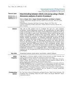
Initial steps during the infection process of cauliflower with seven R. solani AGs and one binucleate Rhizoctonia AGFigure 1
Initial steps during the infection process of cauliflower with seven R. solani AGs and one binucleate Rhizoctonia
AG. A, Microscopic observations of trypan blue stained Rhizoctonia hyphae growing along anticlinal cell walls of cauliflower and
branching in T-shaped angles (upper photograph) and formation of infection cushions (lower photograph). Scale bars = 100
μm. B, Time point (hours post inoculation) of first observation of directed growth of Rhizoctonia hyphae along anticlinal cell
walls and formation of T-shaped branches, formation of infection cushions and penetration through stomata.
A
B
Time point (hpi) of first observation Rank
a
Anastomosis
Group
Directed growth and
T-shaped branches
Infection
cushion
Stomatal
penetration
Directed growth and
T-shaped branches
Infection
cushion
Stomatal
penetration
AG 1-1B 18 18 18 2 2 1
AG 1-1C 12 12 18 1 1 1
AG 2-1 12 12 24 1 1 2
AG 2-2 IIIb 18 24 24 2 3 2
AG 3 24 30 30 3 4 3
AG 4 HGII 18 18 24 2 2 2
AG 5 18 42 24 2 5 2
AG K 36 84 36 4 6 4
a
Dense ranking based on infection rate with rank 1 corresponding with the isolates showing the fastest
development.
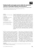
Degradation of cauliflower cell walls by extracellular produced pectic enzymes of seven R. solani AGs and one binucleate Rhizoctonia AGFigure 2
Degradation of cauliflower cell walls by extracellular produced pectic enzymes of seven R. solani AGs and one
binucleate Rhizoctonia AG. Microscopic observations of pectic components in cauliflower cotyledones visualized with
ruthenium red (I, III, V & VII) and toluidine blue (II, IV, VI & VIII) staining after 24 h incubation in sterile culture filtrate of liquid
pectin medium inoculated with a sterile PDA plug as control treatment (I & II), inoculated with R. solani AG 3 (III & IV), inocu-
lated with R. solani AG 4 HGII (V & VI), after 24 h incubation in sterile culture filtrate of liquid cauliflower medium inoculated
with R. solani AG 4 HGII (VII & VIII). Scale bars = 50 μm.
I
II
III
IV
V
VI
VII
VIII
BMC Plant Biology 2009, 9:95 />Page 6 of 12
(page number not for citation purposes)
Role of pectin degrading enzymes in pathogenicity
Under the in vitro conditions tested in this study, all
Rhizoctonia AGs were capable of producing pectic enzymes
which reduced the staining intensity of ruthenium red
and toluidine blue (Fig. 2). Compared with the cotyle-
dons of the control treatment, for which both staining
protocols resulted in a specific coloration of the cell walls,
the cotyledons incubated in the culture filtrate of the eight
Rhizoctonia AGs showed a clear degradation of the cell
walls, including degradation of pectic compounds as indi-
cated by the absence of ruthenium red staining and pink
or purple staining by toluidine blue. No differences were
observed between isolates of pathogenic and non-patho-
genic AGs, suggesting all isolates produced pectinolytic
enzymes that could degrade pectin of cauliflower. Because
the extracellular production of pectin degrading enzymes
depends upon the growth medium [29], two different liq-
uid media were tested; one which contained citrus pectin
and one with cauliflower cell walls. Only the isolate of AG
4 HGII yielded different results for the two media. The cul-
ture filtrate of the AG 4 HGII isolate grown on pectin
medium did not cause a degradation of pectin, while for
the culture filtrate of the cauliflower cell wall medium a
clear degradation of the cotyledonous cell walls was
detected.
Aggressiveness assays
Symptom evaluation at 3 dpi resulted for the aggressive
isolates of AG 1-1B, AG 1-1C, AG 2-1, AG 2-2 IIIb and AG
4 HGII in a disease severity index (DSI) exceeding 3, indi-
cating all symptoms observed showed a susceptible reac-
tion zone (Table 1). For these AGs, resistant reactions
were never observed. At 6 dpi, all AGs were evaluated and
only susceptible reactions were observed for the aggressive
isolates of AG 1-1B, AG 1-1C, AG 2-1, AG 2-2 IIIb and AG
4 HGII. For the weak aggressive isolates of AG 5 and AG 3,
the majority part of the plants showed HR-like lesions,
although some susceptible reactions were also observed
(DSI = 1.6 and 1.4 respectively). Infection with the binu-
cleate isolate of AG K only resulted in HR-like lesions (DSI
= 0.9). To check whether the symptoms caused by the
weak and non-aggressive isolates would shift towards sus-
ceptible reactions, an extra time point at 12 dpi was
included. This was the case for the AG 5 isolate, for which
at 12 dpi all lesions were from the susceptible type and
HR-like lesions were no longer observed (DSI = 3.8). For
the AG 3 isolate, a higher proportion of plants showed
small susceptible reactions combined with HR-like
lesions (DSI = 1.6) and in the case of AG K, all plants
showed HR-like lesions, while susceptible reactions were
absent (DSI = 1).
Histopathological observations
For the aggressive isolates (AG 1-1B, AG 1-1C, AG 2-1, AG
2-2 and AG 4 HGII) samples from 3 and 6 dpi were stud-
ied, while the weak and non-aggressive isolates (AG 3, AG
5 and AG K) were studied at 6 and 12 dpi. Penetration of
epidermal cells by fungal hyphae occurred both by sto-
matal penetration and formation of infection cushions
under which several penetrating hyphae were observed
(Fig. 3). Hyphal penetration was found to be associated
with different levels of cell wall modifications. For
safranin O and aniline blue stain, cellular responses were
classified into three distinct categories (Fig. 4A). In type I
and type II, cell wall fortifications were detected at pene-
tration sites of Rhizoctonia. In the case of type I, hyphae
were completely surrounded by fortified cell walls,
thereby restricting further colonization of the host tissue;
whereas for type II cell wall depositions were detected
although they could not stop the fungal growth and
hyphae were observed beyond the fortified cell walls.
Type III reactions, on the other hand, were characterized
by the absence of cell wall depositions. Staining of the sec-
tions with ruthenium red coloured the pectic compounds
red. At several interaction sites, pectic compounds were
degraded as indicated by the absence of the red stain (Fig.
5A). An overview of the quantitative analysis of the host
cell wall responses observed at the interaction sites of the
eight Rhizoctonia AGs obtained with the three different
stains is presented in Figures 4B, 4C and 5B. The majority
of the type I and type II reaction sites was, besides the wall
Table 1: Disease severity index and average rank of seven
different R. solani AGs and one binucleate Rhizoctonia AG
Disease severity index
Anastomosis Group* 3dpi 6dpi 12 dpi Average rank
AG 1-1B 3.3 ab 3.7 ab nd 1.5
AG 1-1C 3.6 a 3.9 a nd 1.0
AG 2-1 3.2 ab 3.6 ab nd 1.2
AG 2-2 IIIb 3.0 b 3.4 b nd 2.3
AG 3 nd 1.4 c 1.6 b 3.8
AG 4 HGII 3.3 ab 3.6 ab nd 2.2
AG 5 nd 1.6 c 3.8 a 3.2
AG K nd 0.9 d 1.0 c 5.0
Cauliflower plants were grown in potting soil and possessed three
true leaves at the time of artificial inoculation with wheat kernels
colonized by Rhizoctonia. Evaluation was performed at three different
time points using a 0-to-4 scale: 0 = healthy, no symptoms; 1 = HR-
like spots or resistant lesions; 2 = HR-like spots + small susceptible
reactions (< 2 mm); 3 = small susceptible reactions (< 2 mm) and 4 =
large susceptible reactions (> 2 mm). Statistical analysis was
performed on pooled data from two experiments, because
interaction between AG and experiment was not significant and
variations were homogeneous. Different letters indicate statistically
significant differences between AGs according to non-parametric
Kruskal-Wallis and Mann-Whitney tests (α = 0.05). *Negative
significant correlation between average rank and disease severity
index at 6 dpi according to Spearman's rho coefficient of -0.958 (p =
0.01). nd = not determined
BMC Plant Biology 2009, 9:95 />Page 7 of 12
(page number not for citation purposes)
thickening, also associated with granulation of the cyto-
plasm in neighbouring cortical cells. These granules prob-
ably contain phenolic compounds since they stained with
toluidine blue and safranin O. Eventually, these cortical
cells crumpled and collapsed; all these reactions are con-
sistent with a hypersensitive response [30].
Sections of cauliflower stems infected with AG 1-1B, AG 1-
1C, AG 2-1, AG 2-2 IIIb and AG 4 HGII stained with tolu-
idine blue showed the abundant and early formation of
infection cushions and penetration pegs, resulting in a
complete colonization of the cortical cells and vascular
tissue at 3 dpi. For the isolates of AG 3 and AG 5, coloni-
zation occurred slower and only at 12 dpi hyphae of AG 5
were detected in all parts of the cortex and in the vascular
tissue. At that time, hyphae of AG 3 also colonized the cor-
tex and the vascular tissue, although to a lesser extent. The
only isolate that was unable to colonize the cauliflower
cortex was the binucleate AG K isolate; penetrating
hyphae of this AG were limited to substomatal cavities or
the first cortical cell layers underneath the penetration
site.
Results obtained for the safranin O stain and the aniline
blue stain appeared to be very similar (Figs. 4B &4C). For
the majority of the interactions at 3 and 6 dpi, infection
with AG 1-1B, AG 1-1C, AG 2-1 and AG 2-2 IIIb did not
result in the deposition of phenolic compounds or callose
as shown by the high percentage of type III interactions.
These isolates were closely followed by the isolate of AG 4
HGII for which at 3 and 6 dpi approximately 75% of the
interactions were classified as type III for the safranin O
stain and 48.4% at 3 dpi increasing to 61.4% at 6 dpi of
type III interactions for the aniline blue stain. Between the
isolates of AG 3 and AG 5, no significant differences were
found at 6 dpi. However, for the isolate of AG 3 at 12 dpi
a higher percentage of interactions exhibit type I reactions
for safranin O stain (30.2%) and aniline blue stain
(17.6%) compared to the AG 5 isolate (6.0% and 4.1%,
respectively). The highest induction of phenolic com-
pounds and callose deposition was observed for the binu-
cleate isolate of AG K and at 12 dpi all sites of attempted
pathogen entry were associated with an increase in
safranin O and aniline blue staining intensity.
Pectin breakdown, studied by ruthenium red staining, was
already observed at 3 dpi for all the interaction sites of AG
1-1B, AG 1-1C and AG 2-1 (Fig. 5B). For AG 4 HGII, the
majority of the interaction sites also showed pectin degra-
dation. At 6 dpi, around one third of the interaction sites
of AG 3 and AG 5 were associated with a fainter ruthe-
nium red staining, although at 12 dpi significantly more
pectin breakdown was detected for the AG 5 isolate. For
the isolates of AG 2-2 IIIb and AG K, no or only a very low
pectin degradation was observed.
Ranking of AGs and correlation with disease severity
To summarize the results obtained during this research, a
ranking was created for the eight Rhizoctonia AGs. Follow-
ing criteria for ranking were included: directed growth,
infection cushion formation, stomatal penetration,
absence of phenolic compound deposition, absence of
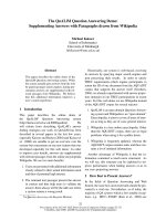
Toluidine blue staining of transversal sections of cauliflower hypocotylsFigure 3
Toluidine blue staining of transversal sections of cauliflower hypocotyls. Stomatal penetration at 6 dpi by binucleate
Rhizoctonia AG K and toluidine blue positive granulation of some adjacent cells (left). Penetration underneath an infection cush-
ion at 3 dpi by R. solani AG 2-1 (right). Scale bars = 50 μm.
BMC Plant Biology 2009, 9:95 />Page 8 of 12
(page number not for citation purposes)
callose deposition and pectin breakdown. The first three
criteria, collectively referred to as infection rate, were
ranked based on the developmental rate of the infection
process with rank 1 corresponding with the isolates show-
ing the fastest development (Fig 1B). The other three cri-
teria dealing with the level of induced defence responses
and pectin degradation were ranked based on the statisti-
cal classes given at 6 dpi for the aggressive isolates and at
12 dpi for the weak and non-aggressive isolates (Figs. 4B,
4C &5B). Based on the average ranking, isolates were
ordered starting from AG 1-1C to AG 2-1, AG 1-1B, AG 4
HGII, AG 2-2 IIIb, AG 5, AG 3 and ending with AG K
(Table 1). Moreover, a significant negative correlation (p
= 0.01) was found between the average ranking and the
DSI caused by the different AGs. The Spearman's rho coef-
ficient equals to -0.958, which should be interpreted as
the first ranked isolates corresponding with the highest
DSI and the isolate with the highest rank corresponding
with the lowest DSI.
Discussion
Although several papers have already been dedicated to
the penetration and colonization process of Rhizoctonia,
the mechanisms involved in the interaction with weak or
non-aggressive isolates remain poorly understood. There-
fore, a study to compare the interaction between cauli-
flower and eight Rhizoctonia AGs with different levels of
aggressiveness was performed. Our observations indicated
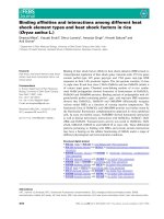
Safranin O and aniline blue staining of cauliflower hypocotyl cells after infection by seven different R. solani AGs and one binu-cleate Rhizoctonia AGFigure 4
Safranin O and aniline blue staining of cauliflower hypocotyl cells after infection by seven different R. solani
AGs and one binucleate Rhizoctonia AG. A, Cellular responses observed with safranin O and aniline blue staining were
classified into three categories; photographs I-III depict representative examples. (I) Rhizoctonia hyphae are completely sur-
rounded by cells fortified with safranin O positive material located in the cell walls or in granules observed in the cytoplasma,
restricting further fungal growth. (II) Fortification of cell walls and presence of safranin O positive granules in the cytoplasma is
observed for some adjacent cells, although colonization by Rhizoctonia hyphae is not stopped. (III) Absence of safranin O posi-
tive host responses in cells neighbouring Rhizoctonia hyphae. Scale bars = 50 μm. B, Frequency distribution of cellular response
categories at 3, 6 and 12 dpi for different Rhizoctonia AGs. The three values within each cell represent the relative proportion
of interaction sites designated as type I, II and III as detected after safranin O staining, respectively. C, Frequency distribution of
cellular response categories at 3, 6 and 12 dpi for different Rhizoctonia AGs. The three values within each cell represent the rel-
ative proportion of interaction sites designated as type I, II and III as detected after aniline blue staining, respectively. At each
time point, at least 50 interaction sites per AG were studied originating from 10 different cauliflower hypocotyls. Within one
column, values followed by the same letter are not significantly different according to Kruskal-Wallis and Mann-Whitney tests
(α = 0.05).
A
B
I II III
Anastomosis Frequency of cellular response category
Rank
a
Group I II III I II III I II III
3 dpi 6 dpi 12 dpi
AG 1-1B 0.0 0.0 100.0 a 0.0 8.3 91.7 ab 1
AG 1-1C 0.0 5.9 94.1 ab 0.0 5.9 94.1 ab 1
AG 2-1 0.0 0.0 100.0 a 0.0 0.0 100.0 a 1
AG 2-2 IIIb 0.0 5.3 94.7 ab 0.0 3.0 97.0 a 1
AG 3 5.7 37.1 57.1 c 30.2 41.5 28.3 b 4
AG 4 HGII 0.0 25.0 75.0 b 0.0 26.1 73.9 bc 2
AG 5 7.7 30.8 61.5 c 6.0 32.0 62.0 a 3
AG K 78.8 18.2 3.0 d 98.2 1.8 0.0 c 5
a
Dense ranking based on statistical classes given at 6 dpi for aggressive isolates and at 12 dpi for weak and non-aggressive isolates.
Anastomosis Frequency of cellular response category
Rank
a
Group I II III I II III I II III
3 dpi 6 dpi 12 dpi
AG 1-1B 0.0 12.5 87.5 a 0.0 16.7 83.3 bc 2
AG 1-1C 0.0 23.5 76.5 ab 0.0 5.9 94.1 ab 1
AG 2-1 0.0 6.3 93.8 a 0.0 0.0 100.0 a 1
AG 2-2 IIIb 0.0 10.5 89.5 a 0.0 1.5 98.5 a 1
AG 3 6.1 36.4 57.6 cd 17.6 51.0 31.4 b 5
AG 4 HGII 0.0 51.6 48.4 b 0.0 38.6 61.4 cd 3
AG 5 11.1 38.9 50.0 d 4.1 36.7 59.2 a 4
AG K 74.2 25.8 0.0 e 100.0 0.0 0.0 c 6
C
I II III
BMC Plant Biology 2009, 9:95 />Page 9 of 12
(page number not for citation purposes)
striking differences among Rhizoctonia AGs during the
early stages of the infection and colonization process and
in the nature and extent of host responses. Moreover, a
highly significant correlation was found between disease
severity rating and ranking of the AGs based on micro-
scopic observations of the infection process, the level of
defence responses and the grade of pectin breakdown.
The pathogenic cauliflower-Rhizoctonia interaction, as
observed for the first ranked isolates of AG 1-1C, AG 2-1
and AG 1-1B, closely followed by AG 4 HGII, was charac-
terized by a high rate of directed growth, formation of
infection cushions and stomatal penetration accompa-
nied with the absence of defence responses and a strong
degradation of pectin. The early observation of the differ-
ent steps in the infection process is in concordance with
previous studies concerning pathogenic Rhizoctonia AGs
on several hosts [9,18,31,32] and the faster and more
abundant stomatal penetration of the AG 1-1B and AG 1-
1C isolates is probably correlated with the aerial nature of
these AGs [33], since isolates from foliage have been
reported to penetrate stomata more frequently [10]. Dur-
ing these pathogenic interactions, pectin degrading
enzymes seemed important and diffused ahead of the fun-
gus, as pathogen ingress was coupled with extensive host
cell deformation and pectin breakdown at locations not
in direct contact with hyphae. For many plant pathogens,
including Rhizoctonia, the role of pectin degrading
Ruthenium red staining of cauliflower hypocotyl cells after infection by seven different R. solani AGs and one binucleate Rhizoc-tonia AGFigure 5
Ruthenium red staining of cauliflower hypocotyl cells after infection by seven different R. solani AGs and one
binucleate Rhizoctonia AG. A, Cellular responses were classified into two categories (I) Representative example of pectin
breakdown as indicated by faint red colour. (II) Uniform red stain of the cell walls indicating absence of pectin breakdown as
observed during the interaction with R. solani AG 2-2 IIIb. Scale bars = 50 μm. B, Relative proportion of interaction sites at
which pectin degradation is observed at 3, 6 and 12 dpi during the interaction with different Rhizoctonia AGs. At each time
point, at least 50 interaction sites per AG were studied originating from 10 different cauliflower hypocotyls. Within one col-
umn, values followed by the same letter are not significantly different according to Kruskal-Wallis and Mann-Whitney tests (α
= 0.05).
A
B
I II
Anastomosis
Group
3 dpi 6 dpi 12 dpi Rank
a
AG 1-1B 100.0 a 100.0 a 1
AG 1-1C 100.0 a 100.0 a 1
AG 2-1 100.0 a 100.0 a 1
AG 2-2 IIIb 0.0 b 0.0 d 5
AG 3 34.5 c 34.0 b 4
AG 4 HGII 85.0 a 59.5 b 2
AG 5 37.2 c 67.4 a 3
AG K 0.0 d 1.8 c 5
a
Dense ranking based on statistical classes given
at 6 dpi for aggressive isolates and at 12 dpi for
weak and non-aggressive isolates.
BMC Plant Biology 2009, 9:95 />Page 10 of 12
(page number not for citation purposes)
enzymes in plant cell wall degradation is well established
[11,12,23,34]. Pectin degradation of the plant cell wall
plays a crucial role in pathogen spread and providing
nutriments to the pathogen and therefore, pectin degrad-
ing enzymes are potentially important for pathogenicity
[35,36].
The small decrease in disease severity, observed for the fol-
lowing ranked isolate of R. solani AG 2-2 IIIb, can be
ascribed to the later formation of infection cushions and
to the remarkable disability to degrade pectic compounds.
Notwithstanding pectin degrading enzymes were pro-
duced during the in vitro experiments and pectic com-
pounds of cauliflower cotyledons were degraded after
incubation in the culture filtrate, the degradation of pectin
was never observed during the histopathological experi-
ments. A conceivable explanation might involve the dif-
ferent composition of pectin present in different plant
parts, such as cotyledons and hypocotyls [37], resulting in
a different susceptibility to degradation by the enzymes
produced by the AG 2-2 IIIb isolate. Another possibility
suggests the involvement of a plant response leading to
the production of plant protein inhibitors which prevent
cell wall degradation and retard fungal growth and colo-
nization [38]. The slower rate of disease development
observed for this isolate further supports this hypothesis.
Inhibitory activity of pectin degrading enzymes by plant
proteins is considered a part of the plants' immune system
and depends on the specific recognition of the pathogen
[39]. In the case of R. solani AG 2-2, a protein inhibiting
pectin lyase activity in sugar beet has already been
described [16]. From this point of view, the specific recog-
nition of R. solani AG 2-2 IIIb by cauliflower might explain
why infection cushion formation and disease develop-
ment was slower and why this AG was never found in
association with cauliflower under field conditions [21].
However, despite the inability to degrade pectic compo-
nents from the cell wall of cauliflower as observed in this
study, AG 2-2 IIIb is generally considered aggressive
towards Brassica crops [21,40] and as a consequence, dur-
ing the interaction with cauliflower pectin degrading
enzymes are not considered essential for the pathogenic-
ity of this AG.
A slower development of the infection process coinciding
with the induction of plant defence responses and a lower
level of pectin breakdown was detected for the weak
aggressive isolates of R. solani AG 5 and AG 3 which
ranked next. Possibly the later penetration of the plant tis-
sue allows the plant to build up a defence reaction, as
observed by the deposition of phenolic compounds and
callose. This defence reaction was more pronounced for
AG 3 compared to AG 5. Furthermore, the frequently
observed pectin degradation by the AG 5 isolate might
help the fungus to overcome the defence responses and to
colonize the plant tissue, resulting in a significantly higher
disease severity at 12 dpi for AG 5 compared to AG 3. Dur-
ing this study the experimental conditions were in favour
of the pathogen, because a relatively high infection pres-
sure was used towards young cauliflower plants. This
might explain why during our experiments isolates of AG
5 and AG 3 could provoke such high levels of damage and
colonize the complete hypocotyl; while under natural
conditions, these AGs are considered not aggressive
towards cauliflower [21]. Probably, under field condi-
tions the plant's defence reactions are sufficient to arrest
fungal colonization.
A non-pathogenic interaction was identified for the last
ranked isolate of binucleate Rhizoctonia AG K and was typ-
ified by a slow infection rate, resulting in only few infec-
tion cushions formed. Contrastingly, Keijer et al. [8]
reported that non-pathogenic isolates could not adhere to
the plant surface preventing further formation of infection
structures. Here, we corroborated the penetration of cauli-
flower by AG K through stomata and infection cushion
formation, indicating the passive defence barriers present
in cauliflower can be overcome by this non-pathogenic
Rhizoctonia AG. Furthermore, a very strong induction of
phenolic compounds and callose was observed in associ-
ation with the absence of pectin breakdown. However,
extracellular pectinolytic enzymes produced by AG K
could degrade the pectin present in cauliflower cotyle-
dons and the lack in pectin breakdown, as observed dur-
ing the histopathological experiments, is probably due to
the restriction of the fungal growth by the local deposition
of cell wall components. Deposition of cell wall fortifica-
tions, is a widely observed phenomenon in preventing
fungal penetration and colonization [41] and at all the
interaction sites with AG K we detected densely stained
cells enriched in phenolic compounds and callose sur-
rounding the penetrating hyphae. Phenolic compounds
not only form physical barriers for the pathogen, they are
also known to have direct antimicrobial activities [42].
The granules present in the cortical cells adjacent to the
penetration site as observed in this study are assumed to
contain phenolic defence compounds synthesized by cau-
liflower in response to the attack by weak and non-aggres-
sive Rhizoctonia isolates. At the reinforced cell walls,
callose accumulation was also detected. Callose, a 1,3-β-
glucan, may provide a physical barrier and has been
described as a key component of penetration resistance in
several plant-pathogen interactions [5,43,44]. Until now,
strengthening of the cell wall in response to Rhizoctonia
attack has only been reported for a non-pathogenic binu-
cleate isolate of AG G. Jabaji-Hare et al. [17] described an
increase in phenolic compounds, but not in callose dur-
ing the interaction of bean and AG G, while Wolski et al.
[45] showed an increase in both lignin and callose using
a purified 1,3-α-glucan elicitor from AG G in potato
BMC Plant Biology 2009, 9:95 />Page 11 of 12
(page number not for citation purposes)
sprouts. In this study, we reported the role of callose and
phenolic compound deposition in the prevention of col-
onization of cauliflower by binucleate Rhizoctonia AG K
and to a lesser extent by R. solani AG 3 and AG 5. The
strong induction of cell wall fortifications with phenolic
compounds and callose, leading to the arrest of the path-
ogen shortly after entrance of AG K implicates a state of
high induced resistance. This might be linked with the
biological control capacity ascribed to several non-patho-
genic isolates of binucleate Rhizoctonia AGs [46]. The pro-
tection of several plant species by binucleate Rhizoctonia
strains against infection by pathogenic R. solani isolates is
a frequently studied topic [47-51]. Moreover, protection
against fungal pathogens of other genera was observed
[52-54]. The potential of the binucleate Rhizoctonia isolate
of AG K used in this study as a biological control agent is
still unclear and requires further research.
Conclusion
In summary, we have shown that during the cauliflower-
Rhizoctonia interaction different levels of disease severity
are correlated with differences in infection rate, differ-
ences in host response and the ability to degrade pectic
compounds. Highly pathogenic interactions were found
for isolates of R. solani AG 1-1C, AG 2-1, AG 1-1B and AG
4 HGII and were characterized by a high infection rate in
association with the absence of host responses and a
strong pectin degradation. The slightly lower disease
severity observed for AG 2-2 IIIb was due to a slower for-
mation of infection cushions and the inability to degrade
pectin. Furthermore, we detected that weak aggressive iso-
lates of R. solani AG 3 and AG 5 and the non-pathogenic
binucleate isolate of Rhizoctonia AG K entered the plant
tissue both by the formation of infection cushions and by
stomatal penetration, indicating all constitutive defence
barriers present in cauliflower were defeated and differ-
ences in aggressiveness were caused by inducible defence
responses. In addition, these defence responses were
shown to include the deposition of phenolic compounds
and callose of which different levels were detected at the
interaction sites of the Rhizoctonia AGs, resulting in differ-
ences in disease severity.
Authors' contributions
The studies were conceived and planned by JP and MH. JP
carried out all experimental work and wrote the draft
manuscript in consultation with MH. The manuscript was
edited and prepared for submission by JP and MH. Both
authors read and approved the final manuscript.
Acknowledgements
This work was supported by a specialization fellowship of the Flemish Insti-
tute for the stimulation of Scientific-Technological Research in Industry
(IWT, Belgium) given to Joke Pannecoucque. We thank Ilse Delaere for
technical assistance and Sarah Van Beneden for critical review of this man-
uscript.
References
1. Shetty NP, Jorgensen HJL, Jensen JD, Collinge DB, Shetty HS: Roles
of reactive oxygen species in interactions between plants
and pathogens. Eur J Plant Pathol 2008, 121(3):267-280.
2. Greenberg JT, Yao N: The role and regulation of programmed
cell death in plant-pathogen interactions. Cell Microbiol 2004,
6(3):201-211.
3. Ferreira RB, Monteiro S, Freitas R, Santos CN, Chen Z, Batista LM,
Duarte J, Borges A, Teixeira AR: The role of plant defence pro-
teins in fungal pathogenesis. Mol Plant Pathol 2007, 8(5):677-700.
4. Huckelhoven R: Cell wall – associated mechanisms of disease
resistance and susceptibility. Ann Rev Phytopathol 2007,
45:101-127.
5. Skalamera D, Jibodh S, Heath MC: Callose deposition during the
interaction between cowpea (Vigna unguiculata) and the
monokaryotic stage of the cowpea rust fungus (Uromyces vig-
nae). New Phytol 1997, 136(3):511-524.
6. Carling DE, Kuninaga S, Brainard KA: Hyphal anastomosis reac-
tions, rDNA-internal transcribed spacer sequences, and vir-
ulence levels among subsets of Rhizoctonia solani
anastomosis group-2 (AG-2) and AG-BI. Phytopathology 2002,
92(1):43-50.
7. Sharon M, Kuninaga S, Hyakumachi M, Naito S, Sneh B: Classifica-
tion of Rhizoctonia spp. using rDNA-ITS sequence analysis
supports the genetic basis of the classical anastomosis
grouping. Mycoscience 2008, 49(2):93-114.
8. Keijer J, Korsman MG, Dullemans AM, Houterman PM, de Bree J, Van
Silfhout CH: In vitro analysis of host plant specificity in Rhizoc-
tonia solani. Plant Pathol 1997, 46(5):659-669.
9. Keijer J: The initial steps of the infection process in Rhizocto-
nia solani. In Rhizoctonia species: Taxonomy, molecular biology, ecology,
pathology and disease control Edited by: Sneh B, Jabaji-Hare S, Neate
SM, Dijst G. Dordrecht, The Netherlands: Kluwer Academic;
1996:149-162.
10. Dodman RL, Barker KR, Walker JC: Modes of penetration by dif-
ferent isolates of Rhizoctonia solani. Phytopathology 1968,
58:31-33.
11. Weinhold AR, Motta J: Initial host responses in cotton to infec-
tion by Rhizoctonia solani. Phytopathology 1973, 63(1):157-162.
12. Marcus L, Barash I, Sneh B, Koltin Y, Finkler A: Purification and
characterization of pectolytic enzymes produced by virulent
and hypovirulent isolates of Rhizoctonia solani Kuhn. Physiol
Mol Plant Pathol 1986, 29(3):325-336.
13. Yang J, Verma PR, Lees GL: The role of cuticle and epidermal
cell wall in resistance of rapeseed and mustard to Rhizoctonia
solani. Plant and Soil 1992, 142(2):315-321.
14. Marshall DS, Rush MC: Infection cushion formation on rice
sheaths by Rhizoctonia solani. Phytopathology 1980,
70(10):947-950.
15. Bateman DF, Lumsden RD: Relation of calcium content and
nature of pectic substances in bean hypocotyls of different
ages to susceptibility to an isolate of Rhizoctonia solani. Phy-
topathology 1965, 55(7):734-738.
16. Bugbee WM: A pectin lyase inhibitor protein from cell walls of
sugar beet. Phytopathology 1993, 83(1):63-68.
17. Jabaji-Hare S, Chamberland H, Charest PM: Cell wall alterations in
hypocotyls of bean seedlings protected from Rhizoctonia
stem canker by a binucleate Rhizoctonia isolate. Mycol Res
1999, 103:1035-1043.
18. Yang J, Verma PR, Tewari JP: Histopathology of resistant mus-
tard and susceptible canola hypocotyls infected by Rhizocto-
nia solani. Mycol Res 1992, 96:171-179.
19. Bassi A, Moore EL, Batson WE: Histopathology of resistant and
susceptible tomato fruit infected with Rhizoctonia solani. Phy-
topathology 1979, 69(6):556-559.
20. Ruppel EG: Histopathology of resistant and susceptible sugar
beet roots inoculated with Rhizoctonia solani. Phytopathology
1973, 63(1):123-126.
21. Pannecoucque J, Van Beneden S, Hofte M: Characterization and
pathogenicity of Rhizoctonia isolates associated with cauli-
flower in Belgium. Plant Pathol 2008, 57(4):737-746.
22. Schneider JHM, Schilder MT, Dijst G: Characterization of Rhizoc-
tonia solani AG 2 isolates causing bare patch in field grown
tulips in the Netherlands. Eur J Plant Pathol 1997, 103(3):265-279.
23. Bugbee WM: Purification and characteristics of pectin lyase
from Rhizoctonia solani. Physiol Mol Plant Pathol 1990, 36(1):15-25.
Publish with Bio Med Central and every
scientist can read your work free of charge
"BioMed Central will be the most significant development for
disseminating the results of biomedical research in our lifetime."
Sir Paul Nurse, Cancer Research UK
Your research papers will be:
available free of charge to the entire biomedical community
peer reviewed and published immediately upon acceptance
cited in PubMed and archived on PubMed Central
yours — you keep the copyright
Submit your manuscript here:
/>BioMedcentral
BMC Plant Biology 2009, 9:95 />Page 12 of 12
(page number not for citation purposes)
24. Ruzin SE: Plant microtechnique and microscopy. New York:
Oxford University Press; 1999.
25. Mellersh DG, Foulds IV, Higgins VJ, Heath MC: H2O2 plays differ-
ent roles in determining penetration failure in three diverse
plant-fungal interactions. Plant J 2002, 29(3):257-268.
26. Scholten OE, Panella LW, De Bock TSM, Lange W: A greenhouse
test for screening sugar beet (Beta vulgaris) for resistance to
Rhizoctonia solani. Eur J Plant Pathol 2001, 107(2):161-166.
27. Lucena MA, Romero-Aranda R, Mercado JA, Cuartero J, Valpuesta V,
Quesada MA: Structural and physiological changes in the
roots of tomato plants over-expressing a basic peroxidase.
Physiol Plantarum 2003, 118(3):422-429.
28. Hood ME, Shew HD: Applications of KOH-aniline blue fluores-
cence in the study of plant-fungal interactions. Phytopathology
1996, 86(7):704-708.
29. Alghisi P, Favaron F: Pectin-degrading enzymes and plant-para-
site interactions. Eur J Plant Pathol 1995, 101(4):365-375.
30. Heath MC: Hypersensitive response-related death. Plant Mol
Biol 2000, 44(3):321-334.
31. Matsuura K: Scanning electron microscopy of the infection
process of Rhizoctonia solani in leaf sheaths of rice plants. Phy-
topathology 1986, 76(8):811-814.
32. Armentrout VN, Downer AJ: Infection cushion development by
Rhizoctonia solani on cotton. Phytopathology 1987, 77(4):619-623.
33. Sneh B, Burpee L, Ogoshi A: Identification of Rhizoctonia spe-
cies. St. Paul, Minnesota, USA: The American Phytopathology Soci-
ety; 1991.
34. Jayasinghe CK, Wijayaratne SCP, Fernando THPS: Characteriza-
tion of cell wall degrading enzymes of Thanatephorus
cucumeris. Mycopathologia 2004, 157(1):73-79.
35. Collmer A, Keen NT: The role of pectic enzymes in plant
pathogenesis. Ann Rev Phytopathol 1986, 24:383-409.
36. Walton JD: Deconstructing the cell wall. Plant Physiol 1994,
104(4):1113-1118.
37. Ridley BL, O'Neill MA, Mohnen DA: Pectins: structure, biosyn-
thesis, and oligogalacturonide-related signaling. Phytochemistry
2001, 57(6):929-967.
38. Juge N: Plant protein inhibitors of cell wall degrading
enzymes. Trends Plant Sci 2006, 11(7):359-367.
39. Federici L, Di Matte A, Fernandez-Recio J, Tsernoglou D, Cervone F:
Polygalacturonase inhibiting proteins: players in plant innate
immunity? Trends Plant Sci 2006, 11(2):65-70.
40. Verma PR: Oilseed rape and canola diseases incited by Rhizoc-
tonia species. In Rhizoctonia species: Taxonomy, molecular biology, ecol-
ogy, pathology and disease control Edited by: Sneh B, Jabaji-Hare S,
Neate SM, Dijst G. Dordrecht, The Netherlands: Kluwer Academic;
1996:249-258.
41. Hardham AR, Jones DA, Takemoto D: Cytoskeleton and cell wall
function in penetration resistance. Curr Opin Plant Biol 2007,
10(4):342-348.
42. Bennett RN, Wallsgrove RM: Secondary metabolites in plant
defense mechanisms. New Phytol 1994, 127(4):617-633.
43. Aist JR: Papillae and related wound plugs of plant cells. Ann Rev
Phytopathol 1976, 14:145-163.
44. Asselbergh B, Hofte M: Basal tomato defences to Botrytis cine-
rea include abscisic acid-dependent callose formation. Physiol
Mol Plant Pathol 2007, 71(1–3):33-40.
45. Wolski EA, Maldonado S, Daleo GR, Andreu AB: A novel alpha-1,3-
glucan elicits plant defense responses in potato and induces
protection against Rhizoctonia solani AG-3 and Fusarium
solani f. sp. eumartii.
Physiol Mol Plant Pathol 2006, 69(1–3):93-103.
46. Sneh B: Non pathogenic isolates of Rhizoctonia spp. (np-R)
and their role in biological control. In Rhizoctonia species: Taxon-
omy, molecular biology, ecology, pathology and disease control Edited by:
Sneh B, Jabaji-Hare S, Neate SM, Dijst G. Dordrecht, The Nether-
lands: Kluwer Academic; 1996:473-483.
47. Khan FU, Nelson BD, Helms TC: Greenhouse evaluation of binu-
cleate Rhizoctonia for control of R. solani in soybean. Plant Dis
2005, 89(4):373-379.
48. Poromarto SH, Nelson BD, Freeman TP: Association of binucle-
ate Rhizoctonia with soybean and mechanism of biocontrol
of Rhizoctonia solani. Phytopathology 1998, 88(10):1056-1067.
49. Burpee LL, Goulty LG: Suppression of brown patch disease of
creeping bentgrass by isolates of nonpathogenic Rhizoctonia
spp. Phytopathology 1984, 74(6):692-694.
50. Hwang J, Benson DM: Biocontrol of Rhizoctonia stem and root
rot of poinsettia with Burkholderia cepacia and binucleate
Rhizoctonia. Plant Dis 2002, 86(1):47-53.
51. Villajuan-Abgona R, Kageyama K, Hyakumachi M: Biocontrol of
Rhizoctonia damping-off of cucumber by non-pathogenic
binucleate Rhizoctonia. Eur J Plant Pathol 1996, 102(3):227-235.
52. Olson HA, Benson DM: Induced systemic resistance and the
role of binucleate Rhizoctonia and Trichoderma hamatum 382
in biocontrol of Botrytis blight in geranium. Biol Control 2007,
42(2):233-241.
53. Cardinale F, Ferraris L, Valentino D, Tamietti G: Induction of sys-
temic resistance by a hypovirulent Rhizoctonia solani isolate
in tomato. Physiol Mol Plant Pathol 2006, 69(4–6):160-171.
54. Jabaji-Hare S, Neate SM: Nonpathogenic binucleate Rhizoctonia
spp. and benzothiadiazole protect cotton seedlings against
Rhizoctonia damping-off and Alternaria leaf spot in cotton.
Phytopathology 2005, 95(9):1030-1036.