báo cáo khoa học: " Analysis of a c0t-1 library enables the targeted identification of minisatellite and satellite families in Beta vulgaris" ppt
Bạn đang xem bản rút gọn của tài liệu. Xem và tải ngay bản đầy đủ của tài liệu tại đây (3.8 MB, 14 trang )
RESEARC H ARTIC LE Open Access
Analysis of a c
0
t-1 library enables the targeted
identification of minisatellite and satellite families
in Beta vulgaris
Falk Zakrzewski
1†
, Torsten Wenke
1†
, Daniela Holtgräwe
2
, Bernd Weisshaar
2*
, Thomas Schmidt
1
Abstract
Background: Repetitive DNA is a major fraction of eukaryotic genomes and occurs particularly often in plants.
Currently, the sequencing of the sugar beet ( Beta vulgaris) genome is under way and knowledge of repetitive DNA
sequences is critical for the genome annotation. We generated a c
0
t-1 library, representing highly to moderately
repetitive sequences, for the characterization of the major B. vulgaris repeat fami lies. While highly abundant
satellites are well-described, minisatellites are only poorly investigated in plants. Therefore, we focused on the
identification and characterization of these tandemly repeated sequences.
Results: Analysis of 1763 c
0
t-1 DNA fragments, providing 442 kb sequence data, shows that the satellites pBV and
pEV are the most abundant repeat families in the B. vulgaris genome while other previously described repeats
show lower copy numbers. We isolated 517 novel repetitive sequences and used this fraction for the identification
of minisatellite and novel satellite families. Bioinformatic analysis and Southern hybridization revealed that
minisatellites are moderately to highly amplified in B. vulgaris. FISH showed a dispersed localization along most
chromosomes clustering in arrays of variable size and number with exclusion and depletion in distinct regions.
Conclusion: The c
0
t-1 library represents major repeat families of the B. vulgaris genome, and analysis of the c
0
t-1
DNA was proven to be an efficient method for identification of minisatellites. We established, so far, the broadest
analysis of minisatellites in plants and observed their chromosomal localization providing a background for the
annotation of the sugar beet genome and for the understanding of the evolution of minisatellites in plant
genomes.
Background
Repetitive DNA makes up a large proportion of eukar-
yotic genomes [1]. Major findings in t he last fe w years
show that repetitive DNA is involved in the regulation
of heterochromatin formation, influences gene expres-
sion or contributes to epigenetic regulatory processes
[2-7]. Therefore, understanding the role of repetitive
DNA and the characterization of their structure, organi-
zation and evolution is essential. A rapid procedure to
identify repetitive DNA is based on c
0
t DNA isolation
[8], which is an efficient method for the detection o f
major repetiti ve DNA fractions as well as for the identi-
fication of novel repetitive sequences in genomes [9].
The c
0
t DNA isolation is based on the renaturation of
denaturated genomic DNA within a defined period of
time and concen tration. The rate at which the fragmen-
ted DNA sequences reassociate is proportional to the
copy number in the genome [8] and therefore , c
0
t DNA
isolated after short reassociation time (e.g. c
0
t-1)repre-
sents the repetitive fraction of a genome. Recently, ana-
lyses of c
0
t DNA we re performed in plants e.g. for Zea
mays, Musa acuminata, Sorghum bicolor and Leymus
triticoides [8,10-12].
Satellite DNA consisting of tandemly organized repeat-
ing units (monomers) of relatively conserved sequence
motifs is a major class of repetitive DNA. Depending on
monomer size, tandem repeats are subdivided into satel-
lites, minisatellites and microsatellites and tandem
repeats with specific functions such as telomeres and
ribosomal genes. The monomer size of minisatellites
* Correspondence:
† Contributed equally
2
Institute of Genome Research, University of Bielefeld, D-33594 Bielefeld,
Germany
Zakrzewski et al. BMC Plant Biology 2010, 10:8
/>© 2010 Zakrzewski et al; licensee BioMed Central Ltd. This is an Open Access article distributed under the terms of the Creative
Commons Attribution License ( enses/by/2.0), which permits unrestricted use, distribution, a nd
reproduction in any medium, provided the original work is properly cited.
varies between 6 to 100 bp [13] and those of microsatel-
lites between 2 to 5 bp [14]. Most plant satellites have a
monomer length of 160 to 180 bp or 320 to 370 bp [15].
Satellite DNAs are non-coding DNA sequences, which
are predominantly located in subterminal, intercalary and
centromeric regions of plant chromosomes. The majori ty
of typical plant satellite arrays are several megabases in
size [15]. In c ontrast, arra ys of minisatellites vary in
length from 0.5 kb to several kilobases [13]. Minisatellites
are often G/C-rich and fast evolving [13] and thought to
originate from s lippage replication or recombination
between short direct repeats [16] or slipped-strand mis-
pairing replication at non-contiguous repeats [17]. Minis-
atellites are poorly investigated in plants. So far, o nly a
few minisatellites were described, for example i n Arabi-
dopsis thaliana, O. sativa, Triticum aestivum, Pisum sati-
vum and some other plant species [18-26]. Moreover,
only two minisatellite families were physically mapped on
plant chromosomes using fluorescent in situ hybridiza-
tion (FISH) [19].
The sequencing of the sugar beet (Beta vulgaris)gen-
ome, which is about 758 Mb in size [27] and has been
estimated to contain 63% repetitive sequences [28], is
under way and the first draft of genome sequence is
currently established [29]. Knowledge about repetitive
DNA and their p hysical localization is essential for the
correct annotation of the sugar beet genome. Therefore,
we detected and cl assified the repeated DNA fraction of
B. vulgaris using sequence data from cloned c
0
t-1 DNA
fragments. We focused on the investigation of novel
tandem repeats and characterized nine minisatellite and
three satellite families. Their chromosomal localization
was determined by multicolor FISH and the organiza-
tion within the genome of B. vulgaris was analyzed by
Southern hybridization.
Results
c
0
t-1 analysis reveals the most abundant satellite DNA
families of the B. vulgaris genome
In order to analyze the composition of the repetitive
fraction of the B. vulgaris genome, we prepared c
0
t-1
DNA from genomic DNA a nd generated a library con-
sisting of 1763 clones with an average insert size
between 100 to 600 bp providing in total 442 kb (0.06%
of the genome) sequence data. For the characterization
of the c
0
t-1 DNA s equences we performed homology
search against nucleotide sequences and proteins in
public databases and classified all clones based on their
similarity to described repeats, telomere-like motifs,
chloroplast-like sequences as well as novel sequences
lacking any homology (Figure 1). More than half of the
c
0
t-1 fraction (60%) belongs to known repeat classes
including mostly satellites. In order to determine the
individual proportion of each repeat family we applied
BLAST analysis using representative query sequences of
each repeat. We observed that the relative frequency of
repetitive sequence motifs found in the c
0
t-1 library cor-
relates with its genomic abundance in B. vulgaris:The
most frequently occurring repeat is pBV (32.8%, 579
clones), [EMBL:Z22849], a highly repetitive satellite
family that is amplified in l arge arrays in centromeric
and pericentromeric regions of all 18 chromosomes
[30,31]. The next repeat in row has been observed in
19.5% of cases (343 clones) and belongs to the highly
abundant satellite family pEV [EMBL:Z22848] that
forms large arrays in intercalary heterochromatin of
each chromosome arm [32]. The c
0
t-1 DNA library also
enabled the detection of moderately amplified repeats.
Telomere-like motifs of the Arabidopsis-type were
detected in 1.1% (20 clones) while a smaller proportion
of sequences belong to the satellite family pAv34 (0.9%,
16 clones), [EMBL:AJ242669] which is organized in tan-
dem arrays at subtelomeric regions [33]. Only 0.1% (2
clones) belong t o the satellite families pHC28 [EMBL:
Z22816] [34] and pSV [EMBL:Z75011] [35], respectively,
which are distributed mostly in in tercalary and pericen-
tromeric chromosome regions. Furthermore, microsatel-
lite motifs were found in 1.7% of c
0
t-1 sequences [36].
Miniature inverted-repeat transposable elements
(MITEs) [EMBL:AM231631], d erived from the Vulmar
family of mariner transposons [37], were identified in
0.3% (6 clones) of the c
0
t-1 sequences, while Vulmar
[EMBL:AJ556159] [38] was detected in a single clon e
only. The repeat pRv [EMBL:AM944555] was found in a
relatively low number of c
0
t-1 sequences (0.4%, 7 clones)
indicating lower abundance than the satellite pBV. pRv
is only amplified within pBV monomers and forms a
complex structure with pBV [31]. Surprisingly, the
homology search enabled the detection of a large
amount of c
0
t-1 sequences (13.6%) that show similarities
to chloroplast DNA.
The identification of novel repetitive sequences was an
aim of the c
0
t-1 analysis. Altogether, we identified 29.3%
(517 clones) of the c
0
t-1 sequences lacking homology to
previously described B. vulgaris repeats. However, to
verify the repetitive character of each sequence motif we
performed BLAST search against available B. vulgaris
sequences. 56582 BAC end sequences (BES) [39], (Holt-
gräwe and Weisshaar, in preparation) covering 5.2% of
the genome were used for analysis. 360 c
0
t-1 sequences
showed hits in BES ranging from 11 to 300 while 39
sequences showed more than 300 hits and 118
sequences less than 10 hits. This observation indicates
that many of these yet uncharacterized c
0
t-1 clones con-
tain sequence mo tifs t hat are h ighly to moderately
amplified in the genome.
We performed an assembly of the 517 uncharacterized
c
0
t-1 clones to generate contigs, which contain
Zakrzewski et al. BMC Plant Biology 2010, 10:8
/>Page 2 of 14
sequences belonging to an individual r epeat family. In
total, 37 contigs ranging in size from 149 bp to 1694 bp
(average size 555 bp) were established. The largest con-
tig in s ize and clone number (1694 bp, 20 sequences)
was used for BLAST search against available sequences.
Analysis of the generated alignment revealed a LTR of a
retrotransposon. The full-length element designa ted
Cotzilla was classified as an envelope-like Copia LTR
retrotransposon related to sirevirus es [40]. The internal
region of Cotzilla showed similarity to 40 sequences of
118 c
0
t-1 clones categorized as retrotransposon-like
(Figure 1C) showing that Cotzilla is the most abundant
retrotransposon within the c
0
t-1 library. Analysis of a
further contig (1081 bp, 4 clones) resulted in the identi-
fication of the LTR of a novel Gypsy retrotransposon
(unpublished) that shows 13 hits within the c
0
t-1 library.
Three further clones displayed similarities to transpo-
sons. The remaining uncharacterized c
0
t-1 clones (396
sequences) were used for the identification of tandemly
arranged repeats.
Figure 1 Classification of isolated c
0
t-1 DNA sequences. A: Absolute and relative distributi on of 1763 c
0
t-1 sequences of the B. vulgaris
genome. B: Number of clones (known repeats in A) with similarities to previously described B. vulgaris repeats. C: Classification of novel
repetitive sequences.
Zakrzewski et al. BMC Plant Biology 2010, 10:8
/>Page 3 of 14
Targeted isolation of minisatellites and satellites using the
c
0
t-1 library
Plant minisatellites do not have typical conserved
sequence motifs, t herefore the analysis of c
0
t DNA is a
use ful method for the targeted isolation of minisatellites.
We scanned the 396 clones of the c
0
t-1 library that show
no similarity to known repeats and detected 35 sequences
that contain tandemly repeated sequences. Based on their
similarity these sequences were grouped into nine minis-
atellite families and three satellite families. The minisatel-
lites were named according to their order of detection
and the satellites according to conserved internal restric-
tion sites (Table 1). A s equence of each tandem repeat
family was used as query and blasted agai nst available
sequences to identify ad ditional B. vulgaris cop ies. Align-
ments of all sequences of each tandem repeat family were
generated and the average monomer size, t he G/C-con-
tent and the identity values of at least 20 randomly
selected monomers determined (Table 1).
In order to investigate the genomic organization and
abundance of the tandem repeats, Southern hybridiza-
tions were carried out. A strong hybridization smear of a
wide molecular weight range was detected in each case
indicating abundance o f the min isatellite fami lies in the
genome o f B. vulgaris (F igure 2A - G). Dis tinct single
bands were observed for the minisatel lite families
BvMSat10 (Figure 2, H) and BvMSat11 (Figure 2, I).
Because of the short length, recognition sites for restric-
tion enzymes are rare or abse nt within minisatellite
monomers. Thus, genomic DNA was restricted with 15
different restriction enzymes to identify restriction
enzymes generating mono- and multimers in minisatel-
lite arrays detectable by Southern hybrid ization. Figu re 2
illustrates the probing of genomic DNA after restriction
with the 5 restriction enzymes generating most ladder-
like patterns in minisatellite and satellite arrays. A typical
ladder-like pattern is detect able for BvMSat04 (Figure
2C,lane1)andBvMSat03(Figure2B,lane2).Multiple
restriction fragments were observed after hybridization of
BvMSat08 (Figure 2 F). The tandem organization o f the
minisatellites lacking restriction sites was confirmed by
sequence analysis or PCR (not shown). Typical ladder-
like patterns were gene rated for each s atellite family. For
example, the tandem organization was verified for the
FokIsatellite,AluI satellite and HinfI satellite after
restriction with AluI (Figure 2, J-L, lane 3,).
To investigate the DNA methylation of the tandem
repeatsinCCGGmotifs,genomicDNAwasdigested
Table 1 Minisatellites and satellites identified in the c
0
t-1 library of B. vulgaris
tandem
repeat
size
[bp]
c
0
t-1
hits
G/C-content
[%]
identity
[%]
EMBL
accession
representative monomere sequence
BvMSat01 10 7 34 40 - 100 ED023089 AACTTATTGG
BvMSat11 15 1 41 36 - 100 DX580797 TAAATAGTCAAGCCC
BvMSat05 21 5 29 38 - 100 ED029002 ACTGAAAAAAAATGAAGACTA
BvMSat07 30 4 32 90 - 100 ED019743 GAAAAAATAAGTTCAGATCAGATCAGATCA
BvMSat08 32 1 48 77 - 100 DX107266 GGGTCGGAATAAATCGGCTTTCGAAATGACTT
BvMSat09 32-39 5 24 46 - 100 FN424406 AGAAGTATACAAGAACATTAATCAAAATATATAAACAAA
BvMSat03 40 3 33 55 - 100 ED024452 GTCTCTAAAGCCATGTATTTAGCGTCACATGAATTTAGTT
BvMSat10 51 3 24 78 - 100 DX980914 GTTTGTTCTTAAAAGGTTGTTCTTGAATTATTATTCAAGTGTTTGGAAAGA
BvMSat04 96 2 41 70 - 100 DX983375 CCTCTAAATGTAAGTGGCTTTAGCAGCACTATAAGTTCTGTGCCTAAAAAA
GGTGGCATTACGGGCAACCAACAATTAGCGACAGGCATATGGTTG
FokI-satellite 130 1 60 81 - 100 DX979624 GGGACTTAGGAGAGTGACCCAACCAAGGAGGGAGACCTCCTTGGGCTGAGT
TGGGTGGACGCGGCTCGGATGAGGGGCCAATGAGCCCCACGCTTGTCCGAG
CCGGTGCCGTCTCTCGCCATGTCAATCT
AluI-satellite 173 1 33 78 - 100 ED022281 ATAATCATACCTCTATGCCTATTCCAAGTTCTAATGGCTAATGCAAGTCCT
AAAATACTCATTTAAACTTTCTACTACATGGTTGTAAGATTCTAAGCAAGT
TTAATACACTTAGCCAATTAAAATGAGAAAAACTAAGCCATTTCGAGCCGT
TTTTTGGGTTTCATGTTCCT
HinfI-satellite 325 2 45 75 - 86 DX982322 TGTGACTTGTAACATTGCGCGGGTGCTTGGCACCATTTGCGTTACCTCAAA
AAGCCTTTGAACACCCCAATTATTCATTTCTCGCGAAATCCAAAATTGCCT
CGAAATGAACGTAAAGGCATCCACATATTTGTTCCAAGCCACATGACTCCT
TTACATTGACCTCCTATGTCCCTAGGAGGCATCCCGTGCCATTTGGAGCTC
GGGCAACGGGAAAGTCCGAAAGCGTGTATAATCTTCAATTTTAGTTGTTTT
TGGGGAATTTTTGGACTACTTCTTCAGGCCCGGTCATATTTTTCTTTCGAA
ACATTCCTAGGAGTGCCGA
The tandem repeats are listed according to their monomer size.
Zakrzewski et al. BMC Plant Biology 2010, 10:8
/>Page 4 of 14
with methylation sensitive isoschizomeres HpaII and
MspI. HpaII only cuts CCGG, whereas MspI cuts CCGG
and C
met
CGG [41]. We detected very large DNA frag-
ments generated by restriction with HpaII and MspI,
which were not resolved by conventional gel electrophor-
esis indicating reduced restriction of DNA in most minis-
atellites and adjacent regions (Figure 2, A - I, lane 4 and
5). The DNA methylat ion of CCGG mo tifs in AluIand
HinfI satellite arrays was observed by the hybridization to
very large DNA-fr agments (Figure 2, K - L, lane 4 and 5).
However, the presence of several small DNA fragments
and signals of mul timers after restrict ion with MspI (Fig-
ure 2J, lane 5) indicates no CNG methylation of some
FokI satellite arrays (Figure 2, J, lane 5).
Physical mapping of tandemly repeated c
0
t-1 clones using
FISH
The physical distribution of the minisatellite and satel-
lite families on mitotic metaphase chromosomes of B.
vulgaris was investigated by fluorescent in sit u
hybridization (FISH) (Figure 3). For the visualization of
chromosome morphology and structure, metaphase
nuclei were stained with DAPI (blue fluorescence in Fig-
ure 3). Euchromatin is detect able by less DAPI staining,
while stronger intensity indicates heterochromatic
regions such as centromeres and pericentromeres. In
order to identify chromosome pair 1, metaphase chro-
mosomes were hybridized w ith 18S-5.8S-25S-rRNA
genes (green signals in Figure 3) that show strong sig-
nals in terminal regions on one pair of chromosomes.
The still decondensed rDNA is displaced or disrupted in
some metaphases resulting in additional signals (e.g. Fig-
ure 3, K and 3J).
Using minisatellites as probes, similarities in the chro-
mosome distribution patterns were preferentially
observed in the intercalary heterochromatin and for
some minisatellites in terminal regions as dispersed sig-
nals. Only weak signals were detectable in centromeric
or pericentromeric regions. Different chromosomes
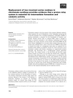
Figure 2 Southern hybridization of genomic B. vulgaris DNA with probes of tandem repeats identified in the c
0
t-1 library.Genomic
DNA was restricted with NdeI (1), BsmAI (2), AluI (3), HpaII (4) and MspI (5) and hybridized with BvMSat01 (A), BvMSat03 (B), BvMSat04 (C),
BvMSat05 (D), BvMSat07 (E), BvMSat08 (F), BvMSat09 (G), BvMSat10 (H), BvMSat11 (I) and the FokI-satellite (J), AluI-satellite (K) and HinfI-satellite (L).
Zakrzewski et al. BMC Plant Biology 2010, 10:8
/>Page 5 of 14
Figure 3 Physical mapping of tandem repeats on mitotic metaphase chromosomes and interphase nuclei of B. vulgaris using FISH.
Blue fluorescence (DAPI stained DNA) shows the morphology of chromosomes. Red signals show chromosomal localization of the tandem
repeats and green signals show position of 18S-5.8S-25S rRNA genes on the chromosomes. Hybridization with the minisatellites BvMSat01 (A),
BvMSat03 (B), BvMSat04 (C), BvMSat05 (D), BvMSat07 (E), BvMSat08 (F), BvMSat09 (G), BvMSat10 (H), BvMSat11 (I) on mitotic metaphases and
probes of the FokI-satellite (J), the AluI-satellite (K) and the HinfI-satellite (L) on mitotic metaphases and interphase nuclei reveals characteristic
chromosomal distribution patterns.
Zakrzewski et al. BMC Plant Biology 2010, 10:8
/>Page 6 of 14
show a variation in signal strength and, hence, in copy
numbers or expansion of minisa tellite arrays (e.g. Figure
3, A-C, F and 3G). While some chromosomes show
stronger banding patterns indicating larger arrays or
clustering of multiple arrays, on other chromosomes
weak or no signals were revealed (e.g. Figure 3, F and
3G), which shows that minisatellite arrays are often
small in size. The detection of signals on both chroma-
tids of many chromosomes verifies the hybridization
pattern.
Physical mapping using probes of the minisatellite
families BvMSat08 and BvMSat09 shows particular
hybridization patterns enabling the discrimination of B.
vulgaris chromosomes (Figure 3, F and 3G). A peculiar
hybridization pattern was observed for BvMSat08, which
shows massive amplification of signals in the intercalary
heterochromatin (Figure 3, F), which are localized on
one chromosome arm of a single chromosome pair indi-
cating very large arrays of multiple BvMSat08 copies or
clustering of arrays. Four chromosomes show only
reduced signals indicat ing a lower number of BvMSat08
arrays on these chromosomes. The minisatellite
BvMSat09 shows massive accumulation of clusters in
the intercalary heterochromatin on twelve chromosomes
(Figure 3, G). Six of them are identifiab le by blocks on
both chromosome arms, whereas the other chromo-
somes a re characterized by blocks on one chromosome
arm only.
For the physical mapping of satellites identified in the
c
0
t-1 library we hybridized metaphase chromosomes and
also interphase nuclei, which enable the detection of sig-
nals at higher resolution (Figure 3, J-L). The FokI-satel-
lite shows a co-loc alization with DAPI-positiv e
intercalary heterochromatin (Figure 3, J). However, the
signals are not uniformly distributed and differ in signal
strength. Hybridization was also detected at terminal
euchromatic chromosome regions, consistent with the
FokI-satellite hybridization pattern in interphase nuclei
in low DAPI-stained euchromatic regions (arrows in
Figure 3, J).
Strong clustering of AluI-satellite arrays was observed
in the intercalary heterochromatin on four chromo-
somes, while eight chromosomes show a weaker hybridi-
zation pattern (Figure 3, K). The remaining six
chromosomes show very weak signals indicating that
AluI-satellites are also present in low copy numbers.
The hybridization pattern in interphase nuclei shows
that m ost AluI-satellite signals are localized within het-
erochromatic chromosome regions adjacent to euchro-
matic regions.
Hybridization with probes of the Hin fI-satellite shows
a different pattern. Signals of the HinfI-satellite are
mostly localized in terminal chromosome regions: twelve
chromosomes show hybridization on both chromosome
arms, while signals only on one chromosome arm are
detectable on the remaining six chromosomes (Figure 3,
L). Hybridization on interphase nuclei revealed the pre-
ferred distribution of HinfI-satellites in euchromatic
regions (arrows in Figure 3, L), while only reduced sig-
nals are notable in heterochromatic blocks.
Minisatellite BvMSat07 consists of a complex microsatellite
array
Among the c
0
t-1 sequences, we identifi ed an a rray of a
microsatellite motif with the consensus sequence
GATCA. Within several c
0
t-1 sequences, three short
imperfect repeats (GAAAA, AATAA and GTTCA) were
interspersed within arrays of GATCA monomers. In
order to examine whethe r this interspersion is con-
served, we analyzed B. vulgaris sequences possessing
GATCA-microsatellite arrays and detected that the min-
isatellite BvMSat07 is derived from the GATCA-micro-
satellite. A t ypical BvMSat07 monomer, which is 30 bp
in size, consists of one GAAAA, one AATAA, one
GTTCA motif conserved in this order and three adja-
cent GATCA monomers, respectively (Figure 4). The
analysis of 20 randomly selec ted minisatellite BvMSat 07
monomers revealed that most monomers show an iden-
tical arrangement of these short subrepeats and that
these monomers share a similarity of 90% to 100%.
Head to head junction is a typical characteristic of
BvMSat05 arrays
The 21 bp minisatellite BvMSat05 varies considerably in
nucleotide composition. Sequence identity analysis of
450 monomers originating from c
0
t-1 and BAC end
sequences revealed that monomers show identities
between 38% and 100%.
BvMSat05 shows a particular genomic organization: In
addition to the head to tail organization, a head to head
junction is detectable within multiple BvMSat05 arrays
(Figure 5). Identity values between 35% and 100% of the
monomers within the inverted arrangement of the two
arrays are s imilar to t he values of head to tail mono-
mers. The tandem arrays of the head to head junction
are flanked one-sided by the conserved sequence motif
GTCGTCCGACCAAAGATTATGGTCGGAC-
GAGTCCGA CACAATACGTTCTCT, which is 50 bp in
size and shows identity of 86% to 100% (Figure 5). Inter-
estingly, this sequence comprises two palindromic
motifs (TCGTCCGACCAAAGATTATGGTCGGACGA
and GTCGGACGAGTCCGAC) (arrows in Figure 5).
Discussion
The aim of this study was the characterization of the
repetitive fraction of the B. vulgaris genome. We gener-
ated and analyzed 1763 highly and moderately repetitive
sequences from a c
0
t-1 DNA library. Our results
revealed that the majority of sequences in the c
0
t-1
library are copies of the satellite families pBV [30] and
Zakrzewski et al. BMC Plant Biology 2010, 10:8
/>Page 7 of 14
pEV [32] while other known repeats of the B. vulgaris
genome are underrepresented. According to the copy
numbers within the c
0
t-1 libr ary, the satellite pBV is the
most abundant s atellite family in the genome of B. vul-
garis followed by the pEV satellite family. This observa-
tion is consistent with the prediction that the number of
copies of a repeat family in c
0
t DNA correlates with its
abundance in the genome [8].
So far, c
0
t DNA isola tion has been perfo rmed in sev-
eral plant genomes. c
0
t DNA libraries representing
highly repetitive sequences were generated from geno-
mic DNA of S. bicolor, M. acuminata and L. triticoides
[8,11,12] while moderately repetitive DNA fractions
were isolated from S. bicolor and Z. mays [8,10]. The c
0
t
analysis enabled the identification of novel repeats, as
well as the detection of most abundant repeat classes
within a plant genome. c
0
t-1 DNA analysis performed in
the L. triticoides genomerevealedahighlyabundant
satellite family [12] which is similar to the observation
that most c
0
t-1 clones of B. vulgaris belong to satellite
Figure 4 BvMSat07 is composed of microsatellite complex repeats. 30 bp monomers of BvMSat07 are typically composed of degenerated
and conserved GATCA-motifs (as example an array of the BAC end sequence FN424407 is shown).
Figure 5 Illustration of the head to head junction of BvMSat05 arrays. A : The BAC end sequence FN424410 contains a head to head
junction of two head to tail BvMSat05 arrays (arrows and double-lined arrows). B: An alignment of ten BAC end sequences illustrates the typical
head to head junction of two head to tail arrays. For each array four monomers, which are separated by a gap, are shown. The number at the
left and right borders of the arrays corresponds to the number of monomers that are not displayed in this illustration. The nucleotides are color-
encoded: Red for adenine, blue for cytosine, yellow for guanine and green for thymine. The tandem arrays are flanked one-sided by a highly
conserved 50 bp motif, which comprises two palindromic sequences (double arrows). Identity values are displayed in percent.
Zakrzewski et al. BMC Plant Biology 2010, 10:8
/>Page 8 of 14
DNA. In contrast, the most abundant repeats detected
in the c
0
t librar ies of S. bicolor, M. acuminata and Z.
mays belong to retrotranspo sons or retrotransposon-
derived sequences. No significant number of tandemly
repeated sequences (except ribosomal genes in the M.
acuminata and S. bicolor genome) has been observed
indicating that retrotransposons constitute the main
repetitive fraction in these genomes [8,10,11].
The detection of the relatively low number of Minia-
ture inverted-repeat transposable elements (MITEs) in
the c
0
t library of B. vulgaris is in contrast to the large
number of MITEs that has been described [37] and indi-
cates a po ssib le bias during library construction. A pos-
sible reason for the low frequency of MITEs in c
0
t-1
DNA might be related to the intramolecule renaturation
via terminal inverted repeats (TIRs) of single stranded
sequences containing MITEs. TIRs of MITEs in B. vul-
garis are relatively short [37] and c
0
t clones containing
inserts l ess than 50 bp have been excluded, hence, short
MITE sequences have been escaped from analysis.
A possible explanation for the differences in the num-
ber of organelle-derived sequences within c
0
t libraries
might be related to plastid and mitochondrial DNA
which was isolated together with nuclear DNA. Hribová
et al. (2007) and Yuan et al. (2003) isolated the c
0
t-0.05
DNA and the c
0
t-100 fraction from the M. acuminata
and Z. mays genome, respect ively, using a similar
approach as in this study [10,11]. The proportion of
chloroplast DNA in the c
0
t-0.05 DNA fraction of M.
acuminata is 4.2%, which is approximately a third com-
pared to the c
0
t-1 DNA fraction of B. vulgaris and the
proportion of organelle-derived DNA in the c
0
t-100
fraction of Z. mays is 1.7% which is much lower as in
c
0
t-1 DNA fraction of B. vulgaris. No chloroplast DNA
was detectable in the highly repetitive c
0
t fraction of S.
bicolor while 10% chloroplast-derived sequences have
been observed in the moderate c
0
t fraction of S. bicolor
[8,10,11]. Another possible scenario explaining these dif-
ferences is that chloroplast DNA was integrated into
nuclear DNA and consequently c
0
t sequences with
homology to chloroplast DNA might also originate from
the nucleus. Chloroplast DNA can be found interspersed
into nuclear DNA in many plant species including B.
vulgaris [42-44]. Moreover, it has been assumed that
chloroplast DNA incorporation into the nucleus is a fre-
quent evolutionary event [44]. However, it is very likely
that the B. vulgaris c
0
t-1 clones containing chloroplast
sequences originate from contaminatio n of the genomic
DNA used for reassociation.
Macas et al. (2007) performed an analysis of genomic
sequence data originating from a single 454-sequencing
run of the Pisum sativum genome to reconstruct the
major repeat fraction and identified retroelements as the
most abundant repeat class within the genome [19].
Similar analyses investigating crop genome compositions
based on next generation sequence technologies have
been reported [45,46]. In our study c
0
t-1 DNA isolation
was used for the classification of the major repeat
families within the B. vu lgaris genome and satellite
DNA was identified as a highly abundant repeat class.
In co ntrast to genome sequencing projects reflecting the
whole genome in its native composition, c
0
t-1 DNA iso-
lation represents only the repetitive fraction and enables
therefore the targeted isolation of major repeats.
Furthermore, less sequence data is necessary for the
detection of major repeats using c
0
t DNA isolation com-
pared with next generation sequence reads. We used
only 442 kB (0.06% of the genome) sequence data for
the detection of the major repeat families of the B. vul-
garis genome while 33.3 Mb (0.77%) of P. sativum [19],
58.91 Mb (1%) of barley [46] and 78.54 Mb (7%) of soy-
bean [45] were analyzed to detect the repeat composi-
tion. Therefore, c
0
t DNA isolation is a very efficient
method for the identification of the repetitive DNA of
genomes not sequenced yet.
Macas et al. (2007) identified 17 novel tandem repeat
families, and two minisatellites were physically mapped
on P. sativum chromosomes [19]. In order to demon-
strate the potential of the c
0
t-1 DNA library for the
detection of novel repeat classes we focused on the
identification of tandemly repeated sequences, particu-
larly on the identification of minisatellites. So far, the
targeted isolation of minisatellites from plant genomes
has not been described and this repeat type is only
poorly characterized. It is not feasible to isolate most
minisatellites as restriction satellites because of their
short length, unusual base composition and hence,
absence of recognition sites. The identification of nine
minisatellite families as described here shows the poten-
tial of c
0
t DNA analysis for th e rapid and targeted isola-
tion of minisatellites from genomes. In addition we
identified three satellite families undiscovered yet
because of their moderate abundance.
In contrast to typical G/C-rich minisatellites [13], all
nine B. vulgaris families show a low G/C content: six of
theninefamilieshaveaG/C-contentbetween24%to
33% (Table 1). Repetitive sequences are often subject to
modification by cytosine methylation. It is known that
deamination converts 5-methylcytosine to thymine,
resulting in an increased AT-content [47]. This m ight
be a possible reason o f the low G/C level of B. vulgaris
minisatellites. Furthermore, the monomers of the B. vul-
garis minisatellite families are different in sequence
length and nucleotide composition from the 14 to 16 bp
G/C-rich core sequence of minisatellites in A. thaliana
or human [25,26].
Most conventional plant satellites show a low G/C
content [48]. However, the FokI-satell ite has a G/C
Zakrzewski et al. BMC Plant Biology 2010, 10:8
/>Page 9 of 14
content of 60% which is in contrast to the HinfI-satellite
and AluI-satellite and other satellites described in B.
vulgaris. Moreover, the monomer size of 130 bp of the
FokI-satellite is different from the typical monomer size
of plant satellites of 160-180 bp or 320 to 370 b p [15],
whereas monomers of HinfI-satellite and AluI-satellite
fall into the typical monomer size range.
Only two of the nine minisatellite families (BvMSat03
and BvMSat04) show the typical ladder-like pattern in
Southern analyses. Dimers of BvMSat03 were detectable
after restriction of genomic DNA with BsmAI (Figure
2B, lane 2). However, partial restriction with BsmAI
generates di- to decamers of BvMSat03 (not shown),
indicating the highly conserved recognition site of
BsmAI in BvMSat03-monomers.
Hybridization of minisatellites to MspIandHpaII
digested DNA indicates cytosine methylation of the
recognition site CCGG. The HinfI-satellite and AluI-
satellite family show also a strong methylation, while a
reduced CNG methylation was detectable for some
FokI-satellite copies. This might be an indication that
some FokI-satellite copies lacki ng CNG methylation
might be linked to the activation of transcription or to
chromatin remodeling [49-52].
Little is known about the lo calization of minisatellites
on plant chromosomes. So far, only two minisatellite
families w ere physically mapped on chromosomes of P.
sativum using FISH [19]. In contrast to minisatellites of
P. sativum detectable only on one and two chromosome
pairs [19], respectively, the B. vulgaris minisatellites
were detectable mostly on all 18 chromosomes with dif-
ferent signal strength, preferentially distributed in the
intercalary heterochromatin and terminal chromosome
regions. This pattern of chromosomal localization shows
similarity to the distribution of microsatellite sequences
on B. vulgaris chromosomes, which show a disp ersed
organization along chromosomes including telomeres
and intercalary chromosomal regions, but are mostly
excluded from the centromere [36]. This is in contrast
to the chromosomal localization of the highly abundant
satellite families pBV and pEV and the satellite family
pAv34 [33], which are detectable in large tandem arrays
in centromeric/pericentromeric, intercalary and subtelo-
meric regions, respectively. Only BvMSat08 and
BvMSat09 can be found in large tandem array blocks
within the intercalary heterochromatin.
The FokI, AluIandHinfI satellite families show dis-
persed localization in smaller arrays with different array
sizes among chromosom es, prefere ntially in the interca-
lary heterochromatin a nd in terminal chromosome
regions, respectively. The HinfI-satellite is predomi-
nantly distributed in terminal chromosome regions. The
pAv34 satellite is also localized in subtelomeric chromo-
some positio ns [33]. However, no copies of pAv34 were
detected within the 13 kb BAC [EMBL:DQ374018] and
the 11 kb BAC [EMBL:DQ374019] that contain a tan-
dem array of the HinfI-satellite consisting of 14 and 26
monomers, respectively, indicating no interspersion of
both satellite families. High resolution FISH on pachy-
tene chromosomes or chromatin fibers using probes of
pAv34 and the HinfI-satellite could be used to gain
information about possible interspersion or physically
neighborhood of both satellite families.
Because of their small size (2-3 μ m) and similar mor-
phology (most chromosomes are meta- to submeta-
centric) FISH karyotype analysis of B. vulgar is has not
been established yet. In contrast to conventional staining
techniques [53], which are not efficient for reliable kar-
yotyping of small chromosomes, FISH is an applicable
method for the discrimination of the B. vulgaris chro-
mosomes. Chromosome 1 can b e identified by strong
signals of terminal 18S-5.8S-25S rRNA genes while
chromosome 4 is detectable by 5S rRNA hybridization
patterns [54]. FISH using probes of BvMSat08 enables
the identification of another chromosome pair, due to
the localization of the large BvMSat08 blocks on both
chromosome arms. Hence, this minisatellite may be an
important cytogenetic marker for future karyotyping
based on FISH. Also, because of their specific chromo-
somal localization, the minisatellite BvMSat09, the AluI
satellite and the HinfI satellite can serve as cytogenetic
markers and support FISH karyotyping in B. vulgaris.
It has been reported that human minisatellites origi-
nated from retroviral LTR-like sequences or fro m the 5’
end of Alu elements [5 5,56] but also other scenarios of
the origin and the evolution were described in human
and in primates [57,58]. In plants, only few data are
available about the origin and the evolution of minisatel-
lite sequences. We propose a possible process which
might describe the origin and/or evolution of minisatel-
lites from microsatellites in the genome of B. vulgaris.
Sequence analysis suggests that BvMSat07 originated
from a microsatellite with the 5 bp monomer sequence
GATCA. During microsatellite evolution complex arrays
of six monomers evolved, which were subsequently tan-
demly arranged. The resulting mini satellite is 30 bp in
size and consists of one GAAAA, AATAA and GTTCA
and three adjacent GATCA monomers. The 5 bp subre-
peats differing from the GATCA monomer sequence
mighthaveoriginatedfromtheGATCA-motifbypoint
mutation. T he complex repeat shows structural similari-
ties to hi gher-order structures of satellites, e.g. the
human alpha satelli te [59]. A satellite higher-order
structure is defined as monomers which form tandemly
arranged highly homogenous multimeric repeat units
[59]. One complex repeat of the m icrosatellite might
have been duplicated and enlarged by replication slip-
page resulting in a BvMSat07 array (Figure 4) and its
Zakrzewski et al. BMC Plant Biology 2010, 10:8
/>Page 10 of 14
copy number might have been increased by recombina-
tion between homologous loci.
Another scenario of minisatellite origin and array
enlargement can be concluded from the minisatellite
family BvMSat05. The palindromic sequences within the
highly conserved 50 bp sequence adjacent t o BvMSat05
arrays may form secondary DNA structures, which may
interfere with the DNA polymerase during DNA replica-
tion. This may result in slippage replication of the DNA
motif upstream, contributing to the generation and
enlargement of BvMSat05 arrays. Moreover, FISH
revealed a subtelomeric localization of BvMSat05 clus-
ters on some chromosomes, he nce, the head to head
junction of head to t ail arrays typical for BvMSat05 may
result from breakage-fusion-bridge cycles as postulated
for tandem repeats near at terminal regions of rye chro-
mosomes [60]. It has been reported that palindromic
sequences may induce genomic instability through pro-
voking double strand breaks and recombination [61].
Therefore, the head to head junction may also be t he
result of DNA repair following possible double strand
breaks within BvMSat05 arrays.
It has also been discussed that ta ndemly repeated
sequences are derived from 3’ UTR regions of retrotran-
sposons [62]. Analysis of retrotransposons in B. vulgaris
[40,63,64] did not reveal any homology to minisatellite
arrays or adjacent regions. However, we detected LTR
sequences of a yet uncharacterized retrotransposon in
the close vicinity of Bv MSat04 arrays (not shown ).
Therefore, the evolution and dispersion of BvMSat04
arrays within the B. vulgaris genome might also be the
result of the activity of this retrotransposon.
In this study we focused in detail on the characteriza-
tion of novel m inisatellites and satellites. Nevertheless,
these tandem repeats make up only 6.8% of the 517
uncharacterized c
0
t-1 sequences indicatin g that the c
0
t-1
library is an efficient source for the identification of
further repeat classes. Examples are the 118 c
0
t-1
sequences possessing motifs of retrotransposon families
as well as the i dentification of the envelope -like Copia
element Cotzilla [40].
Conclusions
We isolated highly to moderately repetitive DNA
sequences from B. vu lgaris originating from a c
0
t-1 DNA
library. Providing the first comprehensive classification of
repeats, we observed that the satellites pBV and pEV
form the most abundant repeat families in B. vulgaris.
We identified nine minisatellite and three previously
unknown satellite families demonstrating that the analy-
sis of c
0
t-1 DNA is an efficient method for the rapid
and targeted isolation of tandemly repeated sequences,
particularly of minisatellites from plant genomes. Minis-
atellites in B. vulgaris display a low G/C content and
deviate strongly from the G/C-rich minisatellite core
sequence observed in A. thaliana and human [25,26]
showing that a minisatellite core motif is not conserved
in plant genomes. Physical mapping of the minisatellites
on chromosomes using FISH revealed a mainly dis-
persed chromosomal distribution pattern. T he possible
origin, enlargement and amplification of minisatellites
arrays were co ncluded for some minisatellite families.
Complex structures of microsatellite arrays may play a
role for the generation of minisatellites. Moreover, DNA
sequences that contain palindromic motifs may be
linked to slippage replication due to interfering with
DNA polymerase during replication and may therefore
be involved in the origin of minisatellites.
Methods
Plant material and DNA preparation
Plants of Beta vulgaris ssp. vulgaris genotyp e KWS 2320
were grown under greenhouse conditions. Genomic
DNA was isolate d from young leaves using the CTAB
(cetyltrimethyl/ammonium bromide) standard protocol
[65].
Construction of the c
0
t-1 DNA library
The c
0
t-1 DNA was prepared with some modifications
according to Zwick et al. [9]. 640 μgofgenomicDNA
was dissolved in 1600 μl water and sheare d at 99°C for
10 minutes followed by sonication at 80°C for 3 minutes
to generate DNA f ragments ranging in size predomi-
nantly between 0.5 to 1.0 kb. Renaturation of DNA frag-
ments was carried out in a 0.3 M NaCl solution at 65°C
after initial denaturation at 92°C for 10 minutes. The
renaturation time was calculated according to Zwick et
al. [9]. S1 nuclease treatment followed to remove single
stranded DNA and single strand overhangs on rena tu-
rated double stranded DNA. The enzyme was inacti-
vated by adding stop solution (3 M Tris pH 8.0, 0.5 M
EDTA) according to Ostermeier et al. [66] and incuba-
tion at 72°C for 20 min. Blunt end c
0
t-1 DNA fragments
were ligated into the SmaI site of dephosphorylated
pUC18 vector. After transformation of XL1Blue cells
(Stratagene), positive clones were identified by blue/
white screening and transferred into 384-well plates,
grown in LB freezing medium and stored at -80°C.
Sequencing of c
0
t-1 clones
Clones were grown in Terrific Broth (TB) medium (1.2%
peptone, 2.4% yeast extract, 72 mM K
2
HPO
4
,17mM
KH
2
PO
4
and 0.4% g lycerol) including 100 μg/ml ampi-
cillin at 37°C. Small-scale plasmid isolation was per-
formed by the TELT procedure [67]. Plasmids were
sequenced on an ABI 3730XL sequencer (Applied Bio-
systems; Fost er City, CA/USA) using BigDye terminator
chemistry, in forward (5’ -CGTTGTAAAACGACG
GCCAGT-3’) and/or reverse (5’-CAGGAAACAGCTAT
GACCATG-3’) directions.
Zakrzewski et al. BMC Plant Biology 2010, 10:8
/>Page 11 of 14
Computational methods
Sequences in c
0
t-1 DNA library, which are homologous
to previously characterized B. vulgaris repeats, were
identif ied using local BLAST option of the BioEdit soft-
ware [68] with a representative query sequence of the
repeat family. Novel c
0
t-1 DNA sequences were charac-
terized using the EMBL database homology search
against nucleotide and amino ac id sequences and an e-
value threshold of 10
-3
. The remaining fraction of t he
c
0
t-1 DNA without homology to EMBL database entries
was used for the identif ication of tan dem repeats using
Tandem Repeats Finder [69]. Subsequently, c
0
t-1
sequences containing tandem repeats were used as
query sequence for the identification of further DNA
copies from BAC end sequences [39], (Holtgräwe and
Weisshaar, in preparation) to reveal their abundance
and array structures. The DNA sequences of each tan-
dem repeat family were aligned manually using the Phy-
logenetic Data Editor [70]. The detection of G/C
content and identity values of each tandem repeat family
was determined by a G/C Content Calculator and C lus-
talX [71] us ing at least 20 randomly selected monomers
of represent ative tandem arrays. Sequences contigs have
been established using DNASTAR Lasergene v8.0.
PCR conditions
Primer pairs were derived from conserved regions of
minisatellite and satellite monomers. The PCR reactions
with 50 ng genomic DNA and a final primer concentra-
tion of 0.5 μM were performed in a 20 μlvolumecon-
taining 0.2 mM dNTPs and 1 unit of GoTaq polymerase
(Promega). The PCR conditions were 94°C for 3 min,
followed by 30 cycles of 94°C for 30 s, 47°C to 65°C
depending on the primer melting temperature of each
repeat family, for 30 s, 72°C for 40 s and a fina l incub a-
tion at 72°C for 5 min. For the generation of probes for
Southern hybridization and fluorescent in situ hybridiza-
tion, the same primers or M13 primers were used to
amplify tandem repeats from c
0
t-1 clones.
Southern hybridization
For Southern hybridization 5 μg of gen omic DNA was
restricted with different enzymes, separated on 1.2%
agarose gels and transferred onto Hybond-XL nylon
membranes (GE Healthcare) using alkaline transfer.
Southern hybridizations using
32
P-labelled probes were
performed using standard protocols [72]. Filters were
hybridized at 60°C and washed at 60°C in 2× SSC/0.1%
SDS for 3 h. The signals were detected by
autoradiography.
FISH
The meristem of young leaves was used for the prepara-
tion of mitotic chromosomes. The maceration of plant
material was performed in an enzyme mixture consist-
ing of 0.3% (w/v) cytohelicase (Sigma), 1.8% (w/v) cellu-
lase fr om Aspergillus niger (Sigma), 0.2% (w/v) cellulase
Onozuka-R10 (Serva) and 20% (v/v) pectinase from A.
niger; followed by spreading of nuclei on slides. Probes
of tandem repeats were labelled with biotin-16-dUTP
(Roche) by P CR according to Schw arzacher et al. [73]
while 18S-5.8S-25S rRNA genes were labelled by nick-
translation with digoxygenin-11-dUTP (Roche). The
hybridization and detection were pe rformed according
to Schmidt et al. [54]. Chromosome preparations were
counterstained with DAPI (4’,6’-diamino-2-phenylindole)
and mounted in antifade solution (CitiFluor). The exam-
ination of slides was carried o ut with a Zeiss Axioplan2
Imaging fluorescent microscope with filters 09 ( FITC),
15 (Cy3) and 02 (DAPI). The images were acquired with
the Applied Spectral Imaging v. 3.3 software coupled
with the high-resolution CCD camera ASI BV300-20A.
The c ontrast of images was optimized using only func-
tions affecting whole image equally by Adobe Photoshop
7.0 software.
Acknowledgements
Falk Zakrzewski acknowledges a fellowship and financial support of the
FAZIT foundation. This work is funded in part by the BMBF grant
“Verbundprojekt GABI-Beet Physical map: Physikalische Genomkarte der
Zuckerrübe zur Nutzung in der Pflanzenzüchtung”, sub-p rojects 0313127B
and 0313127E. We thank Ines Walter for excellent technical assistance.
Author details
1
Institute of Botany, Dresden University of Technology, D-01062 Dresden,
Germany.
2
Institute of Genome Research, University of Bielefeld, D-33594
Bielefeld, Germany.
Authors’ contributions
FZ and TW wrote the paper, participated in the bioinformatic analyses of the
c
0
t-1 library, carried out the alignments of the tandemly repeated c
0
t-1
sequences and performed the molecular genetic studies. DH and BW
performed the sequencing of the c
0
t-1 clones, provided the BAC end
sequence database and helped to draft the manuscript. TS participated in
the design and coordination of the project and has been involved in the
writing of the article. All authors read and approved the final manuscript.
Received: 20 July 2009
Accepted: 11 January 2010 Published: 11 January 2010
References
1. Britten RJ, Kohne DE: Repeated sequences in DNA. Hundreds of
thousands of copies of DNA sequences have been incorporated into the
genomes of higher organisms. Science 1968, 161(841):529-540.
2. Ugarkovic D: Functional elements residing within satellite DNAs. Embo
Reports 2005, 6(11):1035-1039.
3. Lisch D: Epigenetic regulation of transposable elements in plants. Annu
Rev Plant Biol 2009, 60:43-66.
4. Feschotte C: Transposable elements and the evolution of regulatory
networks. Nat Rev Genet 2008, 9(5):397-405.
5. Lippman Z, Gendrel AV, Black M, Vaughn MW, Dedhia N, McCombie WR,
Lavine K, Mittal V, May B, Kasschau KD, et al: Role of transposable
elements in heterochromatin and epigenetic control. Nature 2004,
430(6998):471-476.
6. Weil C, Martienssen R: Epigenetic interactions between transposons and
genes: lessons from plants. Curr Opin Genet Dev 2008, 18(2):188-192.
7. Jurka J, Kapitonov VV, Kohany O, Jurka MV: Repetitive Sequences in
Complex Genomes: Structure and Evolution. Annu Rev Genomics Hum
Genet 2006, 8:241-259.
8. Peterson DG, Schulze SR, Sciara EB, Lee SA, Bowers JE, Nagel A, Jiang N,
Tibbitts DC, Wessler SR, Paterson AH: Integration of Cot analysis, DNA
Zakrzewski et al. BMC Plant Biology 2010, 10:8
/>Page 12 of 14
cloning, and high-throughput sequencing facilitates genome
characterization and gene discovery. Genome Res 2002, 12(5):795-807.
9. Zwick MS, Hanson RE, McKnight TD, IslamFaridi MN, Stelly DM, Wing RA,
Price HJ: A rapid procedure for the isolation of C(0)t-1 DNA from plants.
Genome 1997, 40(1):138-142.
10. Yuan Y, SanMiguel PJ, Bennetzen JL: High-Cot sequence analysis of the
maize genome. Plant J 2003, 34(2):249-255.
11. Hribová E, Dolezelová M, Town CD, Macas J, Dolezel J: Isolation and
characterization of the highly repeated fraction of the banana genome.
Cytogenet Genome Res 2007, 119(3-4):268-274.
12. Anamthawat-Jónsson K, Wenke T, Thórsson Æ Th, Sveinsson S, Zakrzewski F,
Schmidt T: Evolutionary diversification of satellite DNA sequences from
Leymus (Poaceae: Triticeae). Genome 2009, 52:381-390.
13. Vergnaud G, Denoeud F: Minisatellites: mutability and genome
architecture. Genome Res 2000, 10(7):899-907.
14. Chistiakov DA, Hellemans B, Volckaert FAM: Microsatellites and their
genomic distribution, evolution, function and applications: A review
with special reference to fish genetics. Aquaculture 2006, 255(1-4):1-29.
15. Hemleben V, Kovarik A, Torres-Ruiz RA, Volkov RA, Beridze T: Plant highly
repeated satellite DNA: molecular evolution, distribution and use for
identification of hybrids. Systematics and Biodiversity 2007, 5(03):277-289.
16. Haber JE, Louis EJ: Minisatellite origins in yeast and humans. Genomics
1998, 48(1):132-135.
17. Taylor JS, Breden F: Slipped-strand mispairing at noncontiguous repeats
in Poecilia reticulata: a model for minisatellite birth. Genetics 2000,
155(3):1313-1320.
18. Schmidt A, Doudrick RL, Heslop-Harrison JS, Schmidt T: The Contribution
of Short Repeats of Low Sequence Complexity to Large Conifer
Genomes. Theor Appl Genet 2000, 101:7-14.
19. Macas J, Neumann P, Navratilova A: Repetitive DNA in the pea (Pisum
sativum L.) genome: comprehensive characterization using 454
sequencing and comparison to soybean and Medicago truncatula. BMC
Genomics 2007, 8(1):427-443.
20. Gustafson JP, Yano M: Genetic mapping of hypervariable minisatellite
sequences in rice (Oryza sativa L.). Theor Appl Genet 2000, 100(3):447-453.
21. Broun P, Tanksley SD: Characterization of tomato DNA clones with
sequence similarity to human minisatellites 33.6 and 33.15. Plant Mol Biol
1993, 23(2):231-242.
22. Hisatomi Y, Hanada K, Iida S: The retrotransposon RTip1 is integrated into
a novel type of minisatellite, MiniSip1, in the genome of the common
morning glory and carries another new type of minisatellite, MiniSip2.
Theor Appl Genet 1997, 95(7):1049-1056.
23. Martienssen RA, Baulcombe DC: An unusual wheat insertion sequence
(WIS1) lies upstream of an a-amylase gene in hexaploid wheat, and
carries a “minisatellite” array. Mol Gen Genet 1989, 217(2):401-410.
24. Somers DJ, Zhou Z, Bebeli PJ, Gustafson JP: Repetitive, genome-specific
probes in wheat (Triticum aestivum L. em Thell) amplified with
minisatellite core sequences. Theor Appl Genet 1996, 93(5):982-989.
25. Tourmente S: Identification of new minisatellites loci in Arabidopsis
thaliana. J Exp Bot 1998, 49(318):21-25.
26. Tourmente S, Deragon JM, Lafleuriel J, Tutois S, Pélissier T, Cuvillier C,
Espagnol MC, Picard G: Characterization of minisatellites in Arabidopsis
thaliana with sequence similarity to the human minisatellite core
sequence. Nucleic Acids Res 1994, 22(16):3317-3321.
27. Arumuganathan K, Earle ED: Nuclear DNA content of some important
plant species. Plant Mol Bio Reporter 1991, 9(3):186-198.
28. Flavell RB, Bennett MD, Smith JB, Smith DB: Genome Size and Proportion
of Repeated Nucleotide-Sequence DNA in Plants. Biochemical Genetics
1974, 12(4):257-269.
29. PROJECT: Generation of a physical, BAC-based map of the sugar beet
(Beta vulgaris) genome (GABI-BPM). -
bielefeld.de/GF-research/GABI-BPM.html.
30. Schmidt T, Metzlaff M: Cloning and Characterization of a Beta-vulgaris
Satellite DNA Family. Gene 1991, 101(2):247-250.
31. Menzel G, Dechyeva D, Wenke T, Holtgräwe D, Weisshaar B, Schmidt T:
Diversity of a Complex Centromeric Satellite and Molecular
Characterization of Dispersed Sequence Families in Sugar Beet (Beta
vulgaris). Ann Bot (Lond) 2008, 102:521-530.
32. Schmidt T, Jung C, Metzlaff M: Distribution and evolution of two satellite
DNAs in the genus Beta. Theor Appl Genet 1991, 82:793-799.
33. Dechyeva D, Schmidt T: Molecular organization of terminal repetitive
DNA in Beta species. Chromosome Res 2006, 14(8):881-897.
34. Schmidt T, Heslop-Harrison JS: Variability and evolution of highly
repeated DNA sequences in the genus Beta. Genome 1993, 36(6):1074-
1079.
35. Schmidt T, Heslop-Harrison JS: Genomes, genes and junk: the large-scale
organization of plant chromosomes. Trends in Plant Science 1998, 3(5):195-
199.
36. Schmidt T, Heslop-Harrison JS: The physical and genomic organization of
microsatellites in sugar beet. Proc Natl Acad Sci USA 1996, 93(16):8761-
8765.
37. Menzel G, Dechyeva D, Keller H, Lange C, Himmelbauer H, Schmidt T:
Mobilization and evolutionary history of miniature inverted-repeat
transposable elements (MITEs) in Beta vulgaris L. Chromosome Res 2006,
14(8):831-844.
38. Jacobs G, Dechyeva D, Menzel G, Dombrowski C, Schmidt T: Molecular
characterization of Vulmar1, a complete mariner transposon of sugar
beet and diversity of mariner- and En/Spm-like sequences in the genus
Beta. Genome 2004, 47(6):1192-1201.
39. McGrath JM, Shaw RS, de los Reyes BG, Weiland JJ: Construction of a
sugar beet BAC library from a hybrid with diverse traits. Plant Mol Bio
Reporter 2004, 22(1):23-28.
40. Weber B, Wenke T, Frommel U, Schmidt T, Heitkam T: The Ty1-copia
families SALIRE and Cotzilla populating the Beta vulgaris genome show
remarkable differences in abundance, chromosomal distribution, and
age. Chromosome Res , DOI: 10.1007/s10577-009-9104-4.
41. Zilberman D, Henikoff S: Genome-wide analysis of DNA methylation
patterns. Development 2007, 134(22):3959-3965.
42. Cullis CA, Vorster BJ, Vyver Van Der C, Kunert KJ: Transfer of genetic
material between the chloroplast and nucleus: how is it related to stress
in plants?. Ann Bot (Lond) 2009, 103(4):625-633.
43. Blanchard JL, Schmidt GW: Pervasive m igration of organellar DNA to the
nucleus in plants. J Mol Evol 1995, 41(4):397-406.
44. Ayliffe MA, Timmis JN, Scott NS: Homologies to chloroplast DNA in the
nuclear DNA of a number of Chenopod species. Theor Appl Genet 1988,
75(2):282-285.
45. Swaminathan K, Varala K, Hudson ME: Global repeat discovery and
estimation of genomic copy number in a large, complex genome using
a high-throughput 454 sequence survey. BMC Genomics 2007, 8:132.
46. Wicker T, Taudien S, Houben A, Keller B, Graner A, Platzer M, Stein N: A
whole-genome snapshot of 454 sequences exposes the composition of
the barley genome and provides evidence for parallel evolution of
genome size in wheat and barley. Plant J 2009, 59(5):712-722.
47. Montero LM, Filipski J, Gil P, Capel J, Martinez-Zapater JM, Salinas J: The
distribution of 5-methylcytosine in the nuclear genome of plants. Nucleic
Acids Res 1992, 20(12):3207-3210.
48. Macas J, Mészáros T, Nouzová M: PlantSat: a specialized database for
plant satellite repeats. Bioinformatics 2002, 18(1):28-35.
49. Bartee L, Malagnac F, Bender J: Arabidopsis cmt3 chromomethylase
mutations block non-CG methylation and silencing of an endogenous
gene.
Genes Dev 2001, 15(14):1753-1758.
50. Jackson JP, Lindroth AM, Cao X, Jacobsen SE: Control of CpNpG DNA
methylation by the KRYPTONITE histone H3 methyltransferase. Nature
2002, 416(6880):556-560.
51. Johnson LM, Cao XF, Jacobsen SE: Interplay between two epigenetic
marks: DNA methylation and histone H3 lysine 9 methylation. Current
Biology 2002, 12(16):1360-1367.
52. Lindroth AM, Cao X, Jackson JP, Zilberman D, McCallum CM, Henikoff S,
Jacobsen SE: Requirement of CHROMOMETHYLASE3 for maintenance of
CpXpG methylation. Science 2001, 292(5524):2077-2080.
53. Jong JH, Bock TSM: Use of haploids of Beta vulgaris L. for the study of
orcein and giemsa stained chromosomes. Euphytica 1978, 27(1):41-47.
54. Schmidt T, Schwarzacher T, Heslop-Harrison JS: Physical mapping of rRNA
genes by fluorescent in-situ hybridization and structural analysis of 5S
rRNA genes and intergenic spacer sequences in sugar beet (Beta
vulgaris). Theor Appl Genet 1994, 88(6):629-636.
55. Armour JA, Wong Z, Wilson V, Royle NJ, Jeffreys AJ: Sequences flanking
the repeat arrays of human minisatellites: association with tandem and
dispersed repeat elements. Nucleic Acids Res 1989, 17(13):4925-4935.
56. Jurka J, Gentles AJ: Origin and diversification of minisatellites derived
from human Alu sequences. Gene 2006, 365:21-26.
Zakrzewski et al. BMC Plant Biology 2010, 10:8
/>Page 13 of 14
57. Barros P, Blanco MG, Boan F, Gomez-Marquez J: Evolution of a complex
minisatellite DNA sequence. Mol Phylogenet Evol 2008, 49(2):488-494.
58. Boan F, Blanco MG, Quinteiro J, Mourino S, Gomez-Marquez J: Birth and
evolutionary history of a human minisatellite. Mol Biol Evol 2004,
21(2):228-235.
59. Rudd MK, Willard HF: Analysis of the centromeric regions of the human
genome assembly. Trends Genet 2004, 20(11):529-533.
60. Vershinin AV, Schwarzacher T, Heslop Harrison JS: The Large-Scale
Genomic Organization of Repetitive DNA Families at the Telomeres of
Rye Chromosomes. Plant Cell 1995, 7(11):1823-1833.
61. Lisnic B, Svetec IK, Stafa A, Zgaga Z: Size-dependent palindrome-induced
intrachromosomal recombination in yeast. DNA Repair (Amst) 2009,
8(3):383-389.
62. Macas J, Koblizkova A, Navratilova A, Neumann P: Hypervariable 3’ UTR
region of plant LTR-retrotransposons as a source of novel satellite
repeats. Gene 2009, 448(2):198-206.
63. Wenke T, Holtgrawe D, Horn AV, Weisshaar B, Schmidt T: An abundant and
heavily truncated non-LTR retrotransposon (LINE) family in Beta vulgaris.
Plant Mol Biol 2009, 71:585-597.
64. Heitkam T, Schmidt T: BNR - a LINE family from Beta vulgaris - contains a
RRM domain in open reading frame 1 and defines a L1 sub-clade
present in diverse plant genomes. Plant J 2009, 59(6):872-882.
65. Saghai-Maroof MA, Soliman KM, Jorgensen RA, Allard RW: Ribosomal DNA
spacer-length polymorphisms in barley: mendelian inheritance,
chromosomal location, and population dynamics. Proc Natl Acad Sci USA
1984, 81(24):8014-8018.
66. Ostermeier M, Nixon AE, Shim JH, Benkovic SJ: Combinatorial protein
engineering by incremental truncation. Proc Natl Acad Sci USA 1999,
96(7):3562-3567.
67. Ausubel FM, Brent R, Kingston RE, Moore DD, Seidman JG, Smith JAKS:
Current protocols in molecular biology. New York: N.Y: John Wiley & Sons,
Inc 1987.
68. Hall TA: BioEdit: a user-friendly biological sequence alignment editor and
analysis program for Windows 95/98/NT. Nucleic Acids Symposium Series
1999, 41 :95-98.
69. Benson G: Tandem repeats finder: a program to analyze DNA sequences.
Nucleic Acids Res 1999, 27(2):573-580.
70. The Phylogenetic Data Editor. .
71. Larkin MA, Blackshields G, Brown NP, Chenna R, McGettigan PA,
McWilliam H, Valentin F, Wallace IM, Wilm A, Lopez R, et al: Clustal W and
Clustal X version 2.0. Bioinformatics 2007,
23(21):2947-2948.
72. Sambrook J, Fritsch EF, Maniatis T: Molecular Cloning: A Laboratory
Manual. Cold Spring Harbor Laboratory Press, Cold Spring Harbor, NY, 2
1989.
73. Schwarzacher T, Heslop-Harrison P: Practical in situ hybridization. Oxford:
Bios Scientific Publishers 2000.
doi:10.1186/1471-2229-10-8
Cite this article as: Zakrzewski et al.: Analysis of a c
0
t-1 library enables
the targeted identification of minisatellite and satellite families in Beta
vulgaris. BMC Plant Biology 2010 10:8.
Publish with BioMed Central and every
scientist can read your work free of charge
"BioMed Central will be the most significant development for
disseminating the results of biomedical research in our lifetime."
Sir Paul Nurse, Cancer Research UK
Your research papers will be:
available free of charge to the entire biomedical community
peer reviewed and published immediately upon acceptance
cited in PubMed and archived on PubMed Central
yours — you keep the copyright
Submit your manuscript here:
/>BioMedcentral
Zakrzewski et al. BMC Plant Biology 2010, 10:8
/>Page 14 of 14