báo cáo khoa học: " Iron and ferritin accumulate in separate cellular locations in Phaseolus seeds" pdf
Bạn đang xem bản rút gọn của tài liệu. Xem và tải ngay bản đầy đủ của tài liệu tại đây (2.01 MB, 14 trang )
RESEARC H ARTIC L E Open Access
Iron and ferritin accumulate in separate cellular
locations in Phaseolus seeds
Cristina Cvitanich
1*
, Wojciech J Przybyłowicz
2,3
, Dorian F Urbanski
1
, Anna M Jurkiewicz
1
,
Jolanta Mesjasz-Przybyłowicz
2
, Matthew W Blair
4
, Carolina Astudillo
4
, Erik Ø Jensen
1
, Jens Stougaard
1
Abstract
Background: Iron is an important micronutrient for all living organisms. Almost 25% of the wo rld population is
affected by iron deficiency, a leading cause of anemia. In plants, iron deficiency leads to chlorosis and reduced
yield. Both animals and plants may suffer from iron deficiency when their diet or environment lacks bioavailable
iron. A sustainable way to reduce iron malnutrition in humans is to develop staple crops with increased content of
bioavailable iron. Knowledge of where and how iron accum ulates in seeds of crop plants will increase the
understanding of plant iron metabolism and will assist in the production of staples with increased bioavailable
iron.
Results: Here we reveal the distribution of iron in seeds of three Phaseolus species including thirteen genotypes of P.
vulgaris, P. coccineus,andP. lunatus. We showed that high concentrations of iron accumulate in cells surrounding the
provascular tissue of P. vulgaris and P. coccineus seeds. Using the Perls’ Prussian blue method, we were able to detect
iron in the cytoplasm of epidermal cells, cells near the epidermis, and cells surrounding the provascular tissue. In
contrast, the protein ferritin that has been suggested as the major iron storage protein in legumes was only detected in
the amyloplasts of the seed embryo. Using the non-destructive micro-PIXE (Particle Induced X-ray Emission) technique
we show that the tissue in the proximity of the provascular bundles holds up to 500 μgg
-1
of iron, depending on the
genotype. In contrast to P. vulgaris and P. coccineus, we did not observe iron accumulation in the cells surrounding the
provascular tissues of P. lunatus cotyledons. A novel iron-rich genotype, NUA35, with a high concentration of iron both
in the seed coat and cotyledons was bred from a cross between an Andean and a Mesoamerican genotype.
Conclusions: The presented results emphasize the importance of complementing research in model organ isms
with analysis in crop plants and they suggest that iron distribution criteria should be integrated into selection
strategies for bean biofortification.
Background
Iron deficiency is the most prevalent micronutrient
insufficiency worldwide and the leading cause of anemia.
Iron deficiency anemia and its consequences affect
almost 25% of the world population (Report of the UNI-
CEF/Wor ld Health Organization Regional Consult ation,
1999). The diet in resource-poor areas consists in a few
staple crops, which may provide sufficient carbohydrates
but are poor in proteins and micronutrients.
Biofortified micronutrient-rich staple crops can be
developed to improve human nutrition [1,2]. A target
crop for biofortification is the protein-rich common
bean, Phaseolus vulgaris [3]. A high variation in seed
iron content and distribution in P. vulgaris genotypes
has been shown, and is partly due to within-gene pool
and between-gene pool differences [4].
Breeding new bean varieties can be facilitated by the
use of molecular markers linked to high nutritional
values [5]. Establishing which genes are important for
iron uptake, its accumulation in seeds, and its bioavail-
ability, will assist in the design of molecular markers for
genes that are responsible for the high iron trait.
Iro n overload is toxic for plants, while iron deficiency
leads to chlorosis, reduced growth, and eventually death.
Plants have therefore developed mechanisms to tightly
regulate iron metabolism. The responses of non-grami-
naceous plants to iron deficiency include the induction
* Correspondence:
1
Centre for Carbohydrate Recognition and Signalling, Department of
Molecular Biology, University of Aarhus, Aarhus, Denmark
Cvitanich et al. BMC Plant Biology 2010, 10:26
/>© 2010 Cvitanich et al; licensee BioMed C entral Ltd . This is an Open Access article distributed under the terms of the Creative
Commons Attribution License ( which permits unrestricted use, distribution, and
reproduction in any medium, provided the original work is properly cited.
of ferric chelate reductases, an increase in rhizosphere
acidification, and the upre gulation of ZIP type transpor-
ters responsible for iron uptake by the roots (reviewed
by [6-8]). T hese iron deficiency responses are regulated
by transcription factors of the basic helix-loop-helix
family, including FIT1 in Arabidopsis thaliana [9-11].
Once wi thin th e plant , iro n is chelated improving its
mobility and protecting cells from harmful reactive oxygen
species created by ferrous-iron-catalyzed Fenton reactions.
Nicotianamine (NA) , citrate, and the iron transpo rt pro-
tein (ITP) are responsible for binding iron in the xylem
and phloem [12-14], while ferritins were suggested to
store iron in legume seeds and to provide an available iron
pool in leaves [15-19].
Recent findings indicate that vacuoles are important for
seed iron storage. The vacuolar iron transporters,
NRAMP3 and NRAMP4, are important for the mobiliza-
tion of iron during the germination o f A. thaliana seeds
[20]. Furthermore, the vacuolar transporter VIT1 is impor-
tant for the distribution of iron within A. thaliana seeds
[21]. In spite of their effect on iron distribution and seed
germination, loss-of-function mutation of these genes
does not affect seed iron content. In co ntrast, the impor-
tance of NA for seed iron homeostasis was shown by
mutations in N A synthase genes in A. t haliana [22].
Reduction in NA synthase activity did reduce the concen-
tration of iron in seeds.
In addition to significant variations in total iron con-
tent, even within the same species, it was shown that
different legume genotypes accumulate a different pro-
portion of the total seed iron in the seed coat, embryo-
nic axis, and cotyledonary tissues respectively [4,23-25].
This variation in total iron and iron localization must be
explained and ultimately used to advantage in biofortifi-
cation efforts in beans. Therefore specific tissues impor-
tant to iron storage in seeds must be identified and
their iron loading mechanisms revealed.
In this study we identify the cellular localization of
iron in mature seeds of three Phaseolus species includ-
ing 13 genotypes. We have analyzed the iron localization
in mature seeds of P. vulgaris, P. coccineus (runner
bean), and P. lunatus (butter bean). We found that large
concentrations of iron are accumulated in the cytoplasm
of cells surrounding the p rovascular bundles, especially
in iron-rich genotypes of P. vulgaris and P. coccineus
seeds, but not in P. lunatus. Furthermore, we detected
ferritin in the amyloplasts throughout the embryo.
Results
Iron accumulates in defined regions of the cotyledons of
mature P. vulgaris and P. coccineus seeds
We used ICP-AES to measure the iron concentration in
the seed coat, cotyledon, and embryonic axis of beans
from P. coccineus andfromsevenP. vulgaris genotypes
(Table 1). The different tissues were dissected from
mature dry beans. Our results show that the iron con-
centrations in the cotyledons ranged from 43 to 80 μg
g
-1
, and in the embryonic axis from 46 to 103 μgg
-1
.
The largest variation was observed in the seed coats, in
which the iron concentration ranged from 17 to 132 μg
g
-1
, depending on the genotype. This corresponds to 2
to 18% of the total seed iron. Mesoamerican P. vulgaris
genotypes G14519, G4825, DOR364, and the genotype
NUA35 which is derived from a cross between CAL96
and G14519 (CIAT, unpublished), have more iron in
their seed coats than the Andean P. vulgaris genotypes
CAL96, G19833, and G21242. P. coccineus cotyledons
and seed coats had relatively low concent rations of iron
at 45 and 35 μgg
-1
respectively (Table 1) while G14519,
a brown-seeded genotype selected from the CIAT core
germplasm collection for its high-seed-iron content, had
at 132 μgg
-1
the highest concentration of seed coat
iron, and the third highest concentration of cotyledon
iron at 65 μgg
-1
. The concentrations of iron in the
cotyledons and seed coats of G4825, a cream and brown
mottled genotype, were closest to the average of all the
analyzed samples, 56 μgg
-1
in both cases.
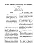
We used micro-PIXE (Particle Induced X-ray E mis-
sion) to investigate the iron distribu tion within the indi-
vidual tissues of seeds from P. coccineus and from the
two P. vulgaris genotypes, G14519 and G4825 (Fig. 1).
This non-destructive technique can detect and quantify
iron, independent of its speciation, present at the surface
of the tissue and in the near-surface layer. In compari-
son to chemical iron staining, t he method does not
require incubation in liquid solution prior to the analy-
sis, reducing the probability of element redistribu tion or
leaching.
Micro-PIXE analysis of cotyledons revealed that speci-
fic regions within this tissue accumulated high concen-
trations of iron (Fig. 1a,c, and 1d). Regions of the
analyzed tissue were selected for quantification and
these are highlighted in the color-coded maps (Fig. 1k
to 1o). Iron-rich regions accumulated between 200 and
500 μgg
-1
of iron, depending on the genotype (Table 2
regions 3 to 7, 15 to 18, and 20 to 21). Differences in
the concentration of iron in these regions within indivi-
dual cotyledons were observed. For example the regions
3, 4, and 6, which have similar sizes, contained 283, 254,
and 341 μgg
-1
of iron respectively. Furthermore, sub-
regions with higher concentrations of iron, 400 to 500
μgg
-1
(regions 5 and 7), could be delineated within the
regions 4 and 6.
Our analyses also show that iron is evenly distributed
in the seed coat of the genotype G14519 (Fig. 1a). The
average iron concentration in the seed coat region 9 was
124 μgg
-1
(Table 2). This value is similar to the 132 μg
g
-1
measur ed for this tissue using ICP-AES (Table 1). In
Cvitanich et al. BMC Plant Biology 2010, 10:26
/>Page 2 of 14
the embryonic axis, iron accumulation was observed
near the radicle meristem (Fig. 1b and 1e). In particular,
high concentr ations of iron, 180 to 190 μgg
-1
,accumu-
latedatthetipoftheradicleofP. coccineus (Table 2
regions 25 to 26). These concentrations are higher than
the 54 μgg
-1
average iron concentration of the analyzed
radicle tissue (region 22). The radicle of G14519 also
contained a higher concentration of iron near the meris-
tematic tissue with an average of 146 μgg
-1
(region 12).
Differences in iron distribution between P. vulgaris
genotypes were detected using the Perl’s Prussian blue
(PPB) method
We used the PPB method to reveal additional details
about the iron distribution within seed tissues. To
address the efficiency of the PPB method, seeds from
the same pool of beans used for ICP-AES analysis
(Table 1) were soaked for 18 hours at room temperature
prior to staining for iron.
In agreement with the ICP-AES and micro-PIXE ana-
lysis, the genotype G14519 showed intense blue staining
in the seed coats (Fig. 2a compared to Table 1 and Fig.
1). The PPB method also shows the presence of seed-to-
seed variations with respect to seed coat iron content in
this genotype (Fig. 2a). A unique distribution of iron
was observed in the seed coats of genot ype G4825. Blue
staining, indicating the presence of high iron concentra-
tion, was observed in the darker brown pigmented areas
of the seed coat, suggesting an association with tannins
located there. In geno type CAL96 which contained only
17 μgg
-1
of iron in the seed coats, only small spots of
blue stain could be detected near the seed hilum.
G19833 and G21242, which are both mottled beans,
also had light staining near the hilum. These results
indicate that there is a positive correlation between iron
concentration in seed coat tissues and the blue stain
achieved by the PPB technique. However, the method
cannot be used to detect iron in darkly pigmented seed
coats, as in the case of DOR364.
The PPB method also detected high iron concentra-
tions in the cotyledonary regions (Fig. 2), a feature pre-
viously revealed using micro-PIXE analysis (Fig. 1).
Most genotypes, with the exception of DOR364, accu-
mulated iron near the provascular tissue (Fig. 2). In
G19833 iron accumulation was evident in the cells sur-
rounding the provascular bundles (Fig. 2n and 2p).
Some genotypes, in particular G14519, NUA35, and
G19833 showed significant iron staining in the region
near the cotyledon epidermis (Fig. 2, arrows).
Stains of the embryonic axis indicated the presence of
higher iron accumulation near the radicle tip (Fig. 2q
and 2u). These results correspond to the observations
from the micro-PIXE analysis (F ig. 1). Strong blue stain
was observed in the endosperm layer of some genotypes,
as for example in NUA35, which is a high-seed-iron
genotype from the Andean gene pool bred at CIAT
through a backcross with G14519 (Fig. 2t). This is in
agreement with ICP-AES analysis, which showed that
the endosperm layer contained 270 ± 2 μgg
-1
and 120
±2μgg
-1
of iron in the related genotypes NUA35 and
G14519, respectively. NUA35 was also high in cotyle-
donary iron and had the highest overall level of seed
iron.
In short, there is good agreement between the obser-
vations using micro-PIXE analysis and the PPB method.
The PPB method is ideal for the detection of seed-to-
seed variations, and to determine the sites of iron accu-
mulation in a cost-effective way.
To address whether different growth conditions
affected the iron distribution in P. vulgaris genotypes,
we perfor med PPB staining on seeds from plants grown
in Darien, Colombia, and on seeds from plants grown in
a greenhouse (Fig. 3). The PPB staining indicate that the
relative iron accumulation in the studied genotypes is
Table 1 Iron concentration in tissues of P. vulgaris (Pv) and P. coccineus seeds
Iron concentration (μgg
-1
) Iron fraction (%) Total iron
Genotype Coty-ledons Axis Seed coats Coty-ledons Axis Seed coats μgg
-1
Gene pool
Pv G4825 56 (sd 9)
a
67 (± 3) 56 (sd 6)
a
91 2 7 57 M
Pv DOR364 43(± 3) 46 (± 1) 71 (± 5) 84 1 15 45 M
Pv G14519 65 (sd 2)
a
70 (± 3) 132 (sd 16)
b
80 2 18 71 M
Pv NUA35 80 (± 4) 103 (± 2) 61 (± 3) 90 1 8 78 N/A
Pv CAL96 57 (± 2) 81 (± 6) 17 (± 2) 96 1 2 54 A
Pv G19833 58 (± 2) 85 (± 4) 30 (± 2) 92 2 5 55 A
Pv G21242 70 (± 9) 97 (± 5) 42 (± 2) 92 2 6 67 A
P. coccineus 45 (sd 5)
a
84(± 10) 35 (sd 3)
a
92 1 7 44 N/A
The iron concentrations in different tissues from mature seeds were obtained by ICP-AES. The iron fraction is the percentage of the total seed iron. The total iron
concentration (Total iron) is based on the measured concentrations in the individual tissues. The genotypes with high seed coat iron are from the Mesoamerican
(M) gene pool, while the genotypes with low seed coat iron are from Andean (A) gene pools. All the P. vulgaris genotypes were grown in one season in Darien,
Colombia. P. coccineus purchased at a market in Denmark shown in Fig. 5 column 1. For some genotypes two
a
or three
b
individual pools of tissue were
measured. N/A: non applicable. Sd: standard deviation.
Cvitanich et al. BMC Plant Biology 2010, 10:26
/>Page 3 of 14
stable in the used growth conditions, for example
DOR364 showed the least staining in all the three envir-
onments while G14519 and NUA35 showed the most
intense blue staining. Only one example of genotype ×
environmental effects was observed. CAL96 accumulated
iron near the provascular bundles when grown in the
greenhouse but not in the field. Seed to seed variations
were observed o ccasionally, even between seeds from a
single harvest from an individual plant. For example
most of the seeds from the genotype DOR364 grown in
the greenhouse in 2009 showed very weak blue staining,
whileafewshowedamoreintensebluecolor(Fig.3,
GH2009).
High concentration of iron accumulates in the first layer
of cells that surround the provascular bundles of the
mature embryos of P. vulgaris
The cellular localization of iron was studied using the
PPB method. Microscopical analysis of tissue sections
allowed us to verify that cells surrounding the provascu-
lar bundles are responsible for iron storage in the coty-
ledons of the G14519 genotype (Fig. 4). In addit ion, the
iron-specific staining is limited to the cytoplasm of these
cells, while no staining was observed in the amyloplasts
(Fig. 4b and 4c). Microscopical analysis confirmed that
the meristematic tissue of the radicle is also rich in iron
(Fig. 4e). In addition, detectable levels of iron were
observed in cells near the radicle epidermis (Fig. 4f), and
in the palisade layer of the s eed coat (Fig. 4g). Due to
the smaller cell size, the subcellular localization of iron
in the radicle and palisade could not be determined in
these sections.
Cells of the provascular region are responsible for iron
storage in P. coccineus, but not in P. lunatus beans
To determine whether iron storage in the provascular
region was a general property of beans from the Phaseo-
lus genus, we used the PPB method to study iron
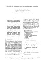
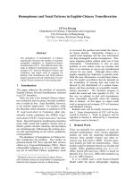
Figure 1 Elemental analysis of mature seeds from P. vulgaris and P. coccineus using micro-PIXE. Elemental maps obtained by Dynamic
Analysis method. Maps of iron (a-e) and phosphorus (p-t). Colour scales showing concentrations are on the left side. f to j show the analyzed
tissue, and the scanned area is marked with an open square. The maps a,b,p, and q represent the iron (Fe) and phosphorus (P) distribution in
the cotyledon (cot) and radicle (rad) of P. vulgaris accession G14519 shown in f and g, respectively. The scanning of a region (shown in h) of the
cotyledon of the P. vulgaris genotype G4825 is represented in the maps c and r. The analysis of the cotyledon and radicle of P. coccineus shown
in i and j resulted in the maps d,e,s, and t. The maps k to o show the regions used for the quantification of elements displayed in Table 2. Each
region has been assigned a unique number. Seed coat: sc. The * is used to mark an area that has been contaminated, and therefore elements
cannot be quantified in the region. Scale bars: 0.5 mm.
Cvitanich et al. BMC Plant Biology 2010, 10:26
/>Page 4 of 14
distribution in P. coccineus and P. lunatus seeds. While
iron is accumulated in the provascular region of the
cotyledons of P. coccineus (Fig. 5a to 5e) and P. vulgaris
(Fig. 2), it was not observed in this region in P. lunatus
(Fig. 5f a nd 5g). Instead, the P. lunatus cotyledons
showed a gradient of increasing iron accumulation
toward the outer epidermal layers, with p rovascular
strands visible as unstained white lines (Fig. 5f and 5g).
These results indicate that the strategies for storing seed
iron are specific for each species.
We studied whether soaking affected seed iron distri-
bution. Our results show that soaking P. coccineus beans
in water for just 2.5 hours had a significant effect on
iron staining (Fig. 5h and 5i compared to 5j and 5k).
Soaked tissue showed stronger iron stain compared to
un-soaked tissue. It is not clear whether this is a result
of increased penetration of the PPB solution into the
soaked tissues or a consequence of iron mobilization.
Iron was detected in the provascular region of soaked
and un-soaked cotyledons, but in particular the radicles
of soaked beans showed significantly stronger staining
(Fig. 5k).
In agreement with the results from P. vulgaris, micro-
scopical analyses of P. coccineus cotyledons show the
accumulation of iron in the cytoplasm of the cells that
surround the provascular bundles (Fig. 5m and 5n). In
sections of P. lunatus cotyledons, iron was only detected
in epidermal cells (Fig. 5p), in cells proximal to the
epidermal layer (Fig. 5p and 5q), and in some cells of
the provascular bundles (Fig. 5o), but not in the cells
surrounding this tissue. With the exception of small
iron staining spots that were found throughout the sec-
tions of cotyledons (indicated by open arrows in Fig. 5o
and 5q), the iron concentration in the examined tissue
was below the detection limit for the method used.
Similar to P. vulgaris and P. coccineus, the iron detected
in the epidermal region was found in the cytoplasm.
TheironintheprovasculartissueofP. lunatus cotyle-
dons was detected when the dry P. lunatus seeds were
dissected and incubated in 70% ethanol prior to staining
for 35 min, but not when the seeds were soaked in
water for 18 to 24 hours before staining. As in P. vul-
garis, P. coccineus and P. lunatus amyloplasts remain
unstained (Fig. 5n and 5q).
Ferritin accumulates in the amyloplasts of mature P.
coccineus seeds
Ferritin has been suggested as the major iron-storing
protein in legume seeds [16]. Therefore we decided to
study whether the iron-rich cells of beans also accumu-
lated more ferritin than the surrounding cells. Ferritin
was detected in tissue sections using immunolocalization
(Fig. 6). The primary antibodies used were raised against
the A. thaliana ferritin1 [26]. Therefore we analyzed
whether these antib odies were able to detect ferritin in
common beans. Western blot an alysis shows that the
Table 2 Average concentrations of iron and phosphorus in selected regions
K
Region 1 2 3 4 5 6 7 8 9
Fe (μgg
-1
) 71 ± 4 60 ± 3 283 ± 21 254 ± 10 411 ± 22 341 ± 20 502 ± 46 80 ± 2 124 ± 5
Det. Lim. (0.6) (2) (5.3) (4.2) (9.4) (6.3) (21) (1.7) (2.7)
P(μgg
-1
) 1170 1080 2490 1900 2390 2400 2720 472 33
± 57 ± 63 ± 180 ± 120 ± 190 ± 220 ± 210 ± 26 ± 13
Det. Lim. (10) (41) (135) (103) (186) (155) (288) (26) (19)
LM
Region 10 11 12 13 14 15 16 17 18
Fe (μgg
-1
) 83 ± 5 125 ± 8 146 ± 6 68 ± 3 49 ± 5 407 ± 22 362 ± 13 257 ± 10 183 ± 11
Det. Lim. (0.5) (0.9) (1.4) (1) (0.5) (7.7) (5.9) (4.4) (6.4)
P(μgg
-1
) 7480 10400 10600 9980 2310 4340 4330 3240 2900
± 200 ± 280 ± 120 ± 120 ± 66 ± 230 ± 180 ± 81 ± 100
Det. Lim. (8.7) (17) (26) (21) (8.4) (158) (90) (66) (129)
NO
Region 19 20 21 22 23 24 25 26
Fe (μgg
-1
) 52 ± 3 232 ± 9 297 ± 14 54 ± 3 35 ± 4 85 ± 4 187 ± 11 192 ± 15
Det. Lim. (0.8) (4.2) (5.4) (0.4) (0.7) (1.1) (4.9) (7.3)
P(μgg
-1
) 3950 5440 5030 6220 3100 6860 6990 9780
± 40 ± 270 ± 230 ± 340 ± 210 ± 190 ± 340 ± 51
Det. Lim. (11) (80) (107) (8.2) (15) (22) (104) (161)
Concentration of iron (Fe) and phosphorus (P) in the regions indicated in Fig. 1k-o. The concentrations were measured using micro-PIXE. The region numbers are
indicated in the table. Minimum detection limits (99% confidence level) are shown in parentheses (Det. Lim.).
Cvitanich et al. BMC Plant Biology 2010, 10:26
/>Page 5 of 14
antibodies detected two bands at approximately 28 kDa
in total-protein extracts from s eeds of P. coccineus (Pc)
and P. vulgaris (Pv) (Fig. 6d and 6e). The presence of
two bands is consistent with previously published results
for other plants [27]. Fluorescence microscopy of the
immunostained tissue from P. coccineus, P. vulgaris, and
P. lunatus cotyledons shows that ferritin accumul ates in
amyloplasts (Fig. 6). Sections that had been stained for
iron using the PPB method were used for the immuno-
detection of ferritin. The pictures illustrate that ferritin
does not accumulate in the cellular compartments
where the iron concentrations are highest. Ferritin was
only detected in the amyloplasts where the concentra-
tions of iron w ere below the levels of detection for the
PPB method (Fig. 4 , 5, and 6). For example, iron was
detected in and near the epidermal cells of P. lunatus,
but ferritin was not detected in these cells (Fig. 5p and
6l). In the same species, iron staining was observed in
the provascular cells while ferritin signals were only visi-
ble in the amyloplasts of the surrounding cells (Fig. 5o
and 6k). A control sample where the primary antibody
was omitted is shown in Fig. 6c. No antibody signals
were observed in the amyloplasts of control sections,
even when longer exposure times were used. Staining of
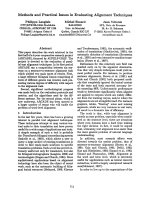
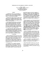
Figure 2 Distribution of iron in seeds from different P. vulgaris genotypes. Perls’ Prussian blue (PPB) staining of P. vulgaris seeds that were
soaked for 18 hours. a: The picture illustrates the variation in the stain intensity among seed coats, cotyledons, and embryonic axis (axis) of the
different genotypes. Stained cross sections (cs) of cotyledons as well as longitudinal sections (ls) are shown. The outer layers of the cotyledons,
proximal to the seed coats, are indicated by arrows. btou:Stereomicroscopy of tissue from a. b to h: Cross sections of cotyledons. i to o:
close-up pictures of b to h near the pro-vascular bundles (pvb). p: longitudinal section of pvb. q: hypocotyl (hyp) and radicle (rad) and attached
cotyledon tissue (cot) showing pvb. r: close-up of the provascular tissue shown in q. s: cotyledon sample where a portion of the epidermis and
proximal cell layers was removed prior staining making the pvb accessible for the Perls’ solution. t: endosperm layer. u: longitudinal section of
bean embryonic axis showing PPB stain of the radicle provascular and meristematic tissue. Scale bars in a:1cmandb to u: 1 mm.
Cvitanich et al. BMC Plant Biology 2010, 10:26
/>Page 6 of 14
the sections using the Lugol solution confirmed that fer-
ritin was only detected where starch (black) was present.
For example, ferritin was not detected in the regions
were the starch crystals were removed during sectioning
(indicated by “*” in Fig. 6i to 6k and 6m to 6o).
These results indicate that the highest iron concentra-
tions are found in epidermal cells, in cells proximal to
the epidermal layer, in provascular cells, or in cells sur-
rounding the provascular bundles of mature Phaseolus
seeds. Iron was primarily detected in the cytoplasm of
these cells while ferritin was always detected in the amy-
lopl asts. Although iron is likely present in ferritin, there
were no correlation between the detection of iron using
the PPB method and the presence of ferritin.
High concentrations of phosphorus accumulate in the
radicles and near the provascular bundles of P. coccineus
and P. vulgaris beans
We used Micro-PIXE ana lysis to study whether there
was a correlation between the accumulation of phos-
phorus and iron. Color maps showing the distribution
of these elements in P. coccineus and P. vulgaris seeds
are shown in Fig. 1. The highest phosphorus concentra-
tionsweremeasuredinradicletissues(Fig.1q,t,l,o
and Table 2, regions 10 to 13, 22, 24 to 26). The regions
of the radicle that accumulated the most iron also had a
hig her concentration of phos phorus (regions 11, 12, 25,
and 26). A similar pattern was observed in the regions
near the provascular bundles of the cotyledons (regions
3,4,6,16,17,18, and 20). However, regions with similar
phosphorus concentrations had significantly different
iron content (for example region 3 and 24 compared to
5 and 25, respectively). Furthermore, low phosphorus
concentrat ions were observed in iron-rich seed coats
(region 9) and in the region near the epidermal surfaces
that accumulated 80 μgg
-1
of iron (region 8). In the
present study we were not able to determine whether
iron and phosphorus co-localized at the sub-cellular
level.
Discussion
Cells surrounding the provascular bundles can store high
concentration of iron
We have discovered that in the cotyledons, the first
layers of cells surrounding the provascular bundles can
accumulate up to 500 μgg
-1
of iron in P. vulgaris and
300 μgg
-1
in P. coccineus genotypes (Fig. 1 and table 2).
In comparison, iron was shown to accumulate in the
provascular tissue of the seeds of A. thaliana [21]. The
same study showed that the disruption of the vacuolar
iron uptake transporter (VIT1) altered the distribution,
but not the total concentration, of iron in seeds. It was
concluded that the accumulation of iron in vacuoles is
critical for seed development. Our microscopical
Figure 3 Effects of different growth con diti ons on seed iron distribution .Perls’ Prussian blue (PPB) staining of seeds from five P. vulgaris
genotypes grown in Darien, Colombia (Darien), or in a greenhouse in Denmark in 2008 (GH2008) or in 2009 (GH2009). The dry seeds were
dissected and stained for 35 minutes using the PPB method. The genotypes names are shown. The scale bar is 1 cm
Cvitanich et al. BMC Plant Biology 2010, 10:26
/>Page 7 of 14
analysis indicates that high concentrations of iron accu-
mulate in the cytoplasm of cells surrounding the provas-
cular bundles of mature P. vulgaris and P. coccineus
seeds (Fig. 4 and 5). Our study does not reveal the cellu-
lar distribution of iron in the provascular tissue. There-
fore it is possible that vacuoles in the provascular tissue
are important for iron storage or for loading iron into
the seeds. It is also possible that vacuolar iron is primar-
ily needed during the early stages of germination and
that the iron stored in the cytoplasm of the cells sur-
rounding the provascular tissue represents an iron
reserve that could be mobilized later during
germination.
The distribution of iron in bean seeds is dependent on
species and genotype
We show that six of the seven studied genotypes of P.
vulgaris accumulate high concentrations of iron in cells
surrounding the provascular bundles of the embryonic
cotyledons (Fig. 2). The distribution of iron in the seeds
of four P. coccineus genotypes was similar to that in P.
vulgaris, while iron was detected throughout the cotyle-
dons of the two P. lunatus genotypes, which showed no
increased accumulation in the cells surrounding the pro-
vascular bundles (Fig. 5).
Our analysis shows that the distribution of iron
between the seed coat and embryo varies between geno-
types within the same species (Table 1 and Fig. 2). Simi-
larly, Ariza-Nieto et al [4] have shown that in common
beans the percentage of iron accumulated in the embryo
is genotype-dependent. They found that seed coat iron
represented 4 to 26% of the seed iron, depending on th e
genotype. In comparison our estimates show that 2 to
18% of the seed iron is present in the seed coats (Table
1). A comparison of the two studies indicates that iron
concentrations in seed coats and the embryonic axis are
slightly higher in the study by Ariza-Nieto et al,even
when the total seed iron concentrations were similar.
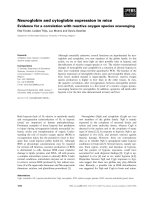
Figure 4 Cellular localization of iron in the P. vulgaris genotype G14519. Beans soaked for 18 hours (a and e to h) or unsoaked (b to d)
stained with Perls’ Prussian blue reagent. a,d,g: Stereomicroscopy. a: Cotyledon tissue, close-up of provascular bundle (pvb). d: Longitudinal
section of embryonic axis (axis) including cotyledon tissue (cot), and the axis attachment zone indicated by a filled arrow. g: Seed coat, showing
the inside (ins) part toward the cotyledons and the stain at the outer side (os) of this tissue. b, c, f and h: light microscopy of thin sections of
PPB stained tissue. b and c: Close-up of cotyledon pvb, surrounded by blue stained bundle-sheath-like cells which contain amyloplasts (am). e:
Close-up of radicle meristem showing the stained meristematic cells (mer). f: Section of hypocotyl tissue showing iron accumulation in the cells
proximal to the epidermis (ep). h: Seed coat section illustrating blue stain in the palisade layer (pl). Scale bars in d and g: 1 mm, in a, b, e, f,
and h: 0.1 mm, and in c: 0.01 mm.
Cvitanich et al. BMC Plant Biology 2010, 10:26
/>Page 8 of 14
These differences could be caused by iron mobilization
during soaking, as the beans used by A riza-Nieto et al
were soaked prior to dissection. Another possibility is
that the endospermal layer, which we have shown can
accumulate 270 μgg
-1
of iron, was still attached to the
seed coat of their soaked beans, while this layer was
mostly removed for our analysis. Other studies of com-
mon beans have shown that 5 to 40% of the seed iron
was found in the seed coats [23,28]. These are signifi-
cantly higher values than found in the present study. It
is possible that the genotypes studied by Moraghan et al
(2002) accumulated more iron than the ones used in
our investigation. Interestingly, Moraghan et al (2002)
analyzed seeds that were harvested at the R7 stage of
growth. It is possible that iron is mobilized to the
embryo during the last stage of maturation, changing
the proportion of seed coat iron between the R7 stage
and maturity. In agreement with that theory, it has been
suggested that iron in pea seeds could temporarily acc u-
mulate in non-vascular cells of the seed coats, thereafter
mobilizing to the embryo apoplast [16].
Among the common bean genotypes analyzed here,
three (CAL96, DOR364, a nd G19833) were in common
with those of Ariza-Nieto et al., while the others
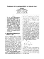
Figure 5 Iron distribution in P. coccineus and P. lunatus seeds.Perls’ Prussian blue staining of P. coccineus and P. lunatus seeds. a to g:
Beans soaked 18 hours prior to staining. a: Stained seed coats and cotyledons from two P. coccineus batches purchased in Denmark (1 and 2),
two P. coccineus genotypes (G35171 and G35172), and two P. lunatus genotypes (G25350 and G25381A). b to g: Close up stereomicroscope
pictures of the samples in a. h to n: P. coccineus batch 1, o: P. lunatus G25381, and p, q: P. lunatus G25350. Stereomicroscopy of stained non-
soaked beans (h and i) and of beans soaked in water for 2.5 hours prior to staining (j and k). h and j: Pictures of cotyledons, the blue stained
provascular region (pvb) is indicated. i and k: Images of embryonic axes. l: Close up picture of the provascular region. m to q are light
microscopy of thin cotyledon sections. m, n, p, and q: seeds were soaked in water for 18-24 hours before dissection and staining. o: dry seeds
were dissected and soaked in 70% ethanol for 24 hours prior to PPB staining. Filled arrows point at iron stained cells and open arrows point at
small iron stained spots. am: amyloplasts, rad: radicles, pvb: provascular bundles, cot: cotyledons. Scale bars in a: 1 cm, in b to k: 1 mm, in l to n:
0.1 mm, and in o to q: 0.01 mm.
Cvitanich et al. BMC Plant Biology 2010, 10
:26
/>Page 9 of 14
represented contrasting genotypes selected for their high
(G14519, G21242, NUA35) and low (G4825) seed iron
content. These previous authors also found variability in
seed coat iron with DOR364 (Mesoamerican) also hav-
ing high seed coat iron relative to CAL96 and G19833
(Andeans) suggesting that gene pool differences affect
this trait. Therefore, our results emphasize the impor-
tance of complementing genetic analy sis with phy siolo-
gical analysis of the gene pools, species, and genotypes
of interest. They also highlight the importance of study-
ing iron metabolism in both model and crop plants.
Indeed, a comparison of the iron accumulation pat-
terns of NUA35 with its parents CAL96 and G14519
suggests that iron distribution criteria should be inte-
grated into selection strategies for bean biofortification.
Ferritin accumulates in the amyloplasts of embryonic cells
It has been suggested that phytoferritin-bound iron is
the principal iron form during early germination [15]. In
peas, ferritin was shown to accumulate in the embryo
and it was suggested that ferritin-iron accounted for
92% of the iron in this tissue [16]. Recent analyses of
legume ferritins, whi ch included soybeans, common
beans, and peas, suggested that ferritin iron can maxi-
mally account for 18 to 42% of the total seed iron
depending on the species [29]. For white and red kidney
beans it was calculated that 20 and 25% of the total
seed iron was bound to ferriti n respectively [29].
Furthermore, it was estimated that up to 5% of the seed
iron in A. thaliana could b e bound to ferritin [30].
However, similar to the findings of Hoppler et al. [29]
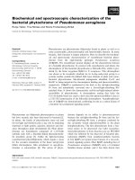
Figure 6 Ferritin immunolocalization. Sections of P. coccineus (a, b, and f to h), P. vulgaris G14519 (i and m), and P. lunatus G25350 (j, k, l, n,
o, and p) cotyledons were immunostained using antibodies raised against A. thaliana ferritin1 (AtFER1) and Alexa 546 secondary antibodies. In
a, b, c, f, g, h, k, l, o, and p the seeds were soaked in water for 18-24 hours prior to PPB staining, while in i, j, m, and n dry seeds were
dissected and soaked in 70% ethanol for 24 hours prior to PPB staining. b, g, i, j, k, and l: fluorescence microscopy of the immunostained tissue
sections shown in a, f, m, n, o, and p respectively. a and m: Light microscopy of Perls’ Prussian blue stained tissue after immunostaining. In f, n,
o, and p the sections were treated with Lugol solution after immunostaining; the starch is stained black. c: Section treated like b, g, and h, but
without primary antibody. h: Confocal microscopy of a single immunostained cell of P. coccineus cotyledon. d: Western blot analysis of the gel
shown in e using anti-AtFER1 serum. e: Coomassie blue stained gel of 6 μg total protein from beans of P. coccineus (Pc) and P. vulgaris (Pv). M:
Protein ladder, pvb: provascular bundle, am: amyloplasts, ep: epidermis, *: site where starch crystals were removed during sectioning. Scale bars
in a to c and f to h: 0.1 mm, and in i to p
: 0.01 mm
Cvitanich et al. BMC Plant Biology 2010, 10:26
/>Page 10 of 14
and Ravet et al. [30], our results indicate the accumu la-
tion of non-ferritin iron in the seeds of the three studied
Phaseolus species. Although it is likely that ferritin con-
tains iron, the concentrations of iron accumulated in the
ferritin-rich amyloplasts was below the level of detection
of the method used (Fig. 6). In contrast, our microscopi-
cal analysis indicates that the cytoplasm of cells sur-
rounding the provascular tissue plays a key role in iron
storage in the bean seeds of the P. vulgaris and P. cocci-
neus species. Iron rich cells were also detected at and
near the epidermal layer in the three st udied Phaseolus
species. In these cells, iron staining was also visible in
the cytoplasm.
The accumulation of ferritin in amyloplasts is in
agreement with the general knowledge that ferritins are
localized in plastids [17,31,32]. A correlation between
starch and ferritin accumulation in legumes is suggested
by their co-localization in amyloplasts and by the knowl-
edge that they both are degraded during seed germina-
tion [33]. Ferritin was also one of the 289 proteins
found in the soluble fraction of amyloplasts from wheat
endosperm [34]. The primary role of ferritin in A. thali-
ana was suggested to be the protection against reactive
oxygen species [30].
Using Western blot analysis, we detected two ferritin
bands in extracts from P. coccineus and P. vulgaris seeds
(Fig. 6). This is in agreement with previous studies of
other seeds. Two ferritin peptides of 28 kDa and 26.5
kDa were detected in extracts from soaked pea, soy-
beans, and maize seeds [35]. The products of at least
two ferritin genes were identified in soybean seeds,
whileonlyoneoftheferritinsfromA. thaliana,
AtFER2, was detected in mature seeds [27,36,30].
Seed regions near the provascular bundles are rich in
iron and phosphorus
The accumulation of ferritin has recent ly been shown to
be affected both by the iron and the phosphate status of
A. thaliana plants [37]. Furthermore, the study showed
that the accumulation of iron in leaves was shifted from
vacuoles to chloroplasts under phosphate deficiency. It
was suggested that the vacuolar iron was found in phos-
phorus complexes that were formed when plants were
grown in phosphate-rich medium, while chloroplast iron
was bound to ferritin [37]. In this study the distribution
of phosphorus was visualized using micro-PIXE analysis
(Fig. 1). The iron-rich regions near the provascular bun-
dles of the cotyledons and of the radicle accumulated a
higher proportion of the seed phosphorus compared to
the surrounding regions. In contrast, iron-rich regions
of the seed coat and near the epidermal cells did not
accumulate considerable amounts of phosphorus.
Therefore, it is possible that some percentage of the
seed iron can be bound to phosphor us-rich compounds ,
but we suggest that the cells near the provascular tissues
have a general role in nutrient storage that explains the
higher concentration of phosphorus. For example iron
and manganese accumulate near the provascular bun-
dles of A. thaliana seeds [21]. Their distribution pat-
terns differ slightly, indi cating that they are not part of
common complexes and emphasizing the role of the
region near the provascular bundles in the storage of
nutrients. Similarly, we suggest that the radicle might
have an important function in storing nutrients that are
needed during the early events of germination.
The iron-rich cells of the provascular region of the
Phaseolus species show similarities to the extended cells
surrounding the provascular bundles of the cotyledons
and leaves of the non- legume Ricinus communis (castor
bean). These cells were shown to accumulate the iron
chelator nicotianamine (NA) [38]. In strategy I plants,
NAissuggestedtobeinvolvedinbindingferrousiron
in the phloem sap [39] and was shown to be important
for the supply of iron t o seeds in A. thaliana [22]. Our
PPB staining indicates that the iron accumulated near
the provascular bundles of beans is primarily ferric iron.
NA can bind both ferrous and ferric iron with similar
affinities [40,41]. Therefore it is possible that NA is
involved in chelating iron in the iron-rich cells of the
provascular region in beans.
Taken together, we show that the distribution of iron
in seeds depends on the species and genotype and that
the cells surrounding t he provascular tissues of the
embryo might play a key role in the storage of minerals
in mature seeds. Furthermore, our results indicate that a
large proportion of the seed iron in the Phaseolus spe-
cies is stored in compounds different from ferritin and
that accumulation of iron in the seed coat is highly vari-
able between P. vulgaris genotypes. In addition, we sug-
gest that the PPB technique is ideal to study iron
distribution in legume seeds during plant breeding and
to detect seed-to-seed variations. This technique may
contribute to a quick and inexpensive method for the
selection of genotypes with more bioavailable iron.
Future studies of the molecular mechanisms behind
the described accumulation of nutrients and their
importance for seed germ ination will shed new light on
how seeds store iron and on the possibility t o improve
the nutritional value of seeds.
Conclusions
Using two different techniques we were able to show
that cells surrounding the provascular tissue contain a
high concentration of iron in P. coccineus and P. vul-
garis seeds. This cell layer appears to be of key impor-
tance for iron storage in the seed or for providing iron
to the germinating seedling. Therefore these cells are an
appropriate target for future molecular biology research.
Cvitanich et al. BMC Plant Biology 2010, 10:26
/>Page 11 of 14
In agreement with previous studies in legumes [29]
and in A. thaliana [30] our studies indicate that high
concentrations of non-ferritin-iron is accumulated in
the seeds of P. coccineus, P. lunatus,andP. vulgaris.
These findings emphasize the need to characterize other
compounds that might be involved in the chelation of
iron in mature seeds and the proportion of iron bound
by the different chelators as well as investigating the
bioavailability of iron from different sources.
Recent research in A. thaliana indicates that vacuolar
transporters are important for seed iron distribution and
for iron loading to the seedling during germination
[20,21]. Our findings suggest that high concentratio n of
iron is accumulated in the cytoplasm of cells surround-
ing the provascular tissue of the P. coccineus and P. vul-
garis seeds. These results indicate either that there are
major differences between A. thaliana and the Phaseolus
species with respect to the subcellular localization of
iron accumulation, or that the vacuolar transporters are
one of several transporters involved in iron mobilization
to and from the seeds. In the latter case, additional
transporters might be involved in the loading of iron to
the cytoplasm of iron-rich cells that surround the pro-
vascular tissue of some legume species.
Methods
Plant materials
Phaseolus vulgaris (7), P. coccineus (2) and P. lunatus (2)
genotypes were obtained from the International Center
for Tropical Agriculture, Cali, Colombia and are main-
tained either with the Genetic Resource Unit (G entries)
or with the Bean Program (CAL, DOR and NUA lines)
at CIAT. All P. vulgaris genotypes were grown both in
Darien, Valle de Cauca, Colombia and in a greenhouse
in Denmark. Darien is 1400 m above sea level, at this
location the growth conditions were: 20°C average yearly
temperature, 1288 mm annual rainfall, Udand soil type,
pH 5.6, and native HCl extractable mineral concentra-
tion for iron in the topsoil a veraged 4.39 μgg
-1
.Inthe
greenhouse, plants w ere grown in soil in five liter pots,
18-23°C daytime/15-20°C nighttime temperatures, 16
hours light/8 hours dark cycle, and 70% relative humid-
ity. The plants were watered with a 0.1% solution of Pio-
neer NPK (14-3-23) + Mg (Blue) (The Broste Group -
Ltd. Reg. No. 39781 ), 50% addi-
tional S was added as NH
4
SO
4
. If not otherwise stated,
the results shown are from seeds grown for one season
in Darien. In the Danish greenhouse, the genotypes
were grown twi ce and seeds were harve sted in August-
September of both 2008 and 2009.
Two batches of P. coccineus beans were purchased at a
retailer in Denmark. Th e P. vulgaris accessions and
breeding lines are from both the Mesoamerican (small-
seeded) and Andean (large-seeded) gene pools of com-
mon bean as indicated in Table 1.
Iron quantification
For each genotype, ten to fifteen dry mature seeds
(depending on seed size) were dissected into embryo
axis, cotyledons, and seed coat tissue s using a razor
blade. The endospermal layer between the cotyledons
and the seed coats was collected separately in order to
avoid contamination between these two tissues. The
iron content of each tissue type was measured in dupli-
cates at the Danish Technological Institute, Kongsvang
Alle 29, Aarhus, Denmark, using inductively coupled
plasma atomic emission spectroscopy (ICP-AES) in axial
mode.
Iron biochemical stains
Perl’ s Prussian blue (PPB) method was used as pre-
viously described [42,43]. In short, 1 volume of 4% HCl
solution was mixed with 1 volume of a fresh 4% solution
of Potassium hexacyanoferrate (II) trihydrate and used
to cover the tissue. Some beans were soaked for 2.5, 18,
or 24 hours i n water at 20°C, sliced, and incubated in
70% ethanol for 1-2 hours, others were sliced without
prior soaking. The tissue was covered with the solution
and allowed to react for 15 to 35 minutes before wash-
ing in pure water at least 5 times. Thereafter the tissue
was kept in 70% ethanol in the dark until analysis.
Preparation of samples for light microscopy
Samples that were stained with PPB method as pre-
viously described were incubated 2 to 5 days in 70%
etha nol prior to fixat ion. The tissue was thereafte r fixed
for 24 to 48 hours at 4°C in 4% paraformaldehyde, 1%
glutaral dehyde, 0.1 M NaHPO
4
pH 7.2. A stepwise
dehydration in ethanol was performed using 30 minutes
incubations in 70%, 80%, 90%, and twice in 96% ethanol.
For embedding and polymerization we used the Techno-
vit 7100 kit (Heraeus Kulzer, Wehrheim, Germany).
Embedding was performed using stepwise increments of
Technovit hardener I in ethanol, and manufacturers’
recommendations were followed for the polymeriza tion.
The specimens were sectioned into 7 to 11 micrometer
thick samples using a Leica RM2045 microtome and
studied by light microscopy.
Immunoassays
Glass slides with sections of PPB stained and unstained
tissue, fixed as previously described, were blocked for an
hour at room temperature (RT) using PBS-blotto solu-
tion (20 mM phosphate pH 7.4, 150 mM NaCl, 0.1% (v/
v) triton X-100, 3% (w/v) nonfat dry milk). Thereafter
the sections were incubated with rabbit serum raised
Cvitanich et al. BMC Plant Biology 2010, 10:26
/>Page 12 of 14
against A. thaliana ferritin 1 [26], diluted 2000 times in
PBS-blotto. For control samples PBS-blotto wit hout
serum was used. The incubation proceeded for 12 to 18
hour s at 4°C. The slides were rinsed twice in PBS- T (20
mM phosphate pH 7.4, 150 mM NaCl, 0.1% (v/v) triton
X-100), and washed 3 times for 5 minutes at room tem-
perature in PBS-T. Thereafter the slides were incubated
with Alexa fluor 546 goat anti-rabbit secondary antibo-
dies (Invitrogen), which was diluted 400 times in PBS-
blotto, for 1 to 3 hours at RT. The slides were rinsed
twice with PBS-T, washed 3 times for 5 minutes in PBS-
T, fixed for 10 minutes in 2% paraformaldehyde in PBS
(20 mM phosphate buffer pH 7.4, 150 mM NaCl), and
washed 3 times in PBS-T. Microscopy was performed
using Zeiss Axioplan fluorescence microscope and Zeiss
LSM510 Meta confocal microscope (excitat ion 543 nm,
emission filter BP 585 to 620 nm).
Western Blots
Cotyledons were dissected from mature dry seeds of P.
coccineus and P. vulgaris and ground usi ng liquid nitro-
gen. Soluble proteins were extracted in 50 mM Tris-
HCl pH 8.0, 2% (w/v) SDS, 1% (v/v) proteinase inhibitor
cocktail (Sigma P9599). A total of 6 μg total protein was
loaded in each lane. The protein was denatured in 77
mM ammediol, 0.057 N HCl, 13% glycerol, 30 mM SDS,
0.03% bromophenolblue, and 15 μMDTTfor10min-
utes at 95°C. The samples and 5 μl pre-stained protein
ladder (Fermentas, SM#0671) were separated in 6 to
12% polyacrylamide gradient gels as previously described
[44,45]. The gels were either stained with Coomassie
Brilliant Blue using standard protocols or electro-blotted
to PVDF membranes. For immuno-detection the mem-
branes were blocked using PBS-blotto as described for
immunoassays, and incubat ed for 12 to 18 hours at 4°C
with rabbit serum raised a gainst A. thaliana ferritin 1
[26], diluted 10,000 times inPBS-blotto.Themem-
branes were washed three times for 15 minutes in PBS-
T, incubated 2 hours at room temperature with peroxi-
dase-conjugated goat anti-rabbit antiserum (Dako
P0448), and washed once for 15 minutes and three
times for 5 minutes with PBS-T. The antibodies were
detected using the ECL technique [46] and Amersham
Hyperfilm™ ECL.
Elemental distribution and quantification by micro-PIXE
Dry seeds were cut with a razor blade and mounted on
a thin Formvar layer. The seed surfaces were coated
with carbon. Microanalyses were performed using the
nuclear microprobe at the Materials Research Depart-
ment, iThemba LABS, South Africa as previously
described [47-49]. I n short, a proton beam of 3.0 MeV
energy and 100 to 150 p A current was focused on a 3 ×
3 μm
2
spot and raster scanned over the areas of interest,
using square or rectangular scan patterns with a variable
number of pixels (up to 128 × 128). Particle-induced X-
ray emission (PIXE) and proton backscattering spectro-
metry (BS) were used simultaneously. Elemental concen-
trations were obtained using GeoPIXE II software [50].
Quantitative elemental images were generated using the
Dynamic Analysis method, an integral part of GeoPIXE
II. Information on atomic ratios of light elements form-
ing organic matrix, necessary for PIXE quantitative ana-
lysis, was found from BS technique. Regions
representing various seed parts were selected within
seed maps on the basis of their morphology from optical
micrographs and of iron distribution images. PIXE and
BS spectra were extracted from these regions to obtain
average concentrations within them.
Abbreviations
ICP-AES: inductively coupled plasma atomic emission spectroscopy; NA:
nicotianamine; PIXE: Particle Induced X-ray Emission; PPB: Perl’s Prussian blue;
ROS: reactive oxygen species; RT: room temperature.
Acknowledgements
Dr. Frédéric Gaymard from Biochimie et Physiologie Moleculaire des Plantes,
UMR 5004 Agro-M/CNRS/INRA/UMII, Montpellier, France for providing the
ferritin antibodies. Technical assistance of Finn Pedersen, Hanne Busk, and
Kirsten Sørensen.
Funding was provided by the Danish Agency for Science, Technology and
Innovation, HarvestPlus/Danida, and the Research Foundation of the
University of Aarhus, Denmark.
Author details
1
Centre for Carbohydrate Recognition and Signalling, Department of
Molecular Biology, University of Aarhus, Aarhus, Denmark.
2
Materials
Research Department, iThemba LABS, Somerset West, South Africa.
3
on leave
from: Faculty of Physics and Applied Computer Science, AGH University of
Science and Technology, Kraków, Poland.
4
International Center for Tropical
Agriculture, Cali, Colombia.
Authors’ contributions
CC drafted the manuscript, responsible for the design and coordination of
experiments, and participated in the execution of the experiments, WJP and
JMP carried out the micro-PIXE analysis and helped to draft the manuscript,
DFU carried out Western blots, AMJ participated in microcopy analysis, MWB
and CA are responsible for breeding the high-iron bean genotypes and
helped to draft the manuscript, EØJ and JS contributed to the design of
experiments and helped to draft the manuscript. All the author s read and
approved the final manuscript.
Received: 15 June 2009
Accepted: 11 February 2010 Published: 11 February 2010
References
1. Nestel P, Bouis HE, Meenakshi JV, Pfeiffer W: Biofortification of staple food
crops. J Nutr 2006, 136(4):1064-1067.
2. Mayer JE, Pfeiffer WH, Beyer P: Biofortified crops to alleviate micronutrient
malnutrition. Curr Opin Plant Biol 2008, 11(2):166-170.
3. Broughton WJ, Hernandez GH, Blair M, Beebe S, Gepts P, Vanderleyden J:
Beans (Phaseolus spp.)- model food legumes. Plant Soil 2003, 252:55-128.
4. Ariza-Nieto M, Blair MW, Welch RM, Glahn RP: Screening of iron
bioavailability patterns in eight bean (Phaseolus vulgaris L.) genotypes
using the Caco-2 cell in vitro model. J Agr Food Chem 2007,
55(19):7950-7956.
5. Hougaard BK, Madsen LH, Sandal N, de Carvalho Moretzsohn M,
Fredslund J, Schauser L, Nielsen AM, Rohde T, Sato S, Tabata S, Bertioli DJ,
Stougaard J: Legume Anchor Markers Link Syntenic Regions Between
Cvitanich et al. BMC Plant Biology 2010, 10:26
/>Page 13 of 14
Phaseolus vulgaris, Lotus japonicus, Medicago truncatula and Arachis.
Genetics 2008, 179(4):2299-2312.
6. Grotz N, Guerinot ML: Molecular aspects of Cu, Fe and Zn homeostasis in
plants. Biochim Biophys Acta 2006, 1763(7) :595-608.
7. Briat JF: Iron Dynamics in Plants. Advances in Botanical Research Academic
PressKader J-C, Delseny M 2007, 46:137-180.
8. Walker EL, Connolly EL: Time to pump iron: iron-deficiency-signaling
mechanisms of higher plants. Curr Opin Plant Biol 2008, 11(5):530-535.
9. Bauer P, Ling HQ, Guerinot ML: FIT, the FER-like iron deficiency induced
transcription factor in Arabidopsis. Plant Physiol Biochem 2007,
45(5):260-261.
10. Yuan Y, Wu H, Wang N, Li J, Zhao W, Du J, Wang D, Ling H-Q: FIT interacts
with AtbHLH38 and AtbHLH39 in regulating iron uptake gene
expression for iron homeostasis in Arabidopsis. Cell Res 18(3):385-397.
11. Wang HY, Klatte M, Jakoby M, Baumlein H, Weisshaar B, Bauer P: Iron
deficiency-mediated stress regulation of four subgroup Ib BHLH genes
in Arabidopsis thaliana. Planta 2007, 226(4):897-908.
12. Durrett TP, Gassmann W, Rogers EE: The FRD3-mediated efflux of citrate
into the root vasculature is necessary for efficient iron translocation.
Plant Physiol 2007, 144(1):197-205.
13. Pich A, Manteuffel R, Hillmer S, Scholz G, Schmidt W: Fe homeostasis in
plant cells: does nicotianamine play multiple roles in the regulation of
cytoplasmic Fe concentration?. Planta 2001, 213(6):967-976.
14. Kruger C, Berkowitz O, Stephan UW, Hell R: A metal-binding member of
the late embryogenesis abundant protein family transports iron in the
phloem of Ricinus communis L. J Biol Chem 2002, 277(28):25062-25069.
15. Tiffin LO, Chaney RL: Translocation of iron from soybean cotyledons. Plant
Physiol 1973, 52:393-396.
16. Marentes E, Grusak MA: Iron transport and storage within the seed coat
and embryo of developing seeds of pea (Pisum sativum L.). Seed Sci Res
1998, 8:367-375.
17. Briat JF, Lobreaux S, Grignon N, Vansuyt G: Regulation of plant ferritin
synthesis: how and why. Cell Mol Life Sci 1999, 56(1-2):155-166.
18. Theil EC: Iron, ferritin, and nutrition. Annu Rev Nutr 2004, 24:327-343.
19. Briat J-F, Lobréaux S:
Iron transport and storage in plants. Trends Plant Sci
1997, 2(5):187-193.
20. Lanquar V, Lelievre F, Bolte S, Hames C, Alcon C, Neumann D, Vansuyt G,
Curie C, Schroder A, Kramer U, Barbier-Brygoo H, Thomine S: Mobilization
of vacuolar iron by AtNRAMP3 and AtNRAMP4 is essential for seed
germination on low iron. EMBO J 2005, 24(23):4041-4051.
21. Kim SA, Punshon T, Lanzirotti A, Li L, Alonso JM, Ecker JR, Kaplan J,
Guerinot ML: Localization of iron in Arabidopsis seed requires the
vacuolar membrane transporter VIT1. Science 2006, 314(5803):1295-1298.
22. Klatte M, Schuler M, Wirtz M, Fink-Straube C, Hell R, Bauer P: The analysis
of Arabidopsis nicotianamine synthase mutants reveals functions for
nicotianamine in seed iron loading and iron deficiency responses. Plant
Physiol 2009, 109, 136374
23. Moraghan JT, Padilla J, Etchevers JD, Grafton K, Acosta-Gallegos JA: Iron
accumulation in seed of common bean. Plant Soil 2002, 246:175-183.
24. Laszlo JA: Mineral content in soybean seed coats and embryos during
development. J Plant Nutr 1990, 13:231-248.
25. Lombardi-Boccia G, De Santis N, Di Lullo G, Carnovale E: Impact of
processing on Fe dialysability from bean (Phaseolus vulgaris L.). Food
Chem 1995, 53(2):191-195.
26. Arnaud N, Ravet K, Borlotti A, Touraine B, Boucherez J, Fizames C, Briat JF,
Cellier F, Gaymard F: The iron-responsive element (IRE)/iron-regulatory
protein 1 (IRP1)-cytosolic aconitase iron-regulatory switch does not
operate in plants. Biochem J 2007, 405(3):523-531.
27. Masuda T, Goto F, Yoshihara T: A novel plant ferritin subunit from
soybean that is related to a mechanism in iron release. J Biol Chem 2001,
276(22):19575-19579.
28. Moraghan JT, Grafton K: Distribution of selected elements between the
seed coat and embryo of two black bean cultivars. J Plant Nutr 2002,
25(1):169.
29. Hoppler M, Schonbachler A, Meile L, Hurrell RF, Walczyk T: Ferritin-iron is
released during boiling and in vitro gastric digestion. J Nutr 2008,
138(5):878-884.
30. Ravet K, Touraine B, Boucherez J, Briat JF, Gaymard F, Cellier F: Ferritins
control interaction between iron homeostasis and oxidative stress in
Arabidopsis. Plant J 2009, 57(3):400-412.
31. Waldo GS, Wright E, Whang ZH, Briat JF, Theil EC, Sayers DE: Formation of
the ferritin iron mineral occurs in plastids. Plant Physiol 1995,
109(3):797-802.
32. Petit JM, Briat JF, Lobreaux S: Structure and differential expression of the
four members of the Arabidopsis thaliana ferritin gene family. Biochem J
2001,
359(3):575-582.
33. Lobreaux S, Briat JF: Ferritin accumulation and degradation in different
organs of pea (Pisum sativum) during development. Biochem J 1991,
274:601-606.
34. Balmer Y, Vensel WH, DuPont FM, Buchanan BB, Hurkman WJ: Proteome of
amyloplasts isolated from developing wheat endosperm presents
evidence of broad metabolic capability. J Exp Bot 2006, 57(7):1591-1602.
35. Laulhere JP, Lescure AM, Briat JF: Purification and characterization of
ferritins from maize, pea, and soya bean seeds. Distribution in various
pea organs. J Biol Chem 1988, 263(21):10289-10294.
36. Dong X, Sun Q, Wei D, Li J, Li J, Tang B, Jia Q, Hu W, Zhao Y, Hua ZC: A
novel ferritin gene, SferH-5, reveals heterogeneity of the 26.5-kDa
subunit of soybean (Glycine max) seed ferritin. FEBS letters 2007,
581(30):5796-5802.
37. Hirsch J, Marin E, Floriani M, Chiarenza S, Richaud P, Nussaume L,
Thibaud MC: Phosphate deficiency promotes modification of iron
distribution in Arabidopsis plants. Biochimie 2006, 88(11):1767-1771.
38. Rutten T, Kruger C, Melzer M, Stephan UW, Hell R: Discovery of an
extended bundle sheath in Ricinus communis L. and its role as a
temporal storage compartment for the iron chelator nicotianamine.
Planta 2003, 217(3):400-406.
39. Briat JF, Curie C, Gaymard F: Iron utilization and metabolism in plants.
Curr Opin Plant Biol 2007, 10(3):276-282.
40. Reichman SM, Parker DR: Revisiting the metal-binding chemistry of
nicotianamine and 2’-deoxymugineic acid. Implications for iron nutrition
in strategy II plants. Plant Physiol 2002, 129(4):1435-1438.
41. Weber G, von Wiren N, Hayen H: Analysis of iron(II)/iron(III)
phytosiderophore complexes by nano-electrospray ionization Fourier
transform ion cyclotron resonance mass spectrometry. Rapid Comm Mass
Spectrom 2006, 20(6):973-980.
42. Choi EY, Graham R, Stangoulis J: Semi-quantitative analysis for selecting
Fe- and Zn-dense genotypes of staple food crops. J Food Compos Anal
2007, 20(6):496-505.
43. Meguro R, Asano Y, Odagiri S, Li C, Iwatsuki H, Shoumura K: Nonheme-iron
histochemistry for light and electron microscopy: a historical, theoretical
and technical review. Arch Histol Cytol 2007, 70(1):1-19.
44. Laemmli U: Cleavage of structural proteins during the assembly of the
head of bacteriophage T4. Nature 1970, 227(5259):680-685.
45. Wyckoff M, Rodbard D, Chrambach A: Polyacrylamide gel electrophoresis
in sodium dodecyl sulfate-containing buffers using multiphasic buffer
systems: properties of the stack, valid Rf- measurement, and optimized
procedure. Anal Biochem
1977, 78(2):459-482.
46. Yang H, Leland JK, Yost D, Massey RJ: Electrochemiluminescence: A New
Diagnostic and Research Tool. Nat Biotech 1994, 12(2):193-194.
47. Prozesky VM, Przybyłowicz WJ, Van Achterbergh E, Churms CL, Pineda CA,
Springhorn KA, Pilcher JV, Ryan CG, Kritzinger J, Schmitt H, Swart T: The
NAC nuclear microprobe facility. Nucl Instrum Meth Phys Res B 1995,
104:36-42.
48. Przybyłowicz WJ, Mesjasz-Przybyłowicz J, Pineda CA, Churms CL,
Springhorn KA, Prozesky VM: Biological applications of the NAC nuclear
microprobe. X Ray Spectrom 1999, 28:237-243.
49. Przybyłowicz WJ, Mesjasz-Przybyłowicz J, Migula P, Nakonieczny M,
Augustyniak M, Tarnawska M, Turnau K, Ryszka P, Orłowska E, Zubek S,
Głowacka E: Micro-PIXE in ecophysiology. X Ray Spectrom 2005,
34:285-289.
50. Ryan C: Quantitative trace element imaging using PIXE and the nuclear
microprobe. Int J Imag Syst Tech 2000, 11:219-230.
doi:10.1186/1471-2229-10-26
Cite this article as: Cvitanich et al.: Iron and ferritin accumulate in
separate cellular locations in Phaseolus seeds. BMC Plant Biology 2010
10:26.
Cvitanich et al. BMC Plant Biology 2010, 10:26
/>Page 14 of 14