Các chẩn đoán phân biệt bệnh lý thần kinh trụ ở khuỷu tay pdf
Bạn đang xem bản rút gọn của tài liệu. Xem và tải ngay bản đầy đủ của tài liệu tại đây (191.17 KB, 7 trang )
Journal of the American Academy of Orthopaedic Surgeons
282
Ulnar nerve compression at the
elbow is commonly accepted as the
second most frequently encoun-
tered nerve entrapment in the upper
extremity, exceeded in prevalence
only by carpal tunnel syndrome.
The incidence of ulnar nerve com-
pression is probably greater if one
includes those individuals who ex-
perience transient numbness and
paresthesias when they lean on the
flexed elbow or when the elbow is
flexed for a prolonged period.
Anatomy and Etiology
The boundaries for potential ulnar
nerve compression begin approxi-
mately 10 cm proximal to the el-
bow and end about 5 cm distal to
the joint. The ulnar nerve can be
compressed anywhere along this
pathway at one or more of five
sites (Fig. 1).
In the middle third of the arm,
the ulnar nerve pierces the medial
intermuscular septum and de-
scends along the medial head of
the triceps muscle. The first area of
potential compression, which is the
widest, begins proximally at the
arcade of Struthers and ends distal-
ly near the medial epicondyle. The
arcade of Struthers is a musculofas-
cial band, 1.5 to 2.0 cm in width,
which is located an average of 8 cm
proximal to the medial epicondyle.
In an anatomic study of cadaver
extremities, it was present in 70%
of specimens.
1
The arcade, which
runs oblique and superficial to the
ulnar nerve, is composed of the
deep investing fascia of the arm,
superficial muscle fibers from the
medial head of the triceps (its most
obvious component), and the
Òinternal brachial ligament,Ó which
arises from the coracobrachialis
tendon. The anterior border of the
arcade is the medial intermuscular
septum. The lateral border is
formed by deep fibers from the
medial head of the triceps.
The arcade of Struthers should
not be confused with the far less
commonly encountered ligament of
Struthers. The ligament of Struthers
is associated with compression of
the median nerve. Although the lig-
ament itself has not been implicated
in compression of the ulnar nerve,
compression by the supracondylar
process has been reported.
2
In the absence of an arcade of
Struthers, the medial intermuscular
septum can cause compression as
the nerve passes over its edge,
which is thicker distally than proxi-
mally. This can occur after anterior
dislocation of the nerve or as a
postoperative complication of
ulnar nerve transposition when the
septum has not been excised. The
medial head of the triceps muscle
can also compress the nerve in this
Dr. Posner is Clinical Professor of Ortho-
paedics, New York University School of
Medicine, New York, NY; and Chief of Hand
Services, New York University Medical
Center/Hospital for Joint Diseases Department
of Orthopaedic Surgery and Lenox Hill
Hospital, New York.
Reprint requests: Dr. Posner, 2 East 88th
Street, New York, NY 10128.
Copyright 1998 by the American Academy of
Orthopaedic Surgeons.
Abstract
Ulnar nerve compression at the elbow can occur at any of five sites that begin
proximally at the arcade of Struthers and end distally where the nerve exits the
flexor carpi ulnaris muscle in the forearm. Compression occurs most commonly
at two sitesÑthe epicondylar groove and the point where the nerve passes
between the two heads of the flexor carpi ulnaris muscle (i.e., the true cubital
tunnel). The differential diagnosis of ulnar neuropathies at the elbow includes
lesions that cause additional proximal or distal nerve compression and systemic
metabolic disorders. A complete history and a thorough physical examination
are essential first steps in establishing a correct diagnosis. Electrodiagnostic
studies may be useful, especially when the site of compression cannot be deter-
mined by physical examination, when compression may be at multiple levels,
and when there are systemic and metabolic problems.
J Am Acad Orthop Surg 1998;6:282-288
Compressive Ulnar Neuropathies at the Elbow:
I. Etiology and Diagnosis
Martin A. Posner, MD
Martin A. Posner, MD
Vol 6, No 5, September/October 1998
283
area. The muscle head can be
hypertrophied, as is commonly
seen in bodybuilders, or it can snap
over the medial epicondyle, caus-
ing a friction neuritis.
The second site of potential com-
pression is the distal end of the
humerus, at or just proximal to the
medial epicondyle. Compression
in this area develops as a conse-
quence of a valgus deformity of the
bone secondary to an old epiphy-
seal injury to the lateral condyle or
a malunited supracondylar frac-
ture. Ulnar neuropathy secondary
to a humeral fracture was first
described by Mouchet in 1914; soon
thereafter it became known on the
European continent as the Òmal-
adie de Mouchet.Ó Two years later,
Hunt introduced the term Òtardy
ulnar palsyÓ in the United States.
The third area of potential com-
pression is the epicondylar or olec-
ranon groove. This is a fibro-
osseous groove, which is bounded
anteriorly by the medial epicondyle
and laterally by the olecranon and
the ulnohumeral ligament; medially,
the groove is covered by a fibro-
aponeurotic band. In its passage
through the groove, the ulnar nerve
is accompanied by an anastomotic
arterial system composed of the
superior and inferior ulnar collateral
arteries from above and the posterior
ulnar recurrent artery from below.
Compression at this site can be
caused by a wide variety of lesions
and conditions, which can be
grouped in three categories: lesions
within the groove, conditions out-
side the groove, and conditions that
predispose the nerve to displace
from the groove. Lesions within the
groove include fracture fragments
and arthritic spurs arising from the
epicondyle or the olecranon, hyper-
trophic bone, soft-tissue tumors,
ganglia, osteochondromas, synovitis
secondary to rheumatoid arthritis,
infections (e.g., tuberculosis), and
hemorrhage due to trauma or bleed-
ing disorders, such as hemophilia.
Nerve compression secondary to
conditions outside the groove is
common among individuals who
lean on the flexed elbow for pro-
longed periods of time, such as
truck drivers who rest their elbows
on the lower edge of the window
frame while driving and patients
confined to bed. External compres-
sion can also occur during surgery
due to improper positioning of the
arm. Many patients in whom symp-
toms develop after surgery are
found to have had preoperative
subclinical nerve compressions that
were simply aggravated, but not
caused, by the operation.
3
Another
condition outside the groove that
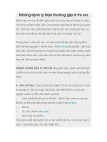
Biceps
Triceps
Arcade of
Struthers
Site 1: Intermuscular septum
Compression caused by
¥ Arcade of Struthers
¥ Medial intermuscular septum
¥ Hypertrophy of the medial head
of the triceps
¥ Snapping of the medial head
of the triceps
Site 2: Area of medial epicondyle
Compression caused by
¥ Valgus deformity of the bone
Site 3: Epicondylar groove
Compression caused by
¥ Lesions within the groove
¥ Conditions outside the groove
¥ Subluxation or dislocation of the nerve
Site 4: Cubital tunnel
Compression caused by
¥ Thickened OsborneÕs ligament
Site 5: Exit of ulnar nerve from flexor carpi ulnaris
Compression caused by
¥ Deep flexor-pronator aponeurosis
Brachialis
Flexor-pronator
muscle group
Flexor carpi ulnaris
Aponeurosis of the flexor carpi ulnaris
Flexor digitorum profundus
Fig. 1 The five sites for potential ulnar nerve compression and the causes of compression at each site. (Adapted with permission from
Amadio PC: Anatomical basis for a technique of ulnar nerve transposition. Surg Radiol Anat 1986;8:155-161.)
Ulnar Neuropathies: Etiology and Diagnosis
Journal of the American Academy of Orthopaedic Surgeons
284
can cause ulnar nerve compression
is the presence of an anomalous
anconeus epitrochlearis muscle that
arises from the medial border of the
olecranon and inserts into the medi-
al epicondyle. In humans, the mus-
cle is probably atavistic and is
replaced by a band passing in the
same direction as the muscle, called
the epitrochleoanconeus ligament.
4
The third category of neuropathy
develops as a consequence of the
nerve shifting out of the epicondylar
groove with elbow flexion and
returning to its normal position with
elbow extension. The nerve can
either subluxate onto the tip of the
epicondyle or dislocate anterior to
the epicondyle. Either situation can
occur as a consequence of congenital
laxity of the fibroaponeurotic cover-
ing over the epicondylar groove or a
traumatic tear in the covering. It can
also result from congenital hypopla-
sia of the trochlea or posttraumatic
deformity of the medial epicondyle.
Subluxation or dislocation of the
ulnar nerve, both pathologic condi-
tions, should not be confused with
asymptomatic hypermobility of the
nerve, which is usually bilateral and
is found in approximately 20% of
the population.
5
However, hyper-
mobile nerves are predisposed to
become inflamed by constant fric-
tion over the medial epicondyle.
They are also at risk to be com-
pressed, when the elbow is flexed,
by external forces such as tight casts
or splints applied for conditions
unrelated to the ulnar nerve. A
hypermobile nerve can also be inad-
vertently injured by an injection
administered to treat medial epi-
condylitis.
6
The fourth site of potential com-
pression is where the nerve passes
through a tunnel between the
humeral and ulnar heads of the
flexor carpi ulnaris muscle. This
site and the epicondylar groove are
the most common sites for ulnar
nerve compression. The floor of the
tunnel is the medial collateral liga-
ment of the elbow. Its roof is a
fibrous band that is a continuation
of the fibroaponeurotic covering of
the epicondylar groove. The fi-
brous band has been referred to as
OsborneÕs ligament, the triangular
ligament, the arcuate ligament, and
the humeroulnar arch. In 1958,
Feindel and Stratford named this
area the Òcubital tunnel.Ó Although
the term Òcubital tunnel syndromeÓ
is often used to describe compres-
sion of the ulnar nerve anywhere in
the elbow, it more accurately refers
to a neuropathy at this specific
anatomic location.
The nerve is vulnerable to com-
pression within the cubital tunnel
during elbow flexion, because the
tunnel normally narrows as Os-
borneÕs ligament stretches and
becomes taut, and the medial collat-
eral ligament relaxes and bulges
medially (Fig. 2). OsborneÕs liga-
ment stretches 5 mm for every 45
degrees of elbow flexion; from full
extension to full flexion, it elongates
40%.
7
The cross-sectional contour of
the tunnel changes from an oval in
elbow extension to a flattened
ellipse in elbow flexion.
8
Pressure
within the tunnel increases 7-fold
with elbow flexion and more than
20-fold when contraction of the flex-
or carpi ulnaris muscle is added.
9
These increases in pressure cause
mechanical deformation of the
nerve and, more important, com-
promise its intraneural circulation.
Animal studies have demonstrat-
ed the vascular effects of pressure.
At a pressure of 20 to 30 mm Hg,
there is impairment in flow in the
epineurial venules and slowing of
intracellular axonal support. How-
ever, capillary flow in the endo-
neurium and arteriolar flow in the
epineurium and perineurium re-
main unchanged. As pressure
increases, its effects become more
profound. At 60 to 80 mm Hg, cir-
culation ceases in the venules, arte-
rioles, and capillaries, and the nerve
becomes ischemic. If pressure is
relieved within 2 hours, intraneural
circulation is rapidly restored,
although the nerve remains edema-
tous for hours due to increased per-
meability of the epineurial vessels.
Prolonged compression, which
mimics many clinical situations,
leads to permanent nerve damage.
The fifth site of potential com-
pression is where the ulnar nerve
leaves the flexor carpi ulnaris.
Normally, the nerve enters the
muscle at the cubital tunnel, re-
mains intramuscular for a distance
of approximately 5 cm, and then
penetrates a fascial layer to lie be-
tween the flexor digitorum superfi-
cialis and flexor digitorum profun-
dus muscles. The nerve can be
constricted by this fascia, which
Fig. 2 Anatomy of the cubital tunnel in elbow extension and flexion. (Adapted with per-
mission from Adelaar RS, Foster WC, McDowell C: The treatment of the cubital tunnel
syndrome. J Hand Surg [Am] 1984;9:90-95.)
Elbow Extension Elbow Flexion
Medial epicondyle
Ulnar nerve
OsborneÕs ligament
Medial collateral
ligament
Olecranon
OsborneÕs ligament
becomes taut
Medial collateral
ligament relaxes
and bulges
medially
Martin A. Posner, MD
Vol 6, No 5, September/October 1998
285
has been referred to as the Òflexor
pronator aponeurosis.Ó
10
Scarring anywhere along the
course of the nerve can restrict its
excursion and result in a traction
injury. Normal excursion of the
nerve with elbow motion is as high
as 10 mm proximal to the medial
epicondyle and 6 mm distal to the
epicondyle.
11
The nerve itself
stretches as much as 4.7 mm with
elbow flexion, and additional
stretching occurs with abduction
and external rotation of the shoul-
der and extension of the wrist.
Diagnosis
Clinical Findings
A complete history, including
assessment of work or leisure-time
activities that aggravate the condi-
tion, and a physical examination
are essential first steps in arriving
at a correct diagnosis. Symptoms
can vary from mild numbness and
paresthesias in the ring and little
fingers to severe pain on the medial
aspect of the elbow and dysesthe-
sias radiating distally into the hand
and sometimes proximally to the
shoulder and neck. The occurrence
of mild paresthesias as an isolated
symptom is not necessarily cause
for concern, as it commonly occurs
in individuals who keep their el-
bows flexed for prolonged periods
of time during the day or at night
while sleeping. Patients with early
stages of nerve compression may
not complain of any actual weak-
ness, although they may be aware
of some deterioration in hand func-
tion. They may report difficulty in
carrying out certain tasks, such as
opening bottles and jars, or may
simply state that their hands fa-
tigue quickly with repetitive activi-
ties.
The physical examination should
always start at the neck. Any limi-
tation of motion, particularly when
accompanied by pain, may indicate
cervical disk disease or arthritis.
Axial compression of the spine may
reproduce radicular pain. When
compression in the brachial plexus
is suspected, the presence of tender-
ness or a Tinel sign with percussion
in the supraclavicular and infra-
clavicular areas should be checked.
Compression can also be due to
thoracic outlet syndrome. There
are a number of provocative tests
for this condition, which are aimed
primarily at obliterating the radial
pulse. These tests include AdsonÕs
maneuver, WrightÕs maneuver, and
RoosÕs test (also referred to as the
overhead exercise test). There is
also the costoclavicular maneuver,
which involves scapular retraction
into a military brace posture. All
these tests are frequently positive
in normal individuals; they are
therefore nonspecific in the patient
whose complaints are predomi-
nantly neurogenic. For a positive
test to be considered relevant, it
should reproduce the patientÕs
symptoms and not simply obliter-
ate the radial pulse.
The elbow is then inspected for
deformity, and the normal carrying
angle and active ranges of joint
motion are measured. The ulnar
nerve is palpated along its course
for any enlargement or mass and in
the epicondylar groove during
elbow flexion for any subluxation
or dislocation. Local tenderness
anywhere along the course of the
nerve aids in identifying sites of
compression. A provocative test
analogous to PhalenÕs test for carpal
tunnel syndrome is the elbow flex-
ion test, which involves maintain-
ing the elbow in full flexion with
the wrist in full extension for 1
minute (up to 3 minutes is consid-
ered by some to be a more ap-
propriate duration). The test is con-
sidered positive if paresthesias or
numbness occurs in the ulnar nerve
distribution. As with PhalenÕs test,
the elbow flexion test is more sensi-
tive than specific, and false-positive
results have been reported in 10%
of normal individuals.
12
Numbness in the ulnar nerve
distribution of the hand is a com-
mon finding, which can vary in
severity depending on the degree
and duration of nerve compression.
The sensory deficits usually in-
clude both sides of the little finger
and the ulnar half of the ring fin-
ger, although normal variations in
the sensory distribution of the
ulnar nerve may extend the numb-
ness to the middle finger or restrict
it to the little finger. A sensory
deficit over the dorsoulnar aspect
of the hand and the dorsum of the
little finger aids in differentiating a
neuropathy at the elbow from one
at the wrist. When nerve compres-
sion is at the wrist in the canal of
Guyon (ulnar tunnel syndrome),
dorsal sensibility remains intact
because that area is innervated by
the dorsal sensory branch of the
ulnar nerve, which leaves the main
body of the nerve at a more proxi-
mal level. Generally, it is 5 to 6 cm
proximal to the ulnar styloid, but
occasionally it is at the level of the
ulnar head. Simultaneous com-
pressive ulnar neuropathies at the
elbow and wrist are common; in
that instance, the Tinel sign will be
positive at both locations.
Sensibility can be tested in sev-
eral ways. Because the initial
changes in nerve compression af-
fect threshold, testing for vibratory
perception and light touch with the
use of Semmes-Weinstein monofil-
aments is more important than
measuring static and moving two-
point discrimination, which reflect
innervation density. Innervation
density is compromised only after
there is axonal degeneration, which
is more likely to occur with chronic
nerve compression of at least sever-
al yearsÕ duration.
Muscle weakness generally oc-
curs later than numbness, although
occasionally inability to adduct the
little finger (positive Wartenberg
Ulnar Neuropathies: Etiology and Diagnosis
Journal of the American Academy of Orthopaedic Surgeons
286
sign) is an early presenting sign.
Weakness affects the intrinsic mus-
cles in the hand more commonly
than the extrinsic muscles in the
forearm, which can be readily
explained by SunderlandÕs study of
intraneural topography.
13
The
motor fascicles to the intrinsic mus-
cles, as well as the sensory fasci-
cles, are situated more medial or
superficial in the ulnar nerve at the
elbow than the motor fascicles to
the extrinsic muscles, and are
therefore more vulnerable to com-
pression (Fig. 3).
Comparing the strength of the
ulnar nerveÐinnervated first dorsal
interosseous muscle with that of the
median nerveÐinnervated abductor
pollicis brevis muscle is important.
However, anomalous intrinsic mus-
cle innervation is common, occur-
ring in approximately 20% of the
population.
14
The most common
anomalous neural pathway is the
Martin-Gruber communication in
the proximal forearm, which carries
motor fibers from the median nerve
to the ulnar nerve. A similar but far
less common connection between
the two nerves exists in the distal
forearm. In the hand, there is the
Riche-Cannnieu connection be-
tween the motor branch of the
ulnar nerve and the recurrent
motor branch of the median nerve.
These anomalous neural communi-
cations in the forearm and hand
explain how the intrinsic muscles
can be completely innervated by
just one nerve, resulting in the so-
called ulnar hand or median hand.
More commonly, one or more
intrinsic muscles have dual inner-
vations.
In addition to these anomalous
muscle innervations, the examining
physician must also be aware of the
various Òtrick movementsÓ where-
by intact muscles mimic move-
ments normally provided by weak-
ened muscles. Common examples
of trick movements for the ulnar
nerveÐinnervated intrinsic muscles
are abduction of the index finger
by the extensor indicis proprius,
adduction of the thumb by the
extensor pollicis longus, and ab-
duction and adduction of the fin-
gers by the extrinsic digital exten-
sors and flexors, respectively.
Trick movements are always weak
movements, which can be detected
by careful observation and by pal-
pating the muscle being tested. A
useful test for ulnar nerve function
that is difficult to duplicate by any
trick movement is the Òcrossed fin-
gersÓ test. This test is based on the
ability to cross oneÕs middle finger
over the index finger, the supersti-
tious Ògood luckÓ gesture learned
in early childhood.
15
When intrinsic weakness is
severe and associated with muscle
wasting, it is indicative of chronic
nerve compression of many monthsÕ
or yearsÕ duration. Muscle weak-
ness in these cases is commonly
associated with clawing of the ring
and little fingers and weakness of
thumb pinch, characterized by a
positive FromentÕs sign (flexion of
the interphalangeal joint of the
thumb) and a positive JeanneÕs sign
(hyperextension of the metacarpo-
phalangeal joint of the thumb).
When extrinsic weakness occurs,
it always involves the flexor digito-
rum profundus to the little finger.
The flexor digitorum profundus to
the ring finger may also be weak,
but usually not to the same degree
because its muscle fibers are fre-
quently dually innervated by both
the ulnar nerve and the anterior
interosseous branch of the median
nerve. Weakness of the flexor carpi
ulnaris muscle is rarely encountered.
Imaging Studies
Radiographic examination of the
elbow is always necessary. In
addition to routine anteroposterior,
oblique, and lateral views, a view
profiling the epicondylar groove is
useful in patients with arthritic and
traumatic conditions in the elbow.
Osteophytes or bone fragments
from the medial trochlear lip are
often seen in these patients.
The role of magnetic resonance
imaging is limited. Although this
modality is capable of visualizing
swelling or enlargement of the ulnar
nerve in the epicondylar groove as
well as space-occupying lesions,
its value is primarily academic.
Magnetic resonance imaging is not
essential for either diagnosing a
neuropathy or determining appro-
priate treatment. Perhaps in the
future, with continuing technical
advancements, it will become more
useful for detecting early nerve
damage.
Electrodiagnostic Studies
Electrodiagnostic studies are
never a substitute for a complete
history and thorough physical
examination. Although these stud-
ies are usually obtained when
nerve compression is suspected,
they are not essential when the
diagnosis is obvious on clinical
examination. Electrodiagnostic
Motor to FCU
Motor to
intrinsic
muscles
Sensory to hand
Motor to FCU
and FDP
Fig. 3 The intraneural topography of the
ulnar nerve in the epicondylar groove.
Both sensory fascicles and motor fascicles
to the intrinsic muscles are situated medial-
ly or superficially in the nerve. The motor
fascicles to the extrinsic muscles, except for
a small fascicle to the flexor carpi ulnaris
(FCU), are situated laterally or deeper in
the nerve and are therefore less vulnerable
to compression. FDP = flexor digitorum
profundus.
Martin A. Posner, MD
Vol 6, No 5, September/October 1998
287
studies can sometimes be mislead-
ing, and they have a false-negative
rate similar to that in patients with
carpal tunnel syndrome. False-
negative studies occur when non-
compressed nerve fibers are tested
rather than the compressed fibers
that are causing sensory symptoms
or muscle weakness. Electrodiag-
nostic studies are important when
clinical symptoms and findings are
equivocal, when the site of nerve
compression is uncertain or is
thought to be at multiple levels, or
when a polyneuropathy or motor
neuron disease is suspected.
Electrodiagnostic studies include
motor and sensory conduction
velocity measurements and elec-
tromyography. Motor conduction
is measured over a 10- to 12-cm
segment of the ulnar nerve where it
crosses the elbow. The skill and
experience of the physician per-
forming the test are important
because anatomic variations can be
encountered. The test should al-
ways be carried out with the elbow
flexed, because conduction times
are as much as 7 to 9 m/sec slower
when the test is performed with the
elbow in full extension.
16
The rea-
son for this is that the true length of
the ulnar nerve is frequently under-
estimated with the elbow in exten-
sion because the nerve is lax in that
position. Slowing of motor conduc-
tion is absolute when it is less than
50 m/sec. Slowing can be relative
when it is more than 10 m/sec
slower across the elbow than it is
farther distally in the forearm (from
below the elbow to the wrist) or far-
ther proximally in the upper arm
(from the axilla to above the elbow).
The age of the patient must be con-
sidered when evaluating conduc-
tion velocities because they can be
as much as 10 m/sec slower than
average in the elderly.
When nerve conduction is slowed,
it is often accompanied by a drop in
amplitude of compound muscle
action potentials (CMAPs). When
present, short-nerve-segment stimu-
lation (the ÒinchingÓ technique) can
be used to localize the lesion.
17
This
technique involves stimulating the
nerve at 2-cm intervals across the
elbow. When the points of maxi-
mum conduction delay and drop in
amplitude are at or just proximal to
the medial epicondyle, compression
is probably in the epicondylar
groove; when they are 2 cm distal to
the epicondyle, compression is prob-
ably at the cubital tunnel.
A Martin-Gruber communica-
tion in the forearm can also lead to
confusing results, as the hypo-
thenar and first dorsal interosseous
muscles are dually innervated by
fibers from both nerves. Conse-
quently, the CMAP amplitude for
these intrinsic muscles will normal-
ly be greater when the ulnar nerve
is stimulated at the wrist rather
than at the elbow, because at the
wrist the ulnar nerve also contains
fibers from the median nerve. The
amplitude at the elbow will nor-
mally be decreased, which may be
misinterpreted as a conduction
block. When ulnar nerve compres-
sion is present, weakness of the
ulnar intrinsic muscles may be
masked by the innervation they
receive from the median nerve.
Awareness of a Martin-Gruber
communication is also important
when planning surgery, as the
point of connection is located 3 to
10 cm distal to the medial epi-
condyle.
18
When the connection is
close to the epicondyle, there is a
potential risk of damage during
ulnar nerve transposition.
Sensory conduction studies are
similar to motor studies in that the
nerve is stimulated and a distant
action potential is recorded. How-
ever, unlike motor fibers, sensory
fibers can be stimulated in two
directions: in the physiologic direc-
tion of conduction (from distal to
proximal [orthodromic]) and in the
opposite direction (from proximal
to distal [antidromic]). For the
ulnar nerve at the elbow, anti-
dromic responses are easier to elicit,
and are recorded by a ring elec-
trode placed around the little fin-
ger. Sensory conduction of the dor-
sal cutaneous nerve of the hand can
also be carried out to distinguish
compression at the elbow from
compression at the wrist.
Electromyographic studies dem-
onstrate the presence of axonal
degeneration in muscles. Because
these changes occur with chronic
neuropathies, electromyography is
not as useful as conduction studies
for the diagnosis of early compres-
sions. When abnormalities are
noted, they are initially seen in the
first dorsal interosseous muscle,
followed in frequency by the mus-
cles in the hypothenar eminence.
Differential Diagnosis
The differential diagnosis includes
any lesion that affects the origins of
the ulnar nerve in the cervical spine
(C8-T1 nerve roots) and/or the
brachial plexus (medial cord). The
most common spinal lesions are
those due to cervical disk disease,
followed by spinal tumors and
syringomyelia. In the brachial
plexus, the medial cord can be com-
pressed by thoracic outlet syndrome
or a Pancoast tumor. Electromy-
ography of median nerveÐ and
ulnar nerveÐinnervated intrinsic
muscles (C8-T1) is helpful in differ-
entiating lesions in the spine and
brachial plexus from distal com-
pressive neuropathies. While ulnar
nerveÐinnervated intrinsic muscles
may be abnormal with an ulnar
neuropathy, the median nerveÐ
innervated abductor pollicis brevis
should be normal.
Not infrequently, the ulnar nerve
is compressed at more than one site.
In 1973, Upton and McComas noted
that many patients with peripheral
compressive neuropathies had con-
comitant nerve damage at the cervi-
Ulnar Neuropathies: Etiology and Diagnosis
Journal of the American Academy of Orthopaedic Surgeons
288
cal roots.
19
They observed that
when neural function was compro-
mised at one level, the axons of that
nerve were more susceptible to
damage at another level, probably
because of impaired axoplasmic
flow. They aptly termed this condi-
tion Òdouble crush.Ó Occasionally,
the nerve can be compressed at
three sites (Òtriple crushÓ).
The differential diagnosis of
ulnar neuropathies should also
include systemic and metabolic dis-
orders, such as diabetes mellitus,
hypothyroidism, alcoholism, ma-
lignant neoplasms, and vitamin
deficiencies. However, the pres-
ence of any of these problems does
not exclude the possibility of a con-
comitant compressive neuropathy.
Classification Systems
Classification of ulnar nerve func-
tion was introduced in 1950 by
McGowan, who proposed a three-
grade system.
20
Grade I lesions are
classified as minimal, with symp-
toms of paresthesias and numbness
but no weakness. Grade II lesions
are intermediate, with wasting of
the interosseous muscles. Grade III
lesions are severe, with complete
intrinsic muscle paralysis. Al-
though both grade II and III lesions
are characterized by numbness, the
difference between the two grades
is based solely on the degree of
muscle weakness. McGowanÕs sys-
tem is, therefore, essentially a pre-
operative rating of intrinsic muscle
function.
Currently, there is no consensus
on any scoring system. Available
systems either rate subjective symp-
toms, which are difficult to quanti-
tate, or fail to compare preoperative
and postoperative conditions.
Summary
Compressive neuropathy of the
ulnar nerve at the elbow is a com-
mon problem and can result in
severe disability. Considering the
anatomic course of the ulnar nerve
through confined spaces and poste-
rior to the axis of elbow flexion,
Lundborg
21
concluded that the
ulnar nerve was Òasking for trou-
ble.Ó Normally, the nerve is sub-
jected to stretch and compression
forces that are moderated by its
ability to glide in its anatomic path
around the elbow. When normal
excursion is restricted, irritation
ensues. This results in a cycle of
perineural scarring, further loss of
excursion, and progressive nerve
damage. Not uncommonly, a com-
pressive neuropathy at the elbow is
associated with additional com-
pression proximally in the neck or
brachial plexus and/or distally in
the canal of Guyon. Multiple sites
of compression can usually be
identified from the history and
physical examination. While elec-
trodiagnostic studies may be help-
ful, their results must be correlated
with the clinical picture for proper
interpretation.
References
1. Spinner M, Kaplan EB: The relation-
ship of the ulnar nerve to the medial
intermuscular septum in the arm and
its clinical significance. Hand 1976;8:
239-242.
2. Fragiadakis EG, Lamb DW: An
unusual cause of ulnar nerve compres-
sion. Hand 1970;2:14-16.
3. Alvine FG, Schurrer ME: Postopera-
tive ulnar-nerve palsy: Are there pre-
disposing factors? J Bone Joint Surg
Am 1987;69:255-259.
4. Masear VR, Hill JJ Jr, Cohen SM: Ulnar
compression neuropathy secondary to
the anconeus epitrochlearis muscle. J
Hand Surg [Am] 1988;13:720-724.
5. Childress HM: Recurrent ulnar-nerve
dislocation at the elbow. Clin Orthop
1975;108:168-173.
6. Idler RS: General principles of patient
evaluation and nonoperative manage-
ment of cubital syndrome. Hand Clin
1996;12:397-403.
7. Vanderpool DW, Chalmers J, Lamb
DW, Whiston TB: Peripheral compres-
sion lesions of the ulnar nerve. J Bone
Joint Surg Br 1968;50:792-803.
8. Apfelberg DB, Larson SJ: Dynamic
anatomy of the ulnar nerve at the el-
bow. Plast Reconstr Surg 1973;51:79-81.
9. Werner CO, Ohlin P, Elmqvist D:
Pressures recorded in ulnar neuropa-
thy. Acta Orthop Scand 1985;56:404-406.
10. Amadio PC, Beckenbaugh RD: En-
trapment of the ulnar nerve by the
deep flexor-pronator aponeurosis. J
Hand Surg [Am] 1986;11:83-87.
11. Wilgis EF, Murphy R: The significance
of longitudinal excursion in peripheral
nerves. Hand Clin 1986;2:761-766.
12. Rayan GM, Jensen C, Duke J: Elbow
flexion test in the normal population.
J Hand Surg [Am] 1992;17:86-89.
13. Sunderland S: Nerves and Nerve In-
juries, 2nd ed. New York: Churchill
Livingstone, 1978, pp 780-795.
14. Rowntree T: Anomalous innervation
of the hand muscles. J Bone Joint Surg
Br 1949;31:505-510.
15. Earle AS, Vlastou C: Crossed fingers
and other tests of ulnar nerve motor
function. J Hand Surg [Am] 1980;5:
560-565.
16. Kincaid JC: AAEE minimonograph
#31: The electrodiagnosis of ulnar neu-
ropathy at the elbow. Muscle Nerve
1988;11:1005-1015.
17. Miller RG: The cubital tunnel syn-
drome: Diagnosis and precise localiza-
tion. Ann Neurol 1979;6:56-59.
18. Uchida Y, Sugioka Y: Electrodiagnosis
of Martin-Gruber connection and its
clinical importance in peripheral nerve
surgery. J Hand Surg [Am] 1992;17:54-59.
19. Upton AR, McComas AJ: The double
crush in nerve entrapment syndromes.
Lancet 1973;2:359-362.
20. McGowan AJ: The results of transpo-
sition of the ulnar nerve for traumatic
ulnar neuritis. J Bone Joint Surg Br
1950;32:293-301.
21. Lundborg G: Surgical treatment for
ulnar nerve entrapment at the elbow
[editorial]. J Hand Surg [Br] 1992;17:
245-247.