Báo cáo y học: " Highly pathogenic avian influenza A virus H5N1 NS1 protein induces caspase-dependent apoptosis in human alveolar basal epithelial cells" docx
Bạn đang xem bản rút gọn của tài liệu. Xem và tải ngay bản đầy đủ của tài liệu tại đây (1.04 MB, 6 trang )
RESEA R C H Open Access
Highly pathogenic avian influenza A virus H5N1
NS1 protein induces caspase-dependent
apoptosis in human alveolar basal epithelial cells
Chuanfu Zhang
1,2†
, Yutao Yang
1,4†
, Xiaowei Zhou
1†
, Xuelin Liu
2
, Hongbin Song
2*
, Yuxian He
3*
, Peitang Huang
1*
Abstract
Background: It is widely considered that the multifunctional NS1 protein of influenza A viruses contributes
significantly disease pathogenesis by modulating a number of virus and host-cell processes, but it is highly
controversial whether this non-structural protein is a proapoptotic or antiapoptotic factor in infected cells.
Results: NS1 protein of influenza A/chicken/Jilin/2003 virus, a highly pathogenic H5N1 strain, could induce
apoptosis in the carcinomic human alveolar basal epithelial cells (A549) by electron microscopic and flow
cytometric analyses. NS1 protein-triggered apoptosis in A549 cells is via caspase-dependent pathway.
Conclusions: Influenza A virus NS1 protein serves as a strong inducer of apoptosis in infected human respiratory
epithelial cells and plays a critical role in disease pathogenesis.
Background
The influenza A virus, which contai ns eight segmen ted
and negative-stranded RNAs as its genome, is a globally
important human and animal respiratory pathogen
responsible for both seasonal “flu” outbreaks and peri-
odic world-wide pandemics. In recent years, the highly
pathogenic avian influenza A virus H5N1 has been fre-
quently transmitted to human and caused a mortality
rate of >30%, raising serious worldwide concern about a
severe influenza pandemic. Although considerable
efforts, the mechanism accounting for the severity of
human H5N1 infection remains elusive. It has been
demonstrated that influenza viruses can induce apopto-
sis in numerous cell types, both in vivo [1-3] and in
vitro [4-12]. Recently, the apoptosis was observed
among the alveolar epithelial cells of two patients who
died of H5N1 infection, suggesting a possible role of
apoptosis in H5N1 pathogenesis in humans [13].
Several viral factors, including neuraminidase, M1,
NS1, and PB1-F2, from different strains of human
influenza viruses could induce or inhibit apoptosis in
human cells [7,14-18]. The multifunctional NS1
protein is widely considered as a virulence factor and
contributes significantly disease pathogenesis by modu-
lating a number of virus and host-cell processes
[19-22]. Prominently, it is hotly debated that whether
the NS1 protein is a proapoptotic or antiapoptotic
factor in infected cells [23]. For example, the NS1
proteins derived from H5N9 or H5N1 could induce
apoptosis in MDCK, HeLa cells or human airway
epithelial cells [8,24]; in sharp contrast, the NS1
proteins from H1N1 or H3N2 were reported to down-
regulate apoptosis in MDCK and Vero cells [18,25].
Furthermore, while it was shown that the H5N1 NS1
protein was capable of inducing caspase pathway-
dependent apoptosis [26], the NS1 from H1N1 could
activate PI3K/Akt pathway to mediate antiapoptotic
signaling responses [27]. It is possible that these
diverse observations might be resulted from the differ-
ences of virus subtypes and strains, as well as the host
cell system being used, highlighting that further
characterization of the NS1 protein and its mechanism
involved in the induction of apoptosis is highly essen-
tial for understanding th e pathogenesis of influenza
* Correspondence: ; ;
† Contributed equally
1
Institute of Biotechnology, Academy of Military Medical Sciences, Beijing
100071, PR China
2
Institute of Disease Control and Prevention, Chinese Academy of Military
Medical Sciences, Beijing 100071, PR China
3
Institute of Pathogen Biology, Chinese Academy of Medical Sciences and
Peking Union Medical College, Beijing 100730, PR China
Zhang et al. Virology Journal 2010, 7:51
/>© 2010 Zhang et al; licensee BioMed Central Ltd. This is an Open Access article distributed under the terms of the Creative Commons
Attribution License ( 2.0), which perm its unrestricted use, distribut ion, and reproduction in
any medium, provided the original work is properly cited.
A viruses. In this study, we demonstrated that the
expression of NS1 proteins of influenza A/chicken/
Jilin/2003(H5N1)could induce apoptosis in the carci-
nomic human alveolar basal epithelial cells (A549) via
the caspase-dependent pathway, providing further
evidences to support that the H5N1 NS1 plays a
critical in disease pathogenesis.
Materials and methods
Viruses and cells
Influenza A/chicken/Jilin/2003(H5N1)virus was grown
in the allantoi c cavities of 10-day- old embryonated
chicken eggs. A549 cells were passaged in Dulbecco’s
modified Eagle’s tissue cult ure medium (DMEM) con-
taining 10% fetal calf serum at 37°C in a 5% CO
2
incu-
bator. For immunoblot analysis, confluent cell
monolayers grown in 25-mm dishes were lysed in
immunoprecipitation assay buffer containing 150 mM
NaCl, 1.0% Nonidet P-40 (NP-40), 0.5% deoxycholate,
0.1% sodium dodecyl sulfate (SDS), and 50 mM Tris-
HCl (pH 8.0). Lysates were clarified by centrifugation
for 10 min at 13,000 g and supernatants were used for
immunoblot analysis.
Construction of NS1-expressing plasmid
Total RNA was extra cted from the cell lysates using the
QIAamp viral RNA mini kit (Qiagen, Hilden, Germany).
The full-length NS1 gene from influenza A/chicken/
Jilin/2003 was amplified using the SuperScript III one-
step reverse transcription-PCR (RT-PCR) system with
Platinum Taq high-fidelity polymerase (Invitrogen,
Carlsbad, CA). The construction of plasmid pCMV-
myc/NS1 followed standard cloning procedures. Compe-
tent Escherichia coli DH5a cells were transformed with
the plasmids, and the plasmids were amplified and puri-
fied using a high-purity plasmid purification kit
(Qiagen).
Electron microscopic analysis
A549 cells were transfected with pCMV-myc/NS1, using
Lipofectamine 2000 reagent (Invitrogen). After 24 h,
cells were collected, digested, washed with phosphate
buffered solution ( PBS), fixed with 4% glutaraldehyde
for 2 h, and then fixed with osmium tetroxide for 1 h,
stai ned with uranium acetate, embedded into 6.8
#
epox-
ide resin. After section ing into ultra-thin slices, the cells
were stained with lead citrate and examined under
transmission electron microscopy.
Flow cytometric analysis
To determine the apoptosis rate, an Annexin V-FITC
apoptosis detection kit (BD Pharmingen, San Diego,
CA) was used to detect early apoptotic activity accord-
ing to the manufacturer’ s instructions, with slight
modifications. After 24 h transfected as described
above, the A549 cells were harvested and washed twice
with ice-cold PBS and resuspended in 100 ml of
binding buffer. A total of 5 ml of Annexin V-F ITC and
10mlofpropidiumiodide(PI)wereaddedandthe
mixture was incubated for 30 min in the dark. Finally,
400 ml of binding buffer was added to the cells, the
mixture were analyzed with a Flow cytometer (Becton
Dickinson Co., San Jose, CA), using an FITC signal
detector (FL1) for Annexin V staining and a phycoery-
thrin emission signal detector for PI staining. The
apoptotic percentage of 10,000 cells was determined.
All the experiments reported in this study were
performedthreetimes.Thedatawereanalyzedusing
WinMDI 2.8 software (Scripps Institute, La Jolla, CA)
for calculation of percentage cells with apoptosis
per group.
Expression of caspase-9 and caspase-3
The expression of caspase-9 and caspa se-3 in NS1-
transfected A549 cells were measured by Western-blot
analysis. Briefly, monolayer of cells transfected with
pCMV-myc/NS1 was lysed with ice-cold lysis buffer
(150 mM Tris-HCl, pH 8.0, 50 mM NaCl, 1 mM EDTA,
0.5% Nonidet P-40, 1 tablet Complete Mini protein inhi-
bitor mixture/10 ml (Roche Applied Science) and
0.7 μg/ml pepstatin), and the lysates were clarified by
centrifugation at 20,000 g for 10 min at 4°C. Caspase-9
and caspase-3 activities were determined according to
the supplemental proto cols of the Caspase-9/Mch6 Col-
orimetric Assay kit and Caspase-3/CPP32 Colorimetric
Protease Assay kit (MBL, Nagoya, Japan), respectively.
Substrate cleavage, which resulted in the release of pNA
(405 nm), was measured using a Multiskan Ascent plate
reader.
Results
Influenza A virus H5N1 NS1 protein induced
apoptosis in A549 cells
The biological activities ofinfluenzaAvirusNS1pro-
teins are likely to be strain- and/or cell-type specific
[28]. Here, we studied whether the NS1 of influenza
A/chicken/Jilin/2003(H5N1)virus could induce apopto-
sis in A549 cells. The NS1 gene of this strain was
amplified by RT-PCR and cloned into a mammalian
expression vector to construct pCMV-myc/NS1. The
expression of NS1 protein in A549 cells was assessed
after transfection with the plasmid by Western blot
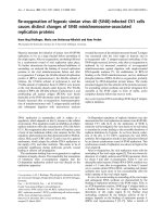
analysis. We examined the morphological changes and
ultrastructural features of the A549 cells transfected
with the NS1-expressing vectors under a transmission
electron microscopy. As shown in Fig. 1C, the NS1-
transfected cells appeared characteristics of apoptotic
cells, including nuclear condensation and chromatin
Zhang et al. Virology Journal 2010, 7:51
/>Page 2 of 6
aggregation to the nuclear membrane. Typicall y, the
apoptotic bodies were found in some cells. In the con-
trast, the cells transfected with the empty vector (Fig.
1B) and normal cells (Fig. 1A) revealed normal
silhouettes.
Further, the A549 cells expressing NS1 protein were
stained with annexin V-FITC and PI and analyzed by
flow cytometry. As showed in Fig. 2B, H5N1 NS1-trans-
fected cells were 2.33% Annexin V
+
/PI
-
(early apoptosis)
and 17.61% Annexin V
+
/PI
+
(latter apoptosis), while
the empty vector-transfected cells were 2.13% Annexin
V
+
/PI
-
and 5.25% Annexin V
+
/PI
+
(Fig. 2A). Collectively,
these results suggested that the NS1 protein of influenza
A virus H5N1 was able to induce apoptosis in A549
cells.
Involvement of caspases in NS1-induced apoptosis
Previous studies suggested that avian influenza virus
A/HK/483/97 (H5N1) NS1 protein-ind uced apoptosis in
a human airway epithelial cells, NCI-H292, was caspase
pathway-dependent [26]. Here, we performed experi-
ments to investigate whether the caspase pathways were
involved in the apoptosis induced by NS1 proteins
derived from the influenza A/chicken/Jilin/2003 virus.
To this end, A549 cells were transfected with the NS1-
expressing plasmid and the cell lysate was prepared as
described in Materials and Methods. First, the expres-
sion of caspase-9 and caspase-3 were detected by
Western-blotting analysis and found that the apoptosis-
related caspase- 9 and caspase-3 were activated in H5N1
NS1-transfected A549 cells (Fig. 3). The active frag-
ments of caspase-9 and caspase -3 could be detected at
12 h, 24 h, 48 h post-trans fection. Second, we measured
the enzyme activities of caspase-9 and caspase-3 and the
results revealed that both apoptosis-associated enzymes
were activated in the NS1-transfected A549 cells (Fig.
4). Therefore, our data verified that the NS1 protein of
influenza A virus H5N1 can induce caspase-dependent
apoptosis in A549 cells.
Discussion
A number of studies have demonstrated that influenza
virus infection can induce apoptosis in a variety o f cell
lines, but the mechanism of this effect remains to be
characterized [1-12]. Previous studies thought that apop-
tosis is a host defense response that limits the virus
replication [29], but recent evidence showed that the
induction of apoptosis is essential for virus mRNA
synthesis and propagation [16,30,31]. Viral proteins
from different strains of human influenza viruses have
been reported to have proapoptotic or antiapoptotic
functions in human cells [7,14-18]. The importance of
non-structural protein NS1 in the viral pathogenesis,
especially its role in the virus-induced apopto sis, has
been recently underlined [8,17,23,25,27]. A highly con-
troversial question is whether this multifunctional pro-
tein is a proapoptotic or antiapoptotic factor in infected
cells. Schultz-Cherry et al. reported that the expression
of NS1 protein of H5N9 was sufficient to induc e apop-
tosis in MDCK and Hela cells [8] ; Lam et al demon-
strated recently that H5N1 NS1 protein could induce
apoptosis in human airway epithelial cells (NCI-H292)
[26]. It was also reported that a poor-apoptosis-inducer
strain could be converted into a strong-inducer strain
A.
B.
C.
Figure 1 H5N1 NS1 protein induces apoptosis in A549 cells
visualized by transmission electron microscopy. Normal cell (A)
and transfected cell with pCMV-Myc empty vector (B) show that the
nuclear shapes are intact. H5N1 NS1-transfected cell shows
chromatins condensed, shrunk and aggregated along inside the
nuclear membrane, and reveals the apoptotic bodies. (× 5,000).
Zhang et al. Virology Journal 2010, 7:51
/>Page 3 of 6
by the NS1 gene substitution by reverse genetics, and
vice versa [25]. Discordantly, the NS1 protein has been
also shown to inhibit apoptosis. For examples, Zhirnov
et al. found that H1N1 NS1 protein had IFN-dependent
antiapoptotic potential and down-regulated the apopto-
tic response in virus-infected in cultured cells and
chicken embryos [18]; Ehrha rdt et al found that the
NS1 proteins of H1N1 and H7N7 activated the
phosphatidylinositol 3-kinase (PI3K/Akt) pathway to
mediate antiapoptotic signalin g responses [27]. The dis-
crepancy has confused our understanding to the role of
NS1 protein in influenza virus-induced apoptosis, high-
lighting that further characteriza tion i s n eeded to
exclude the possibility resulted from th e different cell
lines and virus strains in each experiment [20]. In the
present study, we cloned the NS1 gene from the
Figure 2 Flow cytometric analysis of H5N1 NS-induced apoptosis. The dot plot diagrams represent typical apoptotic and necrotic cell
populations detected by Annexin V-FITC and PI staining. A. A549 cells transfected with the empty PCMV-Myc vectors. B. A549 cells transfected
with pCMV-Myc/NS1. The lower left quadrants of the panels show viable intact cells, which were negative for Annexin V-FITC binding and
excluded PI staining (FITC
-
/PI
-
); the upper right quadrants show nonviable, necrotic cells, which were positive for Annexin V-FITC binding and PI
uptake (FITC
+
/PI
+
). The lower right quadrants represent apoptotic cells, positive for Annexin V-FITC and negative for PI (FITC
+
/PI
-
).
0 h 12 h 24 h 48 h
12Kd
17Kd
Caspase-3
Caspase-9
-actin
Figure 3 Caspase-3 and caspase-9 activation in NS1-transfected A549 cells. The cells were transfected with the plasmid pCMV-Myc/NS1 for
0 h, 12 h, 24 h, and 48 h. Intracellular caspase-3 and caspase-9 activation were detected by Western blotting. The b-actin was used as a loading
control.
Zhang et al. Virology Journal 2010, 7:51
/>Page 4 of 6
influenza A/chicken/Jilin/2003 virus (H5N1) and
expressed the NS1 protein in the human alveolar basal
epithelial cell A549. With electron microscopic and flow
cytometric analyses we verified that the expression of
H5N1 NS1 protein was capable to induce apoptotic
events posttransfection in the A549 cells. The biologi cal
basis accounting for the severity of H5N1 infection in
humans is still unknown. Our current experimental
data, together with the apoptotic observations in the
alveolar epithelial cells of two patie nts who died of
H5N1 infection [13], have provided convincing evidence
to the critical role of H5N1-encoded NS1 protein in
inducing apoptosis.
Apoptosis, or programmed cell death, involves a series
of biochemical events that lead the cells undergo charac-
teristic morphological changes, including blebbing, shrink-
age, nuclear fragmentation, chromatin condensation, and
chromosomal DNA fragmentation [32]. Sequential activa-
tion of caspases cas cade plays a central role in the execu-
tion-phase of cell apoptosis. Recently, It has been reported
that avian influenza virus A/HK/483/97(H5N1) NS1 pro-
tein-induced apoptosis in human lung epithelial cells is
mainly via the caspase-dependent pathway [26], which
enco urages further investigation into the potential of the
NS1 as a novel therapeutic target. To delineate the apop-
totic pathway, we measured the expression of caspase-3
and caspase-9 by Western blotting and their enzyme activ-
ities by colorimetric Assay. The data demonstrated that
these two apoptosis markers were significantly activated in
H5N1 NS1-transfected A549 cells, consistent with the
previous studies. Therefore, we conclude that the NS1
protein encoded by avian influenza A virus H5N1 can
induc e apoptosis in human respiratory epithelial cells via
the caspase-dependent pathway. Since the apoptotic
destruction of host cells has been thought to contribute
the severe disease, we can predict that drugs that can pre-
vent this specific process may reduce disease severity and
improve clinical outcomes. Therefore, further investiga-
tions to clarify whether the NS1 protein and its apoptotic
pathway is worthwhile therapeutic targets for treating
H5N1 infection in humans should be considered.
Acknowledgements
This study was supported by a grant from the National Key Technology R&D
Program of China (No.2006BAD06A01).
Author details
1
Institute of Biotechnology, Academy of Military Medical Sciences, Beijing
100071, PR China.
2
Institute of Disease Control and Prevention, Chinese
Academy of Military Medical Sciences, Beijing 100071, PR China.
3
Institute of
Pathogen Biology, Chinese Academy of Medical Sciences and Peking Union
Medical College, Beijing 100730, PR China.
4
Beijing Institute for Neuroscience,
Capital Medical University, Beijing, 100069, PR China.
Authors’ contributions
CFZ, YTY and XWZ mainly carried out gene cloning, western blot, Flow
cytometric analysis, and wrote the manuscript. XLL contributed to Electron
microscopic analysis. HBS, YXH and PTH conceived the studies and
participated in experimental design and coordination. All authors read and
approved the final manuscript.
Competing interests
The authors declare that they have no competing interests.
Received: 5 February 2010 Accepted: 3 March 2010
Published: 3 March 2010
References
1. Mori I, Komatsu T, Takeuchi K, Nakakuki K, Sudo M, Kimura Y: In vivo
induction of apoptosis by influenza virus. J Gen Virol 1995, 76:2869-2873.
2. Price GE, Smith H, Sweet C: Differential induction of cytotoxicity and
apoptosis by influenza virus strains of differing virulence. J Gen Virol
1997, 78:2821-2829.
3. Roulston A, Marcellus RC, Branton PE: Viruses and apoptosis. Annu Rev
Microbiol 1999, 53:577-628.
4. Fesq H, Bacher M, Nain M, Gemsa D: Programmed cell death (apoptosis)
in human monocytes infected by influenza A virus. Immunobiology 1994,
190:175-182.
5. Hinshaw VS, Olsen CW, Dybdahl-Sissoko N, Evans D: Apoptosis: a
mechanism of cell killing by influenza A and B viruses. J Virol 1994,
68:3667-73.
6. Lowy RJ, Dimitrov DS: Characterization of influenza virus-induced death
of J774.1 macrophages. Exp Cell Res 1997, 234:249-258.
7. Morris SJ, Price GE, Barnett JM, Hiscox SA, Smith H, Sweet C: Role of
neuraminidase in influenza virus-induced apoptosis. J Gen Virol 1999,
80:137-46.
8. Schultz-Cherry S, Hinshaw VS: Influenza virus neuraminidase activates
latent transforming growth factor beta. J Virol 1996, 70:8624-8629.
9. Schultz-Cherry S, Dybdahl-Sissoko N, Neumann G, Kawaoka Y, Hinshaw VS:
Influenza virus ns1 protein induces apoptosis in cultured cells. J Virol
2001, 75:7875-7881.
10. Shan B, Lee WH: Deregulated expression of E2F-1 induces S-phase entry
and leads to apoptosis. Mol Cell Biol 1994, 14:8166-8173.
11. Takizawa T, Matsukawa S, Higuchi Y, Nakamura S, Nakanishi Y, Fukuda R:
Induction of programmed cell death (apoptosis) by influenza virus
infection in tissue culture cells. J Gen Virol 1993, 74:2347-2355.
0
1
2
3
4
5
6
7
8
9
10
0122448
Time(h)
Capsase activity
Cas pase 3
Cas pase 9
Figure 4 Enzyme activities of caspase-3 and caspase-9 in
NS1-transfected A549 cells. Enzyme activities were detected by
Caspase-3/CPP32 Colorimetric Protease Assay and Caspase-9/Mch6
Colorimetric Assay, respectively.
Zhang et al. Virology Journal 2010, 7:51
/>Page 5 of 6
12. Takizawa T, Tatematsu C, Ohashi K, Nakanishi Y: Recruitment of apoptotic
cysteine proteases (caspases) in influenza virus-induced cell death.
Microbiol Immunol 1999, 43:245-252.
13. Uiprasertkul M, Kitphati R, Puthavathana P, Kriwong R, Kongchanagul A,
Ungchusak K, Angkasekwinai S, Chokephaibulkit K, Srisook K, Vanprapar N,
Auewarakul P: Apoptosis and pathogenesis of avian influenza A (H5N1)
virus in humans. Emerg Infect Dis 2007, 13:708-712.
14. Chanturiya AN, Basanez G, Schubert U, Henklein P, Yewdell JW,
Zimmerberg J: PB1-F2, an influenza A virus-encoded proapoptotic
mitochondrial protein, creates variably sized pores in planar lipid
membranes. J Virol 2004, 78:6304-12.
15. Chen W, Calvo PA, Malide D, Gibbs J, Schubert U, Bacik I, Basta S, O’Neill R,
Schickli J, Palese P, Henklein P, Bennink JR, Yewdell JW: A novel influenza
A virus mitochondrial protein that induces cell death. Nat Med 2001,
7:1306-1312.
16. Stasakova J, Ferko B, Kittel C, Sereinig S, Romanova J, Katinger H, Egorov A:
Influenza A mutant viruses with altered NS1 protein function provoke
caspase-1 activation in primary human macrophages, resulting in fast
apoptosis and release of high levels of interleukins 1beta and 18. JGen
Virol 2005, 86:185-195.
17. Zhirnov OP, Ksenofontov AL, Kuzmina SG, Klenk HD: Interaction of
influenza A virus M1 matrix protein with caspases. Biochemistry (Mosc)
2002, 67:534-9.
18. Zhirnov OP, Konakova TE, Wolff T, Klenk HD: NS1 protein of influenza A
virus down-regulates apoptosis. J Virol 2002, 76:1617-25.
19. Noah DL, Krug RM: Influenza virus virulence and its molecular
determinants. Adv Virus Res 2005, 65:121-145.
20. Krug RM, Yuan W, Noah DL, Latham AG: Intracellular warfare between
human influenza viruses and human cells: the roles of the viral NS1
protein. Virology 2003, 309:181-189.
21. Garcia-Sastre A: Inhibition of interferon-mediated antiviral responses by
influenza A viruses and other negative-strand RNA viruses. Virology 2001,
279:375-384.
22. Garcia-Sastre A, Egorov A, Matassov D, Brandt S, Levy DE, Durbin JE,
Palese P, Muster T: Influenza A virus lacking the NS1 gene replicates in
interferon-deficient systems. Virology 1998, 252:324-330.
23. Hyland L, Webby R, Sandbulte MR, Clarke B, Hou S: Influenza virus NS1
protein protects against lymphohematopoietic pathogenesis in an in
vivo mouse model. Virology 2006, 349:156-163.
24. Daidoji T, Koma T, Du A, Yang CS, Ueda M, Ikuta K, Nakaya T: H5N1 avian
influenza virus induces apoptotic cell death in mammalian airway
epithelial cells. J Virol 2008, 82:11294-11307.
25. Morris SJ, Nightingale K, Smith H, Sweet C:
Influenza A virus-induced
apoptosis is a multifactorial process: exploiting reverse genetics to
elucidate the role of influenza A virus proteins in virus-induced
apoptosis. Virology 2005, 335:198-211.
26. Lam WY, Tang JW, Yeung AC, Chiu LC, Sung JJ, Chan PK: Avian influenza
virus A/HK/483/97(H5N1) NS1 protein induces apoptosis in human
airway epithelial cells. J Virol 2008, 82:2741-2751.
27. Ehrhardt C, Wolff T, Pleschka S, Planz O, Beermann W, Bode JG,
Schmolke M, Ludwig S: Influenza A virus NS1 protein activates the PI3K/
Akt pathway to mediate antiapoptotic signaling responses. J Virol 2007,
81:3058-3067.
28. Hayman A, Comely S, Lackenby A, Murphy S, McCauley J, Goodbourn S,
Barclay W: Variation in the ability of human influenza A viruses to induce
and inhibit the IFN-beta pathway. Virology 2006, 347:52-64.
29. Kurokawa M, Koyama AH, Yasuoka S, Adachi A: Influenza virus overcomes
apoptosis by rapid multiplication. Int J Mol Med 1999, 3:527-530.
30. Stray SJ, Air GM: Apoptosis by influenza viruses correlates with efficiency
of viral mRNA synthesis. Virus Res 2001, 77:13-17.
31. Wurzer WJ, Planz O, Ehrhardt C, Giner M, Silberzahn T, Pleschka S, Ludwig S:
Caspase 3 activation is essential for efficient influenza virus propagation.
Embo J 2003, 22:2717-2728.
32. Nagata S: Apoptosis by death factor. Cell 1997, 88:355-365.
doi:10.1186/1743-422X-7-51
Cite this article as: Zhang et al.: Highly pathogenic avian influenza A
virus H5N1 NS1 protein induces caspase-dependent apoptosis in
human alveolar basal epithelial cells. Virology Journal 2010 7:51.
Submit your next manuscript to BioMed Central
and take full advantage of:
• Convenient online submission
• Thorough peer review
• No space constraints or color figure charges
• Immediate publication on acceptance
• Inclusion in PubMed, CAS, Scopus and Google Scholar
• Research which is freely available for redistribution
Submit your manuscript at
www.biomedcentral.com/submit
Zhang et al. Virology Journal 2010, 7:51
/>Page 6 of 6