Báo cáo y học: "A poxvirus Bcl-2-like gene family involved in regulation of host immune response: sequence similarity and evolutionary history" pptx
Bạn đang xem bản rút gọn của tài liệu. Xem và tải ngay bản đầy đủ của tài liệu tại đây (1.23 MB, 12 trang )
RESEA R C H Open Access
A poxvirus Bcl-2-like gene family involved in
regulation of host immune response: sequence
similarity and evolutionary history
José M González, Mariano Esteban
*
Abstract
Background: Poxviruses evade the immune system of the host through the action of viral encoded inhibitors that
block various signalling pathways. The exact number of viral inhibitors is not yet known. Several members of the
vaccinia virus A46 and N1 families, with a Bcl-2-like structure, are involved in the regulation of the host innate
immune response where they act non-redundantly at different levels of the Toll-like receptor signalling pathway.
N1 also maintains an anti-apoptotic effect by acting similarly to cellular Bcl-2 proteins. Whether there are related
families that could have similar functions is the main subject of this investigation.
Results: We describe the sequence similarity existing among poxvirus A46, N1, N2 and C1 protein families, which
share a common domain of approximately 110-140 amino acids at their C-termini that spans the entire N1
sequence. Secondary structure and fold recognition predictions suggest that this domain presents an all-alpha-
helical fold compatible with the Bcl-2-like structures of vaccinia virus proteins N1, A52 , B15 and K7. We propose
that these protein families should be merged into a single one. We describe the phylogenetic distribution of this
family and reconstruct its evolutionary history, which indicates an extensive gene gain in ancestral viruses and a
further stabilization of its gene content.
Conclusions: Based on the sequence/structure similarity, we propose that other members with unknown function,
like vaccinia virus N2, C1, C6 and C16/B22, might have a similar role in the suppression of host immune response
as A46, A52, B15 and K7, by antagonizing at different levels with the TLR signalling pathways.
Background
Innate immune cells recognize pathogens through pat-
tern-recognition receptors (PRRs) [1]. PRRs include
Toll-like receptors (TLRs), RIG-I-like receptors and
NOD-like receptors. Pathogen recognition activates an
immune response through signal ling pathways that trig-
ger the expression of genes encoding Type I IFNs and
pro-inflammatory cytokines. Poxvirus genomes c ontain
a large number of genes i nvolved in avoiding the host
immune response to viral infection [2,3]. Known exam-
ples are vaccinia virus (VACV) genes coding for proteins
A46, A52, B15, K7 and N 1, which interfere with TLR
signalling pathway at different levels. A46 contains a
putative Toll/Interleukin-1 receptor (TIR) domain and
targets several TIR adaptors like MyD88, MAL (TIRAP),
TRIF and TRAM [4,5], thus blocking MAP kinase acti-
vation and TRIF-mediated IRF3 activation. A52 targets
IRAK2 and TRAF6, and has a greater effect than A46
on inhibiting the activation of NF-kappaB [4,6]. Strik-
ingly, it has been reported that A52 also activates p38
MAPK and potentiates LPS-induced IL-10 [7]. Sequence
relationship between A52 and N1 proteins led to experi-
ments that related N1 with the inhibition of NF-kappa B
activation by several signalling pathways [8]. N1 is an
intrace llular homodim er that has been shown to associ-
ate with several components of the IKK complex and
with TANK-binding kinase 1 (TBK1) thus inhibiting
NF-kappaB and IRF3 activation, respectively [8,9],
although recent experiments could not reproduce these
interactions [10,11]. The crystallographic structure of
N1 reveals a surprising similarity to Bcl-2 family of
apoptotic regulators despite the absence of sequence
homology [11,12]. Moreover N1 binds with high affinity
* Correspondence:
Department of Molecular and Cellular Biology, Centro Nacional de
Biotecnología - CSIC, Darwin 3, 28049 Madrid, Spain
González and Esteban Virology Journal 2010, 7:59
/>© 2010 González and Esteban; licensee BioMed Central Ltd. This is an Open Access article distributed under the terms of the Creative
Commons At tribution License ( which permits unrestricted use, distribution, and
reproduction in any medium, provided the original work is proper ly cited.
to BH3 peptides from pro-apoptotic proteins Bid, Bim
and Bak [12] and even inhibits the increase in mito-
chondrial membrane permeability and caspase 3/7 acti-
vation after apoptotic stimuli [11]. B15 (named B14 in
VACV strain Western Reserve) is an intracellular viru-
lence factor [13 ], and has been found to target the IKK
complex by a voiding IKKbeta phosphorylation and sub-
sequent IKK activation which would lead to degradation
of IkappaB, the inhibitor of NF-kappaB [10]. The crys-
tallographic structures of A52 and B15 have been
recently solved, showing that both are homodimers with
a Bcl-2-like fold similar to that of N1 [14]. But in con-
trast to N1 the BH3-peptide-binding groove in both
structures is occluded, what may explain why they can-
not protect staurosporine-treated cells from apoptosis
[14]. Similarly to A52, K7 inhibits TLR-induced NF-kap-
paB activation and interacts with IRAK2 and TRAF6
[15]. Besides, K7 has been shown t o modulate innate
immune signalling pathways by binding the cellular
DEAD-box RNA helicase DDX3, which forms part of a
complex with TBK1-IKKepsilon that activates IRF3, thus
inhibiting the IRF3-mediated IFNbeta gene transcription.
This interaction was not observed in the case of A52. A
NMR solution structure of K7 reveals a monomer t hat
adopts a Bcl-2 fold, although similarly to A52 and B15
its pro-apoptotic peptide binding groove is predicted
not to be functional [16]. The molecular details of the
K7-DDX3 interaction have recently been unveiled [17].
In the Pfam database of protein families and domains
[18] A46, A52, B15 and K7 are included in a single family
(Pox_A46) together with other poxvirus proteins like
VACV C6 and C16/B22, whereas N1 is classified in the
Orthopox_N1 family. Because of the importance of host
immune response modulation for poxviruses we hypothe-
sized the existence of additional genes involved in this role
among those of still unknown function. Hence, in this
investigation we have searched for homologues of
Pox_A46 family within poxvirus genomes using bioinfor-
matics tools. We hav e foun d a cle ar relati onship of A46
family not only with N1 but also with poxvirus N2 and C1
protein families, suggesting that these proteins probably
adopt a common structural fold. The sequence relation-
ship existing among these four families is presented. These
similarities indicate that VACV C6, C16/B22, N2 and C1,
whose function is currently unknown, may be involved in
suppressing the host immune response through the inhibi-
tion of either apoptosis or the TLR signalling pathway. In
addition we show that this family is present exclusively in
a monophyletic subset of vertebrate poxviruses. The
reconstruction of the evolutionary history of this gene
family indicates numerous gene gain events in more
remote ancestral genomes and a further stabilization of
the gene contents in extant genomes.
Results and Discussion
Poxvirus A46, N1, N2 and C1 protein families share a
common domain
In order to find remote homologues of the proteins
belonging to Pox_A46 family, we used sensitive Hidden
Markov Models (HMM) profile-based searches through
HHpred, a sequence homology search method based on
HMM profile vs. profile comparisons [19]. A Pox_A46
family multiple sequence alignment from Pfam database
was used as input to run HHpred against a database of
all Pfam HMM profiles. The results confirmed the rela-
tionship between the Pox_A46 and Orthopox_N1
families (97.6% probability, e-value 3.4E-06), but also
revealed the homology existing b etween the A46 family
and two other families of poxvirus proteins: Pox_N2L
(98.8% probability, e-value 1.6E-10) and Orthopox_C1
(72.5% probability, e-value 0.026). A similar search,
started with the multiple sequence alignment of
Pox_N2L family extracted from Pfam database, detected
the Pox_A46 (99.9% probability, e-value 2.5E-25),
Orthopox_C1 (97% probability, e-value 2.8E-06) and
Orthopox_N1 families (74.5% probability, e-value 0.4).
To detect every protein sequence related to these
families, an iterative HMM search was started with the
Pox_A46 HMM profile from Pfam database against a
poxvirus protein sequence database. This search
detected with significant e-values not only sequences
containing the Pox_A46 domain, but also proteins
belonging to other three Pfam families: Orthopox_N1,
Pox_N2L and Orthopox_C1 (Additional File 1). Thus
the sequence relationships among the four families were
confirmed and all sequences belonging to any of them
were collected. A multiple sequence alignment (Figure
1A) revealed that despite their size heterogeneity all
these proteins contain a c ommon conserved region of
110-140 residues at their C-terminal ends, leaving N-
terminal ends of diverse lengths outside this region. For
instance, in N1 (VACV-WR_028) the conserved region
spans its whole length, while A46 (VACV-WR_172) has
almost 90 extra N-terminal amino acids. A single HMM
profile was built from the common conserved region of
all these sequences and was used to refine the search. A
HMMer search with this profile vs. UniProt database
[20] found all and only the previously collected
sequences. All the significant hits detected were pox-
virus proteins. This result confirms the validity of the
relationship among the four families (A46, N1, N2 and
C1) and suggests that these four families should be
merged into a single one.
Within this set of related poxvirus families three-
dimensional structures are known for VACV proteins
N1, A52, B15 and K7. They pr esent a similar compact
structure, formed by 6-7 alpha-helices, with outstanding
González and Esteban Virology Journal 2010, 7:59
/>Page 2 of 12
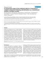
Figure 1 Sequence conservation in A46 and related families. (A) Multiple sequence alignment with the common sequence domain found in
protein families A46, N1, N2 and C1. The alignment is non-redundant at 90% sequence identity. Sequences are identified by species/strain and
gene locus number: SWPV-NEB, swinepox virus strain Nebraska 17077-99; SPPV-TU, sheeppox virus strain TU-V02127; DPV-W848_83, deerpox virus
strain W-848-83; MYXV-LAU, myxoma virus strain Lausanne; RFV-KAS, rabbit fibroma virus strain Kasza; VACV-WR, vaccinia virus strain Western
Reserve; YLDV-Davis, yaba-like disease virus strain Davis; RPXV-UTR, rabbitpox virus strain Utrecht; LSDV-NW_LW, lumpy skin disease virus strain
Neethling Warmbaths LW; ECTV-NAV, ectromelia virus strain Naval. Shading indicates degree of sequence similarity. Conserved motifs are
indicated with horizontal bars on the top of the alignment. Predicted secondary structure is indicated below each block of sequences (orange:
alpha-helix; blue: beta-sheet), except for A46 and N1, for which secondary structures of A52 (PDB:2VVW) and N1 (PDB:2I39), respectively, are
shown. Green arrowheads indicate N1 protein residues putatively involved in BH3 peptide binding [11]. (B) Structural distribution of conserved
motifs. Conserved residues in the multiple sequence alignment were mapped on the N1 structure (PDB:2I39). Secondary structure elements are
depicted in yellow, except conserved residues, in orange. Side chains are coloured in red. Surface is shown in light grey. Structures were
rendered with UCSF Chimera [60].
González and Esteban Virology Journal 2010, 7:59
/>Page 3 of 12
similarity to the Bcl-2 family fold despite their lack of
sequence homology with these cellular proteins. Homol-
ogy at the sequence level with A46 and N1 families
implies that members of the N2 and C1 families will
probablyadoptthesameBcl-2-likefold.Interestingly,
the predicted secondary structure of the conserved
region in N2 and C1 proteins is compatible with this
fold (Figure 1A). To test the hypothesis that these pro-
teins share the common domain of A46 and N1
families, multiple sequence alignments of N2 and C1
families were used to start HHpred searches against a
sequence profile database derived from proteins with
structures in the Protein Data Bank ( PDB) [21]. A
strong relationship was found between N2 and A52
structure (99.0% probability, e-value 1.3E-12). These
results were supported by predicting the structure of
this family with 3D-J ury [22], a fold recognition meta-
server that obtains consensus predictions from different
threading servers. In all cases the best hits were struc-
tures belonging to A46 and N1 families. Only in the
case of C1 the results were not conclusive either with
HHpred (42.5% probability, e-value 0.35) or with 3D-
Jury (not shown). However, given that C1 sequence
homology to N2 is evident from the HHpred searches,
both families will probably share the Bcl-2-like common
domain.
Conserved residues in the common domain of the
poxvirus protein families
Highly conserved amino acids of a multiple sequence
alignment usually indicate that these residues are impor-
tant for protein structure and/or function. In addition,
amino acids that are conserved only in certain subfami-
lies are indicative of importance for specific functions
carried out by these proteins subfamilies. A multiple
sequence alignment of the common domain containing
representative sequences of the four families (A46, N1,
N2 and C1) was analyzed to get an in sight of the con-
served residues. The Proteinkeys web server [23] was
used to find both conserved residues in all families and
specific residues for individual families. Although the
minimum sequence identity between the most divergent
sequences of the four families can be as low as 15%, at
least three conserved motifs could be distinguished in
the multiple sequence alignment (Figure 1A): [LIVM]-x-
x-Y- [IFL]-x- [WY]- [RS] in alpha-helix 1, G-x-x- [FY]-
x-x- [LF]-x-x- [FYL]- [KD]-x-x-A in alpha-helix 2, and
[IV]-G- [LF]-x- [ASG] in alpha-helix 5 (alpha-helices
numbered according to N1). Since a common fold is
assumed for all families, the sequence information was
placed in the context of one of the known three-dimen-
sional structures, that of N1 (PDB:2I39) (Figure 1B).
Interestingly, alpha-helic es 1, 2 and 5 are packed in
close contact to one another in the commo n fold
structure. Most of these conserved residues are hy dro-
phobic and buried inside the protein core, so they are
expected to have an essential role to preserve the
domain structure stability. Because of their level of con-
servation and their position in the structure they might
have been related to the pro-apoptotic peptide binding
site.
Alpha-helix 1 forms part of the dimerization surface
in N1, B15 and A52 proteins [11,12,14]. In the N1
homodimer residues Arg7 and Asp14 of alpha-helix 1 of
different monomers form a potential salt bridge, contri-
buting to dimer stability. This interaction is not found
in A52 and B15 dimers as the relative orientation of
monomers varies. Alpha-helix 2 is an amphipathic helix
whose charged side is exposed and in the case of N1
contain s several residues involved in BH3-peptide bind -
ing like Leu30, Glu32 and Leu33. The C-terminus half
of alpha-helix 5 contains mostly hydrophobic residues
and is buried in the protein core. One pair of amino
acids identified by Proteinkeys as being conserved speci-
fically in one subset of proteins is that of charged resi-
dues Arg12 and Asp31, which are located in conserved
motifs in alpha-helices 1 and 2, respectively. These posi-
tions are highly correlated in the multiple sequence
alignment, where both are present in a large subset of
members of N1 and A46 families and completely absent
in others. These amino acids join alpha-helices 1 and 2
through a potential salt bridge and probably contribute
to the stability of BH3-peptide binding site structure.
The same interaction is also conserved in K7 (Arg37
and Asp61) and A52 (Arg67 and Asp87) proteins. On
the other hand there are a number of charged residues
which are exposed on the surface of the proteins with
known structure and seem relatively conserved in all
families. For instance the pattern of charged residues
alternating with hydrophobic residues in alpha-helix 2 is
observedinN1,K7,B15andA52structuresanditcan
be predicted in other proteins from their sequences. In
N1 protein residues projecting outwards from alpha-
helix 2 include Asp22, Lys25, Lys26 and Glu32, of
which only the last one belongs to the ligand binding
site [11]. Arg81 at the C-terminal end of alpha-helix 5
in N1 is exposed and charged residues at equivalent
positions are conserved in A46 and N2 families. Conser-
vation of these exposed residues may indicate a possible
functionality, for instance an interaction with other pro-
teins. Experimental data revealing detailed poxvirus-host
protein interaction mechanisms are still scarce and
more will be needed to confirm whether any of the con-
served residues is functionally important.
Evolutionary history of A46 and related families
In an attempt to reconstruct the evolutionary history of
the whole family first we built its complete phyletic
González and Esteban Virology Journal 2010, 7:59
/>Page 4 of 12
pattern, meaning by that the distribution of the subfami-
lies or groups of orthologues that integrate the gene
family across all species of chordopoxviruses. Our gene
set was divided into ten o rthologue groups (Figure 2A).
These orthologue groups are exclusively present in a
monophyleticgroupthatincludesthegenusOrthopo x-
virus and a clade comprising five other genera (Yata-,
Capri-, Sui-, Lepori-andCervidpoxvirus), named Clade
II by convention [24]. We could not find any remote
homologueofthisgenefamilyintheremainingtaxo-
nomic groups of the poxvirus phylogeny. The distribu-
tion and number of genes of every orthologue group
varies among different species (Figure 2B and Additional
File 2), although they are always restricted to both term-
inal genome regions, where genes involved in virus- host
interaction are usually located in poxvirus genomes
[25,26]. Eight of the orthologue groups can be found in
orthopoxvirus genomes: N1L, N2L, A52R and B15R can
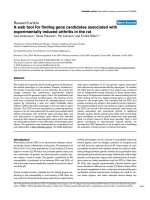
Figure 2 Groups of orthologous genes in A46 and related families. (A) Phylogenetic relationships among the orthologue groups obtained
from A46, N1, N2 and C1 families. A Bayesian phylogenetic tree was constructed from a multiple sequence alignment of proteins encoded by
genes in the ten orthologue groups. For simplicity only a representative species of every poxvirus genus, as depicted in (B), was selected.
Posterior probabilities of every node are shown. (B) Virus genomes representing genera Orthopoxvirus (VACV-COP), Leporipoxvirus (MYXV-LAU),
Capripoxvirus (LSDV-NW_LW), Suipoxvirus (SWPV-NEB), Yatapoxvirus (YLDV-Davis) and Cervidpoxvirus (DPV-W848_83) are depicted, indicating the
relative genome positions of genes included in the orthologue groups. Species/strain names as in Figure 1A; VACV-COP, vaccinia virus strain
Copenhagen. Numbers above every line represent the gene positions in the genome. Symbols below every line represent gene names. Genes
drawn in the same colour belong to the same orthologue group.
González and Esteban Virology Journal 2010, 7:59
/>Page 5 of 12
also be found in the Clade II species, whereas C6L, C1L,
K7R and A46R are unique to orthopoxviruses. On the
other hand two subfamilies are absent in this genus:
those of orthologous genes to myxoma virus m136R and
deerpoxvirus 159R, respectively.
The information provided by the phyletic pattern was
superimposed on a consensusphylogenetictreebuilt
from several single-copy conserved genes in all pox-
viruses.Thetopologyofthistreewassimilartoother
poxvirus phylogenies [27,28]. The family gene content
evolution across the poxvirus phylogeny was recon-
stru cted using the maximum likelihood metho d of Mik-
los and Csuros [29] implemented in the program Count
[30]. This method allows inferring the genome sizes and
gene repertoires of ancestral viruses, along with gene
gain and loss events. The reconstruction of the evolu-
tionary history of the family (Figure 3) suggests that the
common ancestor of orthopoxviruses and the Clade II
would have contained three genes of this family. Which
orthologue group it could have belonged to cannot be
deduced since probabilities are low for all of them (p <
0.5). As a comparison, reconstruction by parsimony sug-
gests that this ancestor would have had four subfamilies
(N1L, N2L, A52R and B15R). Less controversy exists
between both methods for more recent ancestors. The
common ancestor to all orthopoxviruses would have
contained eight genes, what implies five gene gain
events according to the maximum likelihood method. In
this occasion the gene content of the ancestral virus is
more evident as it most likely contained all eight ortho-
logue groups present in practically every extant ortho-
poxvirus (with p = 1). In the branch leading to the
Clade II its common ancestor would have possessed
four genes belonging to this family, implying three gene
gains over the preceding node. The four genes present
in the ancestral genome were with p = 1 N2L, A52R,
m136R and B15R. More recent evolutionary events
include small gene gains and small gene losses in the
branches leading to extant species. Altogether these data
suggest that this gene family originated in the virus line-
age lead ing to the common ancestor of orthopoxviruses
and the Clade II, where between three and four gene
gain events occurred. However it is unlikely that these
gene gains occurred independently in a single ancestral
virus. Furthermore, because of the evident sequence
similarity among the putative genes in the ancestral
virus genome, the most probable hypothesis would be
that a Bcl-2 protein had been acquired from a eukaryo-
tic host by the common ancestor of the subset of verte-
brate poxviruses previously mentioned and probable
events of gene duplication occurred within its genome
before speciation proceeded. After the divergence of
both poxviruses lineages new gene gain events increased
the number of orthologue groups, probably because of
the evolutionary advantage that these proteins conferred
over the host organism in terms of regulation or sup-
pression of antiviral immune response. However in
more recent ancestors the overall number of subfamilies
within poxvirus genomes appears to have stabilized. An
explanation for this stabilization might be that the gene
repertoire of this family was varied enough to accom-
plish its mission.
N1 is the only protein of this family with the s ame
functionality as the putative Bcl-2 ancestor gene so far.
While keeping the same basic tertiary structure these
proteins evolved u ntil they managed to bind a diverse
range of cellular proteins involved in an important path-
way in response to pathogen attacks. As yet the presence
of only other three families of Bcl-2-like genes has been
confirmed in poxviruses. They are vaccinia virus F1L [31]
with orthologues in all orthopoxviruses, myxoma virus
M11L [32,33] with orthologues in all genera of the Clade
II, and fowlpox virus FPV039 [34] with orthologues in
avipoxviruses. These are apparently single-copy genes
and have no sequence similarity with the A46 and related
Bcl-2-like families. Furthermore they lack sequence
homology among them and only the avipoxvirus protein
displays some sequence similarity with cellular Bcl-2 pro-
teins. Very interestingly, these three families carry out
thesamefunction,apoptosis inhibition by binding pro-
apoptotic BH3 peptides, but do not coincide in any pox-
virus genome. Whether the origin of every poxvirus Bcl-
2-like protein is independent or they arose from a gene
present in a common ancestor of chordopoxviruses and
any sequence relationshi p was lost during successive spe-
ciation events is undetermined. Nevertheless it is tempt-
ing t o consider that the presence of other Bcl-2-like
apoptosis inhibitor s in poxvirus genomes offered the A46
and related families the opportunity to freely evolve.
Functional considerations of the four protein families
The common structural core and the sequence homol-
ogy to N1 might suggest that some of the other proteins
belonging to A46, N2 and C1 families could be involved
in an anti-apoptotic role as N1. However this function-
ality has yet to be p roven. On the contrary, it has bee n
discarded for A52 and B15 [14] and probably for K7
[16]. However, the proteins A46, A52, B15, K7 and N1
target diverse host participants of the TLR signalling
pathway (Figure 4) that are apparen tly unrelated among
them, suggesting that the mechanisms of action of these
poxvirus proteins are heterogeneous. We describe below
the information available t hus far on A46, N1, N2 and
C1 families regarding the functional characteristics of
these proteins, which might help to infer the molecular
mechanism of these functionalities and find whether
these functions can be transferred to other proteins in
these families.
González and Esteban Virology Journal 2010, 7:59
/>Page 6 of 12
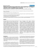
Figure 3 Reconstruction of ancestral gene repertoires in the evolutionary history of A46 and related families. The number in every node
represents the inferred or real number of groups of orthologues present in each genome. This number was inferred for ancestral species by the
maximum likelihood method implemented in the Count program [30]. The background colour of the number indicates the kind of variation in
the gene content since the preceding node: green for nodes with a net gene gain, red for nodes with a net gene loss, and grey if the gene
content remained unchanged. The tree contains a representative strain for every species of the subfamily Chordopoxvirinae with a completely
sequenced genome and is based on a maximum likelihood phylogenetic tree (Additional File 3). Species/strain names as in Figures 1 and 2;
TATV-DAH68, Taterapox virus strain Dahomey 1968; CMLV-CMS, Camelpox virus strain CMS; VARV-IND3_1967, Variola virus strain India 3 Major
1967; CPXV-GRI, Cowpox virus strain GRI-90; MPXV-SLE, Monkeypox virus strain Sierra Leone; YMTV-Amano, Yaba monkey tumor virus strain
Amano; RFV-Kas, Rabbit fibroma virus strain Kasza; SPPV-A, Sheeppox virus strain A; GTPV-G20LKV, Goatpox virus strain G20-LKV; BPSV-AR02,
Bovine papular stomatitis virus strain BV-AR02; ORFV-NZ2, Orf virus strain NZ2; MOCV-st1, Molluscum contagiosum virus strain subtype 1; CNPV-
VR111, Canarypox virus strain ATCC VR111; FWPV-Iowa, Fowlpox virus strain Iowa; CRV-ZWE, Crocodilepox virus strain Zimbabwe.
González and Esteban Virology Journal 2010, 7:59
/>Page 7 of 12
N1 is the only of these families with an experimentally
confirmed anti-apoptotic role. The N1 binding site to
BH3 peptides consists basically of a hyd rophobic groove
flanked by charged residues [11]. Functional N1 residues
are scarcely conserved in the rest of related families
(Figure 1A). However, among the set of N1 residues
which putatively interact with BH3 peptides, there are
three residues (Ile75, Leu30 and Glu32) which belong to
conserved motifs in alpha-helices 2 and 5. Proteins A52
and B15 do not inhib it staurosporine-induc ed apoptosis
and this might be explained because in their surfaces
the BH3-peptide binding groove would be blocked due
to the greater length of alpha-helix 2, about one turn
longer in comparison with that of N1 protein [ 14].
Alpha-helix 2 in N1 has 12 residues while in A52, B15
and K7 it comprises 17 residues. In most members of
the families A46, N2 and C1, the length of alpha-helix 2
can be predicted because two conserved Gly residues
usually delimit it, and in all cases it would have approxi-
mately the same length as in A52. Thus none of these
proteins would be expected to have anti-apoptotic prop-
erties like N1, although experiments should be per-
formed to confirm this hypothesis.
VACV A46 inhibits TLR signalling pathway by bind-
ing to MyD88 and TRIF adaptors, a TIR-like domain
being likely responsible for these interactions. This TIR-
like domain has not yet been found in other VACV pro-
teins or other poxvirus proteins apart from close A46
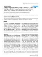
Figure 4 Inhibition of host signalling pathways by VACV members of A46 and related families. TLRs are distributed in the plasma
membrane and endosomes. When a pathogen is recognized by a TLR adaptor proteins are recruited which transmit the signal further
downstream until specific transcription factors are activated and enhance the expression of genes encoding type I IFNs and pro-inflammatory
cytokines. VACV proteins belonging to A46 and N1 families interfere with the TLR signalling pathway at different levels. A46 targets all known
adaptor proteins: MyD88, MAL (TIRAP), TRIF and TRAM. A52 targets IRAK2 and TRAF6, intermediary between adaptors and transcription factors. K7
inhibits IRAK2, TRAF6 and also DDX3, which is part of the complex that activates transcription factor IRF3. B15 targets the IKK complex by
avoiding IKKbeta phosphorylation, what eventually causes the inhibition of NF-kappaB. N1 associates with several components of the IKK
complex and with TBK1, inhibiting NF-kappaB and IRF3 activation, respectively.
González and Esteban Virology Journal 2010, 7:59
/>Page 8 of 12
homologues in orthopoxviruses. Three conserved
sequence motifs of TIR domains were described along
the A46 protein sequence [4,5]: one in its unique N-ter-
minus and the other two in the alpha-helices 1 and 7 of
the common domain with N1. Despite the sequen ce
sim ilarity in these motifs the overall predicted structure
of A46 protein is not coincident with that of TIR
domains, which in the case of TLR1 and TLR2 contain
a central five-stranded parallel beta-sheet surrounded by
five alpha-helices on both sides [35]. In fact we could
not find any relationship of A46 or any other VACV
protein with TIR domains by using tools for remote
sequence homolo gy search or fold recognition (data not
shown). This seems to discard the straightforward expla-
nation that A46 would have acquired its unique role by
grabbing a functional TIR domain from a host cell gen-
ome. In fact, if A46 had really evolved from a remote
Bcl-2-like ancestor and not from a TLR-like ancestor
the origin of the TIR conserved motifs might have prob-
ably been due to mutations which constituted an evolu-
tionary advantage for viruses containing this gene.
A52 inhibits TLR-dependent N F-kappaB activation by
binding to both TRAF6 and IRAK2 [4,6]. Experiments
with different mutant proteins have produced some data
about A52 interaction with these host proteins at the
molecular level. A deletion mutant including its N-term-
inal 144 residues was sufficient for inhibiting NF-kappaB
activation and was able to interact with IRAK2 but not
with TRAF6 [6], although it is not clear whether TRAF6
interacts with the A52 C-terminus. Moreover the N-
terminal 36 residues of A52 were not required to inhibit
IL-1alpha-induced NF-kappaB activation [14]. A small
peptide from VACV A52 has been shown to mimic the
function of the whole protein as it avoids TLR-depen-
dent cytokine secretion [36]. Recent experiments
demonstrating that A52 inhibits NF-kappaB activation
by several TLRs only through its interaction with IRAK-
2 but not TRAF6 [37] support the hypothesis that this
peptide acts on IRAK -2. The sequence corresponding to
the peptide is moderately conserved among A52 ortho-
logues and poorly conserved among other related pox-
virus proteins. On the other hand we could not find in
A52 sequence a canonical TRAF6-binding motif, P-x-E-
x-x-(acidic/aromatic), that was identified in several
TRAF6 cellular interaction partners [38]. This suggests
thatA52mustbindTRAF6throughadifferent
mechanism.
The crystal structure of K7 in complex with a 20
amino-acid DDX3 peptide has determined the precise
details of their interaction [17]. DDX3 binds to a deep
hydrophobic pocket in a negatively charged face of K7
delimited by its N-terminus, alpha-helix 1 and a non-
helical segment equivalent to alpha-he lix 6 in Bcl-2-like
proteins. Interestingly, this region corresponds to the
dimerization interface in A52, which differentiates from
K7 in that it cannot bind DDX3. Like A52, K7 binds the
TRAF domain of TRAF6 [15] but our search did not
find a canonical TRAF6-binding motif in its sequence.
It is striking how proteins of these families evolved
from a common Bcl-2-like domain with anti-apoptotic
role to perform diverse functions always related with the
inhibition of the host immune response, more specifi-
cally the TLR signalling pathway, but at different levels
and using different mechanisms. These poxvirus pro-
teins probably act at the level of subtle protein interac-
tion to sequester a target protein or impede a complex
formation, but their mechanisms of action are mostly
unknown. Although the structures of some of these pro-
teins have been elucidated, as yet only one of them
represents a complex with a host target peptide, what
still hinders the prediction of possible functions for
other members of these families.
Experimental data are scarce or even absent for VACV
proteins C1, C6, N2 and C16/B22. C6 protein has been
found in a very low proportion in vaccinia virus IMV
particles [39], as is the case of A46. One possible reason
for their presence in the virion could be that they are
necessary for the viral cycle early after virus entry. On
the other hand a VACV attenuated strain with a C6L
gene deletion has shown an enhanced immune response
in vivo (manuscript in preparation), indicating that this
protein may also be involved in the regulation of the
host immune response. An early study revealed N2 loca-
tion in the host cell nucleus during virus replication and
discovered that a s ingle nucleotide substitution in the
5’-UTR of N2L gene was responsible for an alpha-ama-
nitin-resistant phenotype [40]. This data could suggest a
possible function of N2 in transcription, although this
hypothesis has not been confirmed yet. An experiment
performed to determine interactions between VACV
and host cell proteins revealed three possible interacting
partnersforC6andotherthreeforN2,asdetermined
by yeast two-hybrid and validated by pull-down [41].
However none of them seems to be directly related with
the host immune response . One of the C6 binding part-
ners was programmed cell death 6 interacting protein
(PDCD6IP/A LIX), which has been involved in apoptosis
regulation, cytokinesis and HIV-1 budding. VACV C6
also interacted with keratin 4 (KRT4) and troponin I,
skeletal, fast (TNNI2). In the same experiment three
possible binding partners were described for N2: karyo-
pherin alpha 2 (KPNA2), t hat may be involved in
nuclear transport of proteins, phospholipid scramblase 4
(PLSCR4), that participates in the regulation of the
movements of phospholipids in membranes, and valosin
containing protein p97/p47 complex interacting protein
1 (VCPIP1), a deubiquitinating enzyme required for
Golgi and ER assembly. These interaction data can help
González and Esteban Virology Journal 2010, 7:59
/>Page 9 of 12
to uncover possible roles of C6 and N2, although they
must be taken cautiously until more specific experi-
ments are performed. To our knowledge, no experimen-
tal data have been published yet about VACV proteins
C1 or C16/B22.
Recent studies on vaccinia virus transcription revealed
the existence of an immediate-early class of genes [42].
This class includes five genes of this family (A52R,
B15R, C6L, K7R and N 2L), while other five (A46R, N1L,
C1L and C16L/B22R) belong to the early class. An
immediate-early or early expression pattern can be char-
acteristic of proteins involved in i mmune response eva-
sion. Thus, those data a gree with the known functions
of A46, A52, B15, K7 and N1, and may support a possi-
ble role in immune response evasion of the members of
these families with still unknown function.
The above findings have implications in the use of
poxviruses as vaccines, in particular vaccinia virus atte-
nuated strains MVA [43,44] and NYVAC [45] that have
been studied extensively [46]. In comparison with strain
WR, MVA lacks A52R and C1L genes while NYVAC
lacks C6L, N1L, N2L and C1L genes. However MVA
contains one (MVA189R) and NYVAC contains two
(C16L/B22R) additional genes with similarity to B15R
which are not present in strain WR. A major difference
in behaviour between these attenuated strains is that
NYVAC provokes greater cytopathic effect, phosphoryla-
tion of EIF2-alpha and apoptosis in infected cells [47].
C6L, N2L and N1L are among the genes present in
MVA and absent in NYVAC and thus could explain this
behaviour.
Conclusions
We have described the sequence relationship among
four families of poxv irus proteins, A46, N1, N2 and C1,
which share a common domain with a Bcl-2-like fold,
and proposed their integration into a single family. The
phylogenet ic distribution and reconstruction of the evo-
lutionary history of this family indicate that it originated
in the common ancestor of orthopoxviruses and a clade
formed by five other poxvirus genera. After initial
increases in the family gene content in the most ances-
tral viruses a balance between gene gains and losses
appears to have stabilized the number of family mem-
bers in extant poxviruses. Their roles determined so far
indicate that these proteins have specialized in regulat-
ing the host immune response, clearly suggesting that
similar functions should be researched for other mem-
bers of this family with still undefined function, like N2,
C1, C6 and C16/B22. The diversity of host targets and
the lack of precise data about what residues are involved
in poxvirus-host protein interactions hamper the predic-
tion of new targets for these families. Nevertheless,
based on secondary structure predictions, our analysis
foresees that practically all members of this family will
be unable to bind pro-apoptotic peptides and inhibit
apoptosis as N1 does. This study highlights the rele-
vance of poxvirus protein families in innate immune
sensing and suggests, from a point of view of t he appli-
cation of atte nuated poxviruses as vacc ines, that to
avoid redundancy in related functions, gene deletions of
entire families should be considered when recombinant
vectors are developed with improved immune capacity.
Methods
Sequence homology analysis
Poxvirus protein sequences were obtained from the Pox-
virus Bioinformatics Resource Center database [48,49].
Multiple sequence alignments of fami lies were
retrieved from Pfam database version 23 [18] when indi-
cated. A global sequence alignment was obtained with
MAFFT [50] using the L-INS-i mode with default para-
meters and including three-dimensional structures to
guide the alignment. The alignment was then manually
adjusted.
Profile versus profile searches were performed with
HHpred [19] in the global alignment mode and scoring
secondary structure. Searches were carried out aga inst
Pfam-A_23 and PDB70 HMM profile databases available
in the same web server.
Iterative searches with HMMer [51], a method based
on HMM profile vs. sequence comparisons, were per-
formed as follows. A single search was started with a
HMM profile against a database of poxvirus protein
sequences. All hit sequences below a threshold e-value
of 0.01 were automatically aligned and from the align-
ment a new HMM profile was built which was used to
start a new search. This was performed several rounds
until the search reached the convergence, i.e. no new
sequences were added.
Secondary structure predictions were performed with
PsiPred [52] starting from multiple sequence alignments
of single families.
Phylogenetic analyses
The Bayesian phylogenetic tree of representative pro-
teins of orthologue groups was obtained by running
MrBayes v3.1.12 [53,54] for 100000 generations in two
rounds of two chains each through the Phylemon web
server [55]. Trees were visualized with Phylodendron
[56].
For the poxvirus phylogenetic tree concatenated align-
ments of proteins encoded by five single-copy conserved
poxvirus genes (E9L, J3R, J6R, H6R and D5R) from
every chordopoxvirus species with at least one fully
sequenced genome were used. An entomopoxvirus spe-
cies was used as an outgroup to root the tree. The max-
imum likelihood phylogenetic tree was buil t with
González and Esteban Virology Journal 2010, 7:59
/>Page 10 of 12
PhyML v3.0 [57] with the LG substitution model, four
substitution rate categories, estimated proportion of
invariable sites and branch support estimated by non-
parametric bootstrap analysis with 100 replicates.
Reconstruction of the family gene content evolution
Groups of orthologous proteins were detected by using
the bidirectional best hit method. Starting with a dataset
containing all poxvirus sequences, a BlastP [58] search
was performed with every sequence within or with
homology to the A46 family against the whole dataset.
Two proteins belonging to different species were consid-
ered orthologues if each was the best hit of the other in
their respective species. The ort hologue groups obtained
were contrasted with the Poxvirus Orthologous Clusters
[59] from the Poxvirus Bioinformatics Resource Center
database. For simplicity, several paralogues were
included in orthologue groups in the cases of orthopox-
virus proteins in the B15R group and Clade II proteins
in the N2L group.
Thegenecontentevolutionwasreconstructedwith
Count [30]. Input data comprised a table with the distri-
bution of the groups of orthologous genes across the
chordopoxvirus genomes (Additional File 2) and the
poxvirus phylogenetic tree (Additional File 3). The
ancestral reconstruction by likelihood maximization
based on a phylogenetic birth-and-death model was cho-
sen [29]. Rate optimization was performed using a gain-
loss-duplication model with a Poisson family size distri-
bution at the root. Family sizes and lineage-spec ific
events (gains, losses, expansions and contractions) were
computed using posterior probabilities in the optimized
gain-loss-duplication model.
Additional file 1: Poxvirus protein sequences detected by an
iterative HMM search. Poxvirus protein sequences detected with an
e-value < 1 in the final round after an iterative HMM search started with
the Pox_A46 HMM profile from Pfam database against a poxvirus protein
sequence database from the Poxvirus Bioinformatics Resource Center
.
Additional file 2: Distribution of orthologue groups across poxvirus
genomes. Table that displays the number of genes of every orthologue
group (rows) across every poxvirus species (columns).
Additional file 3: Maximum likelihood phylogenetic tree of poxvirus
species (Newick format). Maximum likelihood phylogenetic tree built
from concatenated alignments of sequences of proteins encoded by five
single-copy conserved poxvirus genes (E9L, J3R, J6R, H6R and D5R) from
every chordopoxvirus species with at least one fully sequenced genome.
Protein sequences from an entomopoxvirus (AMEV-Moyer) were included
to root the tree.
Acknowledgements
This investigation was supported by grants from the Fundación Marcelino
Botín and the Spanish Ministry of Science and Innovation (SAF2008-02036).
We thank Luis Sánchez-Pulido for helping with sequence searches with
HMMer and Alan Goodman for editorial help.
Authors’ contributions
JMG carried out the bioinformatics analyses, participated in the design of
the study and drafted the manuscript. ME conceived the study, participated
in its design and helped to draft the manuscript. All authors read and
approved the final manuscript.
Competing interests
The authors declare that they have no competing interests.
Received: 21 December 2009 Accepted: 15 March 2010
Published: 15 March 2010
References
1. Kumar H, Kawai T, Akira S: Pathogen recognition in the innate immune
response. Biochem J 2009, 420:1-16.
2. Johnston JB, McFadden G: Poxvirus immunomodulatory strategies:
current perspectives. J Virol 2003, 77:6093-6100.
3. Perdiguero B, Esteban M: The interferon system and vaccinia virus
evasion mechanisms. J Interferon Cytokine Res 2009, 29:581-598.
4. Bowie A, Kiss-Toth E, Symons JA, Smith GL, Dower SK, O’Neill LA: A46R and
A52R from vaccinia virus are antagonists of host IL-1 and toll-like
receptor signaling. Proc Natl Acad Sci USA 2000, 97:10162-10167.
5. Stack J, Haga IR, Schroder M, Bartlett NW, Maloney G, Reading PC,
Fitzgerald KA, Smith GL, Bowie AG: Vaccinia virus protein A46R targets
multiple Toll-like-interleukin-1 receptor adaptors and contributes to
virulence. J Exp Med 2005, 201:1007-1018.
6. Harte MT, Haga IR, Maloney G, Gray P, Reading PC, Bartlett NW, Smith GL,
Bowie A, O’Neill LA: The poxvirus protein A52R targets Toll-like receptor
signaling complexes to suppress host defense. J Exp Med 2003,
197:343-351.
7. Maloney G, Schroder M, Bowie AG: Vaccinia virus protein A52R activates
p38 mitogen-activated protein kinase and potentiates
lipopolysaccharide-induced interleukin-10. J Biol Chem 2005,
280:30838-30844.
8. DiPerna G, Stack J, Bowie AG, Boyd A, Kotwal G, Zhang Z, Arvikar S, Latz E,
Fitzgerald KA, Marshall WL: Poxvirus protein N1L targets the I-kappaB
kinase complex, inhibits signaling to NF-kappaB by the tumor necrosis
factor superfamily of receptors, and inhibits NF-kappaB and IRF3
signaling by toll-like receptors. J Biol Chem 2004, 279:36570-36578.
9. Bartlett N, Symons JA, Tscharke DC, Smith GL: The vaccinia virus N1L
protein is an intracellular homodimer that promotes virulence. JGen
Virol 2002, 83:1965-1976.
10. Chen RA, Ryzhakov G, Cooray S, Randow F, Smith GL: Inhibition of IkappaB
Kinase by Vaccinia Virus Virulence Factor B14. PLoS Pathog 2008, 4:e22.
11. Cooray S, Bahar MW, Abrescia NG, McVey CE, Bartlett NW, Chen RA,
Stuart DI, Grimes JM, Smith GL: Functional and structural studies of the
vaccinia virus virulence factor N1 reveal a Bcl-2-like anti-apoptotic
protein. J Gen Virol 2007, 88:1656-1666.
12. Aoyagi M, Zhai D, Jin C, Aleshin AE, Stec B, Reed JC, Liddington RC:
Vaccinia virus N1L protein resembles a B cell lymphoma-2 (Bcl-2) family
protein. Protein Sci 2007, 16:118-124.
13. Chen RA, Jacobs N, Smith GL: Vaccinia virus strain Western Reserve
protein B14 is an intracellular virulence factor. J Gen Virol 2006,
87:1451-1458.
14. Graham SC, Bahar MW, Cooray S, Chen RA, Whalen DM, Abrescia NG,
Alderton D, Owens RJ, Stuart DI, Smith GL, Grimes JM:
Vaccinia virus
proteins A52 and B14 Share a Bcl-2-like fold but have evolved to inhibit
NF-kappaB rather than apoptosis. PLoS Pathog 2008, 4:e1000128.
15. Schroder M, Baran M, Bowie AG: Viral targeting of DEAD box protein 3
reveals its role in TBK1/IKKvarepsilon-mediated IRF activation. Embo J
2008, 27(15):2147-57.
16. Kalverda AP, Thompson GS, Vogel A, Schroder M, Bowie AG, Khan AR,
Homans SW: Poxvirus K7 protein adopts a Bcl-2 fold: biochemical
mapping of its interactions with human DEAD box RNA helicase DDX3. J
Mol Biol 2009, 385:843-853.
17. Oda S, Schroder M, Khan AR: Structural basis for targeting of human RNA
helicase DDX3 by poxvirus protein K7. Structure 2009, 17:1528-1537.
18. Finn RD, Tate J, Mistry J, Coggill PC, Sammut SJ, Hotz HR, Ceric G,
Forslund K, Eddy SR, Sonnhammer EL, Bateman A: The Pfam protein
families database. Nucleic Acids Res 2008, 36:D281-288.
González and Esteban Virology Journal 2010, 7:59
/>Page 11 of 12
19. Soding J: Protein homology detection by HMM-HMM comparison.
Bioinformatics 2005, 21:951-960.
20. Consortium TU: The Universal Protein Resource (UniProt) 2009. Nucleic
Acids Res 2009, 37:D169-174.
21. Berman HM, Westbrook J, Feng Z, Gilliland G, Bhat TN, Weissig H,
Shindyalov IN, Bourne PE: The Protein Data Bank. Nucleic Acids Res 2000,
28:235-242.
22. Ginalski K, Elofsson A, Fischer D, Rychlewski L: 3D-Jury: a simple approach
to improve protein structure predictions. Bioinformatics 2003,
19:1015-1018.
23. Proteinkeys. [].
24. Hughes AL, Friedman R: Poxvirus genome evolution by gene gain and
loss. Mol Phylogenet Evol 2005, 35:186-195.
25. Seet BT, Johnston JB, Brunetti CR, Barrett JW, Everett H, Cameron C,
Sypula J, Nazarian SH, Lucas A, McFadden G: Poxviruses and immune
evasion. Annu Rev Immunol 2003, 21:377-423.
26. Moss B: Poxviridae: the viruses and their replication. Fields virology
Philadelphia, PA: Wolters Kluwer Health/Lippincott Williams & WilkinsKnipe
D, Howley PM , 5 2007, 2905-2946.
27. Afonso CL, Tulman ER, Delhon G, Lu Z, Viljoen GJ, Wallace DB, Kutish GF,
Rock DL: Genome of crocodilepox virus. J Virol 2006, 80:4978-4991.
28. Bratke KA, McLysaght A: Identification of multiple independent horizontal
gene transfers into poxviruses using a comparative genomics approach.
BMC Evol Biol 2008, 8:67.
29. Csuros M, Miklos I: Streamlining and large ancestral genomes in Archaea
inferred with a phylogenetic birth-and-death model. Mol Biol Evol 2009,
26:2087-2095.
30. Count: analysis of gene content evolution. [ />~csuros/gene_content/count.html].
31. Kvansakul M, Yang H, Fairlie WD, Czabotar PE, Fischer SF, Perugini MA,
Huang DC, Colman PM: Vaccinia virus anti-apoptotic F1L is a novel Bcl-2-
like domain-swapped dimer that binds a highly selective subset of BH3-
containing death ligands. Cell Death Differ 2008, 15:1564-1571.
32. Douglas AE, Corbett KD, Berger JM, McFadden G, Handel TM: Structure of
M11L: A myxoma virus structural homolog of the apoptosis inhibitor,
Bcl-2. Protein Sci 2007, 16:695-703.
33. Kvansakul M, van Delft MF, Lee EF, Gulbis JM, Fairlie WD, Huang DC,
Colman PM: A structural viral mimic of prosurvival Bcl-2: a pivotal role
for sequestering proapoptotic Bax and Bak. Mol Cell 2007, 25:933-942.
34. Banadyga L, Gerig J, Stewart T, Barry M: Fowlpox virus encodes a Bcl-2
homologue that protects cells from apoptotic death through interaction
with the proapoptotic protein Bak. J Virol
2007, 81:11032-11045.
35. Xu Y, Tao X, Shen B, Horng T, Medzhitov R, Manley JL, Tong L: Structural
basis for signal transduction by the Toll/interleukin-1 receptor domains.
Nature 2000, 408:111-115.
36. McCoy SL, Kurtz SE, Macarthur CJ, Trune DR, Hefeneider SH: Identification
of a peptide derived from vaccinia virus A52R protein that inhibits
cytokine secretion in response to TLR-dependent signaling and reduces
in vivo bacterial-induced inflammation. J Immunol 2005, 174:3006-3014.
37. Keating SE, Maloney GM, Moran EM, Bowie AG: IRAK-2 p articipates in
multiple toll-like receptor signaling pathways to NFkappaB via activation
of TRAF6 ubiquitination. J Biol Chem 2007, 282:33435-33443.
38. Ye H, Arron JR, Lamothe B, Cirilli M, Kobayashi T, Shevde NK, Segal D,
Dzivenu OK, Vologodskaia M, Yim M, Du K, Singh S, Pike JW, Darnay BG,
Choi Y, Wu H: Distinct molecular mechanism for initiating TRAF6
signalling. Nature 2002, 418:443-447.
39. Chung CS, Chen CH, Ho MY, Huang CY, Liao CL, Chang W: Vaccinia virus
proteome: identification of proteins in vaccinia virus intracellular mature
virion particles. J Virol 2006, 80:2127-2140.
40. Tamin A, Esposito J, Hruby D: A single nucleotide substitution in the 5’-
untranslated region of the vaccinia N2L gene is responsible for both
alpha-amanitin-resistant and temperature-sensitive phenotypes. Virology
1991, 182:393-396.
41. Zhang L, Villa NY, Rahman MM, Smallwood S, Shattuck D, Neff C,
Dufford M, Lanchbury JS, Labaer J, McFadden G: Analysis of vaccinia virus-
host protein-protein interactions: validations of yeast two-hybrid
screenings. J Proteome Res 2009, 8:4311-4318.
42. Assarsson E, Greenbaum JA, Sundstrom M, Schaffer L, Hammond JA,
Pasquetto V, Oseroff C, Hendrickson RC, Lefkowitz EJ, Tscharke DC, Sidney J,
Grey HM, Head SR, Peters B, Sette A: Kinetic analysis of a complete
poxvirus transcriptome reveals an immediate-early class of genes. Proc
Natl Acad Sci USA 2008, 105:2140-2145.
43. Antoine G, Scheiflinger F, Dorner F, Falkner FG: The complete genomic
sequence of the modified vaccinia Ankara strain: comparison with other
orthopoxviruses. Virology 1998, 244:365-396.
44. Mayr A, Stickl H, Muller HK, Danner K, Singer H: The smallpox vaccination
strain MVA: marker, genetic structure, experience gained with the
parenteral vaccination and behavior in organisms with a debilitated
defence mechanism (author’s transl). Zentralbl Bakteriol B 1978,
167:375-390.
45. Tartaglia J, Perkus ME, Taylor J, Norton EK, Audonnet JC, Cox WI, Davis SW,
Hoeven van der J, Meignier B, Riviere M, Languet B, Paoletti E: NYVAC: a
highly attenuated strain of vaccinia virus. Virology 1992, 188 :217-232.
46. Gomez CE, Najera JL, Krupa M, Esteban M: The poxvirus vectors MVA and
NYVAC as gene delivery systems for vaccination against infectious
diseases and cancer. Curr Gene Ther 2008, 8:97-120.
47. Najera JL, Gomez CE, Domingo-Gil E, Gherardi MM, Esteban M: Cellular and
biochemical differences between two attenuated poxvirus vaccine
candidates (MVA and NYVAC) and role of the C7L gene. J Virol 2006,
80:6033-6047.
48. Lefkowitz EJ, Upton C, Changayil SS, Buck C, Traktman P, Buller RM:
Poxvirus Bioinformatics Resource Center: a comprehensive Poxviridae
informational and analytical resource. Nucleic Acids Res 2005, 33:D311-316.
49. Poxvirus Bioinformatics Resource Center. [].
50. Katoh K, Toh H: Recent developments in the MAFFT multiple sequence
alignment program. Brief Bioinform 2008, 9 :286-298.
51. Eddy SR: Profile hidden Markov models. Bioinformatics 1998, 14:755-763.
52. Jones DT: Protein secondary structure prediction based on position-
specific scoring matrices. J Mol Biol 1999, 292:195-202.
53. Huelsenbeck JP, Ronquist F: MRBAYES: Bayesian inference of phylogenetic
trees. Bioinformatics 2001, 17:754-755.
54. Ronquist F, Huelsenbeck JP: MrBayes 3: Bayesian phylogenetic inference
under mixed models. Bioinformatics 2003, 19:1572-1574.
55. Tarraga J, Medina I, Arbiza L, Huerta-Cepas J, Gabaldon T, Dopazo J,
Dopazo H: Phylemon: a suite of web tools for molecular evolution,
phylogenetics and phylogenomics. Nucleic Acids Res 2007, 35:W38-42.
56. Phylodendron: phylogenetic tree drawing. [ />treeapp/].
57. Guindon S, Gascuel O: A simple, fast, and accurate algorithm to estimate
large phylogenies by maximum likelihood. Syst Biol 2003, 52:696-704.
58. Altschul SF, Gish W, Miller W, Myers EW, Lipman DJ: Basic local alignment
search tool. J Mol Biol 1990, 215:403-410.
59. Upton C, Slack S, Hunter AL, Ehlers A, Roper RL: Poxvirus orthologous
clusters: toward defining the minimum essential poxvirus genome. J
Virol 2003, 77:7590-7600.
60. Pettersen EF, Goddard TD, Huang CC, Couch GS, Greenblatt DM, Meng EC,
Ferrin TE: UCSF Chimera–a visualization system for exploratory research
and analysis. J Comput Chem 2004, 25:1605-1612.
doi:10.1186/1743-422X-7-59
Cite this article as: González and Esteban: A poxvirus Bcl-2-like gene
family involved in regulation of host immune response: sequence
similarity and evolutionary history. Virology Journal 2010 7:59.
Submit your next manuscript to BioMed Central
and take full advantage of:
• Convenient online submission
• Thorough peer review
• No space constraints or color figure charges
• Immediate publication on acceptance
• Inclusion in PubMed, CAS, Scopus and Google Scholar
• Research which is freely available for redistribution
Submit your manuscript at
www.biomedcentral.com/submit
González and Esteban Virology Journal 2010, 7:59
/>Page 12 of 12