Báo cáo y học: " Characterization and frequency of a newly identified HIV-1 BF1 intersubtype circulating recombinant form in São Paulo, Brazil" ppt
Bạn đang xem bản rút gọn của tài liệu. Xem và tải ngay bản đầy đủ của tài liệu tại đây (4.91 MB, 12 trang )
RESEARC H Open Access
Characterization and frequency of a newly
identified HIV-1 BF1 intersubtype circulating
recombinant form in São Paulo, Brazil
Sabri Saeed Sanabani
1,2*
, Évelyn Regina de Souza Pastena
1,2
, Walter Kleine Neto
1
, Vanessa Pouza Martinez
1,2
,
Ester Cerdeira Sabino
1
Abstract
Background: HIV circulating recombinant forms (CRFs) play an important role in the global and regional HIV
epidemics, particularly in regions where multiple subtypes are circulating. To date, several (>40) CRFs are
recognized worldwide with five currently circulating in Brazil. Here, we report the characterization of near full-
length genome sequences (NFLG) of six phylogenetically related HIV-1 BF1 intersubtype recombinants (five from
this stud y and one from other published sequences) representing CRF46_BF1.
Methods: Initially, we selected 36 samples from 888 adult patients residing in São Paulo who had previously been
diagnosed as being infected with subclade F1 based on pol subgenomic fragment sequencing. Proviral DNA
integrated in peripheral blood mononuclear cells (PBMC) was amplified from the purified genomic DNA of all 36-
blood samples by five overlapping PCR fragments followed by direct sequencing. Sequence data were obtained
from the five fragments that showed identical genomic structure and phylogenetic trees were constructed and
compared with previously published sequences. Genuine subclade F1 sequences and any other sequences that
exhibited unique mosaic structures were omitted from further analysis
Results: Of the 36 samples analyzed, only six sequences, inferred from the pol region as subclade F1, displayed BF1
identical mosaic genomes with a single intersubtype breakpoint identified at the nef-U3 overlap (HXB2 position
9347-9365; LTR region). Five of these isolates formed a rigid cluster in phylogentic trees from different subclade F1
fragment regions, which we can now designate as CRF46_BF1. According to our estimate, the new CRF accounts for
0.56% of the HIV-1 circulating strains in São Paulo. Comparison with previously published sequences revealed an
additional five isolates that share an identical mosaic structure with those reported in our study. Despite sharing a
similar recombinant structure, only one sequence appeared to originate from the same CRF46_BF1 ancestor.
Conclusion: We identified a new circulating recombinant form with a single intersubtype breakpoint identified at
the nef-LTR U3 overlap and designated CRF46_BF1. Given the biological importance of the LTR U3 region,
intersubtype recombination in this region could play an important role in HIV evolution with critical consequences
for the development of efficient genetic vaccines.
Background
The immense genetic variability of HIV-1 viruses is con-
sidered the key factor that frustrates efforts to halt the
virus epidemic and poses a serious challenge to the
development and efficacy of vaccines. Like other human
positive-sense RNA viruses, HIV has a high mutation
rate as a result of the error-prone nature of their reverse
transcriptase (3 × 10
-5
mutations per nucleotide per
replication cycle)[1,2]. This high rate of mutation
coupled with the in creased replication capacity of the
virus (10.3 × 10
9
particles per day) [3], allows for the
accumulation and fixation of a variety of advantageous
genetic changes in a virus population, which are selected
for by the host immune response and can resist newly
evolving host defense. Recombination is another poten-
tial evolutionary source that significantly contributes to
* Correspondence:
1
Fundação Pro-Sangue, Hemocentro, São Paulo, Brazil
Sanabani et al. Virology Journal 2010, 7:74
/>© 2010 Sanabani et al; licensee BioMed Central Ltd. This is an Open Access article distributed under the terms of the Creative
Commons Attribution License ( ), whi ch permits unrestricted use, distribution , and
reproduction in any medium, pr ovided the original work is properly cited.
the genetic diversification of HIV by successfully repair-
ing d efective viral genes and by producing new viruses
[4]. To date, HIV-1 viruses are classified into four phy-
logenetic groups: M, O, N and P, which most likely
reflect four independent events of cross-species trans-
mission from chimpanzees [5-7]. The M group (for
main), responsible for the majority of viral infection
worldwide, is further subdivided into nine subtypes (A-
D, F-H, J and K), among which subtypes A and F have
bee n further classified into two sub-subtypes [5]. More-
over, early sequencing studies have provided evidence of
recombination between genomes of different HIV sub-
types [8,9]. Such interclade recombinant strains are con-
sistently r eported from regions where two or more
clades are predominant. Recombinant strains from at
least three unlinked epidemiological sources, w hich
exhibit identical mosaic patterns, have been classified
separately as circulating recombinant forms (CRFs)
[10,11]. Currently , there are more th an 40 defined CRFs
that are epidemiologically
important as subtypes [12]. In addition to the known
CRFs, a large number of unique recombinant viruses,
which are called unique recombinant forms (U RFs),
have been characterized wo rldwide [13]. Together, CRFs
and URFs account for 18% of incident infections in the
global HIV-1 pandemi c [12]. HIV-1 subtypes, CRFs and
URFs show considerably different patterns of distribu-
tion in different geographical regions [12,14].
In Brazil, the number of persons living with HIV
reached an estimated number of 730,000 cases at the
beginning of 2008 (2008 Report on the Glob al AIDS
Epidemic). Like in other European countries and in
North America, HIV-1 subtype B is a major genetic
clade circulating in the country. However, the exis tence
of other subtypes such as F1, C, B/C and B/F, has been
consistently r eported [15-23]. Data from recent studies
of the near full length genom es (NFLG) of HIV have
provided evidence of Brazilian CRF strains desi gnated as
CRF28_BF, CRF29_BF, CRF39_BF, CRF40_BF and
CRF31_BC [17,24-26] />sequence/HIV/CRFs/CRFs.html.
In 2006, Thompson and colleagues [27] published two
NFLG of similar BF1 mosaic viruses from patients in
Rio de Janeiro 9 4BR-RJ-41 (GenBank: AY455781) and
99UFRJ-16 (GenBank: AY455782). Here, we describe the
HIV-1 NFLG of an additional six isolates with similar
BF1 mosaic genomes from patients without evidence of
direct epidemiological linkage.
Methods
Study population
The six samples reported in this study were from indivi-
duals residing in São Paulo in the southeast region of
Brazil and considered the most populous city in South
America. The rationale for selection of these samples
has been previously reported [28]. The data, including
age, gender, number of CD4-positive T cells, and viral
load were obtained from medical records and shown in
Table 1. No evidence of direct epidemiological linkage
could be established.
Amplification and sequencing of HIV-1 DNA
The genomic DNA used for the PCR analyses was
extractedusingtheQIAampbloodkit(Qiagen)accord-
ing to the manufacturer instructions. T he NFLGs from
five overlapping fragments were obtained by PCR using
the Platinum Taq DNA polymerase (5 U/μl) (Invitrogen)
and determined by a previously reported method
[16,17].ToruleoutthepossibilityofTaq-generated
recombinants, an additional PCR product of 670 bp,
which spans most of the viral LTR, was generated in
separate PCR reactions using previously described pri-
mers and conditions [29]. All amplification reactions
were done in duplicate to eliminate PCR artifacts, ensur-
ing that sequenced NFLG were not assembled from het-
erogeneous DNA targets. To test for PCR carry over
contamination, extraction and PCR negatives were run
in each experiment. Both complementary DNA strands
from each a mplicon were directly sequenced by cycle
sequencing using a variety of internal primers, BigDye
terminator chemistry and Taq polymerase on an auto-
mated sequencer (ABI 3130, Applied Biosystems Inc.,
Foster City, CA), essentially according to the protocols
rec ommended by the manufacturer. Fragments for each
amplicon were assembled into contiguous sequences on
a minimum overlap of 30 bp with a 97-100% minimal
mismatch and edited using the Sequencher program 4.7
(Gene Code Corp., Ann Arbor, MI).
Screening for recombination events and identification of
breakpoints
Sequences were screened for the presence of recombina-
tion patterns by the jumping profile Hidden Markov
Model (jpHMM) [30] and further confirmed using the
bootscanning meth od [31] implemented by SimPlot
Table 1 Characteristics of the six patients included in this
study.
Sample ID Age/years Sex CD4 count,
cells/mm
2
Viral load,
copies/mL
06BR_FPS561 44 F
1
81 721
07BR_FPS625 35 F 621 4734
07BR_FPS742 47 F 140 123617
07BR_FPS783 42 M
2
209 80751
07BR_FPS810 38 F 208 49240
07BR_FPS812 45 F 362 5694
F; Female, M; Male
Sanabani et al. Virology Journal 2010, 7:74
/>Page 2 of 12
3.5.1 for Windows [32]. The following parameters were
used in this method: window size, 250 bp; step size, 20
bp; the F84 model of evolution (Maximum likelihood
(ML)) as a model to estimate nucleotide substitution;
transition\transversion ratio, 2.0; and a bootstrap of 100
trees. In addition, the signi ficant threshold for the boot-
scan was set at 90%. The alignment of multiple
sequences, including reference sequences representing
subtypes A-D, F-H, J and K , were
performed by the CLUSTAL X program [33] followed
by manual editing in the BioEdit Sequence Alignment
Editor program [34]. Gaps and ambiguous positions
were removed from alignment. Positions of crossover
sit es were defined based on the distri bution of informa-
tive sites supporting the two incongruent topologies that
maximize the c
2
value [35], a method implemented in
Simplot.
Phylogenetic tree analysis
Phylogenetic relationships between the individual
sequence types were determined by two methods: the
neighbor-joining (NJ) algorithm of MEGA v.4 [36] and
the ML of PHYML v.2.4.4 [37]. For NJ, trees were con-
structed under the maximum composite likelihood sub-
stitution model and bootstrap resampling was carried
out 1000 times for analysis by the MEGA software. ML
phylogenies were constructed using the GTR + I + G
substitution model and a BIONJ starting tree. Heuristic
tree searches under the ML optimality criterion were
performed using the NNI branch-swapping algorithm.
The approximate likelihood ratio test (aLRT) based on a
Shimodaira-Hasegawa-like pro cedure was used as a sta-
tistical test to calculate branch support. Comparison of
tree topologies between subgenomic regions was per-
formed using the algorithm described by Nye et al [ 38].
Trees were displayed using the program MEGA v .4
package. The nucleotide similarities were estimated
using the maximum composite likelihood model im ple-
mented by MEGA v.4 software.
GenBank accession numbers
GenBank accession numbers for the proviral NFLG
sequences reported in this study are (06BR_FPS561:
HM026455, 07BR_FPS625: HM026456, 07BR_FPS742:
HM026457, 07BR_FPS783: HM026458, 07BR_FPS810;
HM026459, 07BR_FPS812: HM026460).
Results
Recombinant Analysis
A total of six strains (06BR FPS561, 07BR FPS625, 07BR
FPS742, 07BR FPS783, 07BR FPS810, and 07BR FPS812)
preliminarily classified as subclade F1 by sequence ana-
lysis of a partial pol region were corroborated by further
phylogenetic analysis of the complete coding sequences
and part of the LTR region. Analysis of the proviral
NFLGs revealed all isolates retain intact reading frames
for a majority of their genes and no gross deletions or
rearrangements were observed. The NFLG sequence
from each strain was initially investigated using jpHMM
which showed them to display identical mosaic struc-
tures with a single intersubtype breakpoint identified at
the nef-U3 overlap (HXB2 position 9347-9365). The
recombinant genomes essentially consisted of subclades
F1 and B as parental sequences. Frag ments identified as
subclade F1 were found to cover al most all o f the gen-
ome coding regions while fragment classified as subty pe
B consisted of a short se quence comprising the last part
of the 3’ LTR. Furthermore, the analysis also revealed
that all the six isolates had a mosaic sequence pattern
nearly identical to the previously published Brazilian
BF1 isolates 94 BR-RJ-41 (GenBank: AY455781) and
99UFRJ-16 (GenBank: AY455782). Based on these preli-
minary analyses, we reanalyzed all six sequences using
the bootscanning method with three different subtype
reference sequences (subtype B, F and C) obtained from
the full-length alignment of the HIV sequence database
. In agreement with the results
obtained by jpHMM, bootscanning analysis confirmed
similar mosaic structures with almost identical break-
point positions within these six isolates (Figure 1). The
BF1 intersubtype transitions were estimated at nucleo-
tides 9347-9365, based on the HIV HXB2 numbering
system , by mapping the informative site and c
2
maximi-
zation. To further test for recombination, ML phyloge-
netic trees were inferred for the regions of nucleotide
sequence on either side of the breakpoints detected by
bootscan method (Figure 1). This analysis corroborates
the results from the boo tscan and thus provided unam-
biguous evidence for a single recombination event sup-
ported by high aLRT values among the six isolates.
To rule out the possibility of Taq-generated recombi-
nant artifacts, an additional PCR product of 670 bp cov-
ering most of the viral LTR was generated in a separate
PCR reaction using previously described primers and
conditions [29]. The results confirmed the recombina-
tion breakpoint obtained using complete viral sequences.
Phylogenetic analysis of regions bounded by the
crossover sites
As shown in Figure 2a, phylogenetic reconst ructions for
F1 specific regions bound by the crossover site, as
defined by bootscan analysis, were compared with repre-
sentatives of all subtype and sub-subtype references
available in the HIV database (year 2008) and with other
subclade F1 published sequences. The result of the ML
tree revea led all our sequences clustered on a branch of
subclade F1 and further into one separate sub-branch
intrinsic to South America, particularly Brazil (100%
Sanabani et al. Virology Journal 2010, 7:74
/>Page 3 of 12
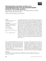
Figure 1 Phylogenetic relations between the parental regions of our six recombinants. The left part of the figure shows Bootscan results
for the recombinants compared to representatives of HIV-1 subtype B (green line), F1 (red line) C (blue line) reference sequences. The right part
refers to the ML phylogenic-based regions between recombination breakpoints as defined by bootscan plot. The recombinants are highlighted
with black circles. For clarity purposes, the trees were midpoint rooted. The scale bar represents 0.01 nucleotide substitution per site.
Sanabani et al. Virology Journal 2010, 7:74
/>Page 4 of 12
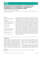
Figure 2 An exploratory ML tree calculated from fragments between breakpoints of sequences identified in this study (indicated by
black circles), published sequences with identical breakpoints (indicated by triangle) and reference sequences of subtype A-D, F-H, J
and K . (A) Tree of the viral genomes corresponding to the subclade F1 segments (HXB2 nucleotides 623-9347). (B) Tree
of the viral genomes corresponding to the subtype B segments in the LTR region (HXB2 nucleotides 9348-9719). For clarity purposes, the tree
was midpoint rooted. The approximate likelihood ratio test (aLRT) values of ≥ 90% are indicated at nodes. The scale bar represents 0.05
nucleotide substitution per site.
Sanabani et al. Virology Journal 2010, 7:74
/>Page 5 of 12
aLRT). During analysis of the tree topology of the F1
region depicted in Figure 2a, all new sequences, except
isolate 06BR FPS561, formed a single cluster with two
previously published Brazilian isolates (F1.
BR.01.01BR125 and F1.BR.01.01BR087) supported by
100% aLRT values. Isolate 06BR FPS561 formed a rigid
subcluster (94% aLRT) with two strains (F1.JP.2004.
DR6190 and F1.JP.2004.DR6082) recently isolated in
Japan and believed to be deri ved from Brazil [Tatsumi
et al, unpublished study]. Additionally, the Brazilian iso-
lates 94BR-RJ-41 and 99UFRJ-16 from Rio de Janeiro
formed a separate branch (<90% aLRT) distinct from
the o ther branches. To test the stability and branching
orders of the F1 fragment in our sequences, ML trees
were independently made from gag-pol an d env
sequences using the same multiple genome alignment
generated for the full length of F1 fragment (Figure 3a
&3b). The phylogenetic trees from both regions received
an overall topologic al score of 78.5% according to the
algorithm of Nye et al [38]. The computed topological
score of the clusters that include all our isolates except
06BR FPS561 in both regions was 100%. Isolate 06BR
FPS561 placed the gag-pol region within the F1.JP.2004.
DR6190, F1.JP.2004.DR6082, 07BR844 and 94BR-RJ-41
cluster with an 84% of aLRT value, while env grouped
with another subcluster that included 06BR564 and
02BR082 (aLRT 94%). The computed topological score
of this cluster in both regions was 30% with a branch
length mismat ch of 53.5%. Similarly, isolates 94BR-RJ-41
and 99UFRJ-16 changed their topo logical positions over
the gag-pol and env regions of their genomes (aLRT
<90%). Thus, the shifting of topological position s of i so-
late 06BR FPS561, 94BR-RJ-41 and 99UF RJ-16 into two
different phylogenetic trees is suggestive evidence of
intrasubtype recombination event o r other factors, such
as convergence. Furthermore, the monophyletic cluster
of isolates F1.JP.2004.DR6190 and F1.JP.2004.DR6082
depicted in Figure 2a was also supported in trees of
both subgenomic regions (Figure 3a &3b).
Thephylogenetictreebasedonthefragmentcharac-
terized as subtype B by bootscan from all of the six iso-
lates is shown in Figure 2b. The resulting tree topology
agrees with the accepted HIV-1 group M phylogeny and
the majority of the internal nodes are supported with
high aLRT values. Despite the fact that B fragments in
these isolates have shorter sequences and some group
M variants cannot resolve some of the internal nodes,
all of them can resolve the terminal nodes.
Molecular rate of CRF46_BF1
Five of the current six BF1 isolates described in this
study (designated as CRF46_BF1 in the Los Alamos
database) were detected in 36 samples selected from 888
samples infected with HIV-1 F1 based on pol
subgenomic fragment sequencing [28]. Based on these
results, the molecular distributio n of the CRF46_BF1
accounts for 0.56% of the HIV-1 circulating strains in
São Paulo.
Identification of Related HIV-1 Strains in the database
A search for similar recombination patterns in a
sequence database revealed the occurrence of three iso-
lates from Brazil (GenBank: AY455781 ; 94BR-RJ-41,
AY455782; 99UFRJ-16 and DQ358801; 01BR087) and
two isolates from Japan (GenBank: AB480299; F1.
JP.2004.DR6082 and AB480301; F1.JP.2004.DR6190). It
is to be noted that, as a result of our current analysis,
the sequences F1.JP.2004.DR6082, F1.JP.2004.DR6190,
and 01BR087, which are characterized as pure subclade
F1 [17] [Tatsumi et al, unpublished study], showed
strong phylogenetic evidence for recombination among
subclade F1 and subtype B, suggesting that a revised
classification of these isolates in the GenBank and the
HIV databases is appropriate.
Next, we aimed to compare the recombinant profiles
of our sequences to other HIV BF1 genomes at the
nucleotide level to illustrate the distribution of their
breakpoints. This was done by retrieving the full-length
genomes from all BF1 and CRF_BF1 isolates available in
the Los Alamos database. The automated jpHMM was
used for mapping breakpoints with significant recombi-
nation signal (Figure 4). Our analysis showed that two
variants (GenBank:DQ085869; BREPM11931 and
DQ085870; BREPM11931) annotated as BF1 recombi-
nants in the database, appear ancestral to subtype B
strains. The recombination mapping of the nef-U3 over-
lap detected in our sequences was also found in
CRF39_BF1 and four other URF BF1 recombinants. In
addition, m ost of the sequences have undergone multi-
ple rounds of recombination events. These data suggest
that this part of the nef-U3 overlap is a possible ‘ hot
spot’ for recombination.
Fragment B from all six isolates shared 96% sequence
identity with the B stretch in the nef-U3 overlap from
the Brazilian 93br029 which was isolated in 1993. Thus,
we assume that the initial recombination event hap-
pened several years before 1993.
Partial LTR nucleotides alignment features
A detailed scrutinization of the partial nucleotide align-
ment of the 3’ LTR regions relative to HXB2 and con-
sensus sequences of other HIV subtypes (Year 2005) is
shown in Figure 5. Conform to the consensus sequence
GGGRNNYYCC, additional NF- Bbindingsiteswere
found in three strains from the current study. A sub-
clade F1 specific insert of 13-15 [39] nucleotides down-
stream of the NF-B
III
binding site was not observed in
our sequences and added further support to our results,
Sanabani et al. Virology Journal 2010, 7:74
/>Page 6 of 12
indicating that our sequences are not genuine F1 sub-
subtypes but BF1 recombinant isolates. Absence of this
nucleotide signature was also observed in isolates F1.
JP.2004.DR6082, F1.JP.2004.DR6190, and 01BR087,
which have previously been classified as pure subclade
F1 sequences.
Discussion
In the present study, we have charac terized six NFLG
sequences that posses mosaic genomic structure identi-
cal to the previously described strains, 94BR_RJ_41 and
99UFRJ_16 with a genome of predominantly subtype F1
and the nef-U3 overlap portion of the LTR of subtype B
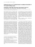
Figure 3 Maximum likelihood tree of sequences identified in this study (indicated by black circles), published sequences with
identical breakpoints (indicated by triangle) and reference strains inferred from full-length gagpol (A) and env (B) reading frames. For
clarity purposes, the tree was midpoint rooted. The approximate likelihood ratio test (aLRT) values of ≥ 90% are indicated at nodes. The scale bar
represents 0.05 nucleotide substitutions per site.
Sanabani et al. Virology Journal 2010, 7:74
/>Page 7 of 12
(Figure 1). Moreover, three additional full-length gen-
ome sequences, which were initially characterized as
pure subclde F1, now clearly appear to harbor a small
fragment derived from subtype B in their LTR in a posi-
tion identical to the breakpoint reported in our
sequences. In phylogenetic tree of the full length and
subgenomic regions of F1 subclade segment, isolates F1.
JP.2004.DR6082 F1.JP.2004.DR6190 (recovered from
japanese patients), 94BR-RJ-41 and 99UFRJ-16
(recovered from patients residing in Rio de Janeiro)
position outside the single cluster formed by isolates
01BR087 and all BF1 recombinants identifie d in this
study, except 06BR FPS561 (recovered from patients
residing in São Paulo) (Figure 2a&3a). The discordant
branching between gag-pol and env sequences of 06BR
FPS561, 94BR-RJ-41 and 99UFRJ-16 isolates can be
explained by the occurrence of a nother recombination
events after the spread of their common ancestor.
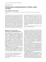
Figure 4 Schematic representation of the NFLG structure and breakpoint profiles of the sequences identified in this study and other
BF1 URF and CRF published sequences. Sequences marked with the symbol (†) were originally classified as pure F1 subclade. Sequences
marked with the symbol (*) were originally classified as pure subtype B. The region of subclade F1 and subtypes B are indicated at the bottom.
Positions of breakpoints are marked with grey arrowhead and numbered according to the HXB2 sequence.
Sanabani et al. Virology Journal 2010, 7:74
/>Page 8 of 12
Generally, our results suggest that the 11 recombinant
sequences w ere not the r esult of one, but at least three
independent recombination events that produce similar
simple recombinant structures. In particular, BF
sequences isolated in Japan and Rio de Janeiro may have
originated from different BF recombinant ancestors than
those sequen ces isolated in São Paulo. Thus, by exclud-
ing all the isolates that branch out of the main c luster,
we provide a total of 6 sequences (01BR087 and 5
sequences described in this study) that meet the formal
requirement for assigning a new CRF46_BF1. Again, in
the phylogenetic tree of the F1 subclade fragment, the
two recently isolated Japanese strains (F1.JP.2004.
DR6190 and F1.JP.2004.DR6082) formed a rigid subclus-
ter with isolate 06BR FPS561 and branch outside the
subcluster formed by the other five viruses described in
this study, but still strongly position within the main
Brazilian subclade F1 sequences. This result suggests
that the viruses found in the Japanese patients share a
distinct common ancestry originating in Brazil. It is pos-
sible that the heavy traffic of people from both countries
across international borders could h ave facilitated t he
spread of these viruses in both countries.
Based on the criteria of inclusion of the samples in
this study, we were able to show that the CRF46_BF1
accounts for 0.56% of the HIV-1 circulating strains in
São Paulo, similar to the frequency of subclade F1
reported from this region [ 28]. The apparently low pre-
valence of the CRF46_BF is ecological and may not be
due t o inherent properties of the virus itself but rather
to the chance results of subtype B (a founder virus in
Brazil), where it is introduced and consequently estab-
lished into our HIV infected population before the new
CRF and other subtypes are introduced.
Our analysis also showed that the recombination of
subclade F1 with subtype B at the nef-U3 overlap por-
tionoftheLTRappearstobearecurrentfinding
because it has also been found i n CRF39_B F1 an d other
unique HIV-1 recombinants [17,25,40,41]. In HIV, the
existence of recombinational hot spots is common given
that they have been described in cell-free systems [42]
and exists in the dimer i nitiation sequence of the HIV-1
Figure 5 Alignment of the nucleotide sequences within the LTR region spanning HXB2 positions -162 to +3 (GenBank accession
number K03455). Dots indicate nucleotide identity to the HXB2 sequence and dashes (-) represent gaps introduced to achieve the best
alignment. Motifs present in the HXB2 strain are underlined. Boxed sequences in subclade F1 isolates indicate the 13-15 nucleotide insertion.
Sanabani et al. Virology Journal 2010, 7:74
/>Page 9 of 12
5’-untranslated region and some preferential sites across
the viral genome [43-46]. Several studies have demon-
strated that RNA hairpin structures strongly correlate
with recombination hotspots in various regions of the
HIV-1 genome[42,43,46,47]. Thus, based on the later
mechanisms, it is possible that hai rpins promote recom-
bination by hamper ing the R T during reverse transcrip-
tion or direct interaction with template [46,48,49].
The HIV-1 LTR region is composed of various cis-act-
ing regulatory components needed for proviral DNA
synthesis, integration of the nascent viral cDNA into the
host cell genome, transcription and modulation o f HIV
genes expression [50,51]. Early reports showed that the
LTR region is made up of three segments designated as
U3, R and U5 [ 52]. The U3 modulatory region entirely
overlaps with nef [53] and is essentially required during
reverse transcription for first template transfer and inte-
gration of the provirus into the host genome. Moreover,
this region seems to regulate the transcription pathway
of HIV viral promoters by directly or indirectly interact-
ing with a large number of cellular proteins, including
NF-AT, Ets-1, USF, AP-1, COUP and Sp1 [54]. Thus,
substitution through recombination of the nef-U3 over-
lap portion of the LTR with that of a genetically differ-
ent subtype, as in our isolates, may affect the binding of
both cellular and viral transcription factors. In turn, this
may influence viral transcription levels, potentially
enhancing the propagation of a recombinant virus lead-
ing to the persistance of a circulating form.
Several studies reported successful results in inhibiting
HIV-1 replication by using synthetic siRNAs targeting
either viral RNA sequences or cellular mRNAs encoding
proteins that are critical for HIV-1 replication [55-58].
The study conducted by Yamamoto and his colleagues
[59] showed a considerable sustainable suppression of
HIV replication and control of CC-chemokine produc-
tion associated with nef expression in HIV-1-infected
macrophages following transfection of short hairpin
RNA (shRNA) by a lentivirus vector system expressing
HIV-specific shRNAs. These results allowed the au thors
to conclude that lentivirus-vector-based RNA interfer-
ence of the U3-overlapping region of HIV-1 nef may
have potential usefulness as a genetic vaccine against
HIV-1 infection. Furthermore, Ludwig and collaborator
[60] proved that HIV-1 contains an antisense gene in
the U3-R regions of the LTR responsible for both an
antisense RNA transcript and p roteins. This antisense
transcript has tremendous potentia l for intrinsic RNA
regulation because of its overlap with the beginning of
all HIV-1 sense RNA transcripts by 25 nucleotides. The
novel HIV antisense proteins encoded in a region of the
LTR that has already been shown to be deleted in some
HIV-infected long-term survivors and represent new
potential targets for vaccine development [60,61].
Given the biological relevance described to the U3
region, it is probable that the intersubtype recombina-
tion in this region could play an important role in HIV
evolution with critical consequences for the develop-
ment of efficient genetic vaccines.
During phylogene tic analysis, the B fragme nts of our
six strains and the other five strains (marked with a tri-
angle symbol in Figure 2b), which showed identical
mosaic genomic structures, were clearly distinct from
available South American subclade F1 sequences, parti-
cularly of Brazilian origin. This result coupled with the
absenceofthe13-15nucleotides insertion downstream
of the NF-B
III
binding site, which is typical for sub-
clade F1, agrees with the interpretation that the segment
at the nef-U3 overlap portion of the LTR of the eleven
isolates originates from subtype B. Unlike the marked
clustering of the eleven isolates in the tree generated
from the F1 fragment, the tree of fragment B depicted
in Figure 2b shows them to fall in different sub-
branches within subtype B reference sequences. This
result is most likely explained by the short lengths of
the fragment B sequences.
Conclusion
In this study, we describe the NFLG sequence analysis
from six HIV-1 isolates sampled from São Paulo and
five other published isolates that had an identical break-
points between subclades F1 and B at the nef-U3 over-
lap portion of LTR. Six of these sequences (five from
this study and one from other published sequences) are
currently classified as a member of the CRF46_BF1
family. Our data is relevant to guide diagnosis and vac-
cine development. We conclude that recombination is a
potentially important mechanism that significantly con-
tributes to HIV genetic variability with serious implica-
tions for diagnosis, drug treatment and optimal vaccine
development.
Acknowledgements
This work was supported by grants 06/50096-0, 2004/15856-9 and 2007/
04890-0 from the Fundação de Amparo a Pesquisa do Estado de São Paulo
(FAPESP).
Author details
1
Fundação Pro-Sangue, Hemocentro, São Paulo, Brazil.
2
Retrovirology
Laboratory, Federal University of São Paulo, Brazil.
Authors’ contributions
SS conceived and designed the study, did the data analysis of the
sequences, and wrote the manuscript. ÉRP, WKN and VPM conducted the
characterization of the full-length genome analysis. ECS designed, wrote the
manuscript and directed the study. All authors read and approved the final
manuscript.
Sanabani et al. Virology Journal 2010, 7:74
/>Page 10 of 12
Competing interests
The authors declare that they have no competing interests.
Received: 28 January 2010 Accepted: 16 April 2010
Published: 16 April 2010
References
1. Mansky LM: The mutation rate of human immunodeficiency virus type 1
is influenced by the vpr gene. Virology 1996, 222:391-400.
2. Mansky LM, Temin HM: Lower in vivo mutation rate of human
immunodeficiency virus type 1 than that predicted from the fidelity of
purified reverse transcriptase. J Virol 1995, 69:5087-5094.
3. Perelson AS, Neumann AU, Markowitz M, Leonard JM, Ho DD: HIV-1
dynamics in vivo: virion clearance rate, infected cell life-span, and viral
generation time. Science 1996, 271:1582-1586.
4. Worobey M, Holmes EC: Evolutionary aspects of recombination in RNA
viruses. J Gen Virol 1999, 80(Pt 10):2535-2543.
5. Robertson DL, Anderson JP, Bradac JA, Carr JK, Foley B, Funkhouser RK,
Gao F, Hahn BH, Kalish ML, Kuiken C, et al: HIV-1 nomenclature proposal.
Science 2000, 288:55-56.
6. Gao F, Bailes E, Robertson DL, Chen Y, Rodenburg CM, Michael SF,
Cummins LB, Arthur LO, Peeters M, Shaw GM, et al: Origin of HIV-1 in the
chimpanzee Pan troglodytes troglodytes. Nature 1999, 397:436-441.
7. Plantier JC, Leoz M, Dickerson JE, De Oliveira F, Cordonnier F, Lemee V,
Damond F, Robertson DL, Simon F: A new human immunodeficiency
virus derived from gorillas. Nat Med 2009, 15:871-872.
8. Sabino EC, Shpaer EG, Morgado MG, Korber BT, Diaz RS, Bongertz V,
Cavalcante S, Galvao-Castro B, Mullins JI, Mayer A: Identification of human
immunodeficiency virus type 1 envelope genes recombinant between
subtypes B and F in two epidemiologically linked individuals from Brazil.
J Virol 1994, 68:6340-6346.
9. Robertson DL, Sharp PM, McCutchan FE, Hahn BH: Recombination in HIV-
1. Nature 1995, 374:124-126.
10. Carr JK, Salminen MO, Albert J, Sanders-Buell E, Gotte D, Birx DL,
McCutchan FE: Full genome sequences of human immunodeficiency
virus type 1 subtypes G and A/G intersubtype recombinants. Virology
1998, 247:22-31.
11. Robertson DL, Anderson JP, Bradac JA, Carr JK, Foley B, Funkhouser RK,
Gao F, BH H, Kalish ML, Kuiken C, et al: HIV-1 nomenclature proposal: a
reference guide to HIV-1 classification. Human retroviruses and AIDS 1999:
a compilation and analysis of nucleic acid and amino acid sequences. Los
Alamos, CA Kuiken CL, Foley B, Hahn B, et al 1999, 492-505.
12. Hemelaar J, Gouws E, Ghys PD, Osmanov S: Global and regional
distribution of HIV-1 genetic subtypes and recombinants in 2004. AIDS
2006, 20:W13-23.
13. McCutchan FE: Global epidemiology of HIV. J Med Virol 2006, 78(Suppl 1):
S7-S12.
14. Taylor BS, Sobieszczyk ME, McCutchan FE, Hammer SM: The challenge of
HIV-1 subtype diversity. N Engl J Med 2008, 358:1590-1602.
15. Barreto CC, Nishyia A, Araujo LV, Ferreira JE, Busch MP, Sabino EC: Trends in
antiretroviral drug resistance and clade distributions among HIV-1–
infected blood donors in Sao Paulo, Brazil. J Acquir Immune Defic Syndr
2006, 41:338-341.
16. Sanabani S, Neto WK, de Sa Filho DJ, Diaz RS, Munerato P, Janini LM,
Sabino EC: Full-length genome analysis of human immunodeficiency
virus type 1 subtype C in Brazil. AIDS Res Hum Retroviruses 2006,
22:171-176.
17. Sanabani S, Kleine Neto W, Kalmar EM, Diaz RS, Janini LM, Sabino EC:
Analysis of the near full length genomes of HIV-1 subtypes B, F and BF
recombinant from a cohort of 14 patients in Sao Paulo, Brazil. Infect
Genet Evol 2006, 6:368-377.
18. Passaes CP, Guimaraes ML, Bello G, Morgado MG: Near full-length genome
characterization of HIV type 1 unique BC recombinant forms from
Southern Brazil. AIDS Res Hum Retroviruses 2009, 25:1339-1344.
19. Soares EA, Santos RP, Pellegrini JA, Sprinz E, Tanuri A, Soares MA:
Epidemiologic and molecular characterization of human
immunodeficiency virus type 1 in southern Brazil. J Acquir Immune Defic
Syndr 2003, 34:520-526.
20. Brindeiro RM, Diaz RS, Sabino EC, Morgado MG, Pires IL, Brigido L,
Dantas MC, Barreira D, Teixeira PR, Tanuri A: Brazilian Network for HIV
Drug Resistance Surveillance (HIV-BResNet): a survey of chronically
infected individuals. Aids 2003, 17:1063-1069.
21. Rodrigues R, Scherer LC, Oliveira CM, Franco HM, Sperhacke RD, Ferreira JL,
Castro SM, Stella IM, Brigido LF: Low prevalence of primary antiretroviral
resistance mutations and predominance of HIV-1 clade C at polymerase
gene in newly diagnosed individuals from south Brazil. Virus Res 2006,
116:201-207.
22. Brennan CA, Brites C, Bodelle P, Golden A, Hackett J Jr, Holzmayer V,
Swanson P, Vallari A, Yamaguchi J, Devare S, et al: HIV-1 strains identified
in Brazilian blood donors: significant prevalence of B/F1 recombinants.
AIDS Res Hum Retroviruses 2007, 23:1434-1441.
23. Eyer-Silva WA, Couto-Fernandez JC, Morgado MG: Molecular epidemiology
of HIV type 1 in inner Rio De Janeiro State, Brazil. AIDS Res Hum
Retroviruses 2007, 23:303-308.
24. De Sa Filho DJ, Sucupira MC, Caseiro MM, Sabino EC, Diaz RS, Janini LM:
Identification of two HIV type 1 circulating recombinant forms in Brazil.
AIDS Res Hum Retroviruses 2006, 22:1-13.
25. Guimaraes ML, Eyer-Silva WA, Couto-Fernandez JC, Morgado MG:
Identification of two new CRF_BF in Rio de Janeiro State, Brazil. AIDS
2008, 22:433-435.
26. Santos AF, Sousa TM, Soares EA, Sanabani S, Martinez AM, Sprinz E,
Silveira J, Sabino EC, Tanuri A, Soares MA: Characterization of a new
circulating recombinant form comprising HIV-1 subtypes C and B in
southern Brazil. AIDS 2006, 20:2011-2019.
27. Thomson MM, Sierra M, Tanuri A, May S, Casado G, Manjon N, Najera R:
Analysis of near full-length genome sequences of HIV type 1 BF
intersubtype recombinant viruses from Brazil reveals their independent
origins and their lack of relationship to CRF12_BF. AIDS Res Hum
Retroviruses 2004, 20:1126-1133.
28. Sanabani SS, Pastena ER, Kleine Neto W, Barreto CC, Ferrari KT, Kalmar EM,
Ferreira S, Sabino EC: Near full-length genome analysis of low prevalent
human immunodeficiency virus type 1 subclade F1 in Sao Paulo, Brazil.
Virol J 2009, 6:78.
29. Gao F, Robertson DL, Morrison SG, Hui H, Craig S, Decker J, Fultz PN,
Girard M, Shaw GM, Hahn BH, Sharp PM: The heterosexual human
immunodeficiency virus type 1 epidemic in Thailand is caused by an
intersubtype (A/E) recombinant of African origin. J Virol 1996,
70:7013-7029.
30. Schultz AK, Zhang M, Leitner T, Kuiken C, Korber B, Morgenstern B,
Stanke M: A jumping profile Hidden Markov Model and applications to
recombination sites in HIV and HCV genomes. BMC Bioinformatics 2006,
7:265.
31. Salminen MO, Carr JK, Burke DS, McCutchan FE: Identification of
breakpoints in intergenotypic recombinants of HIV type 1 by
bootscanning. AIDS Res Hum Retroviruses 1995, 11:1423-1425.
32. Lole KS, Bollinger RC, Paranjape RS, Gadkari D, Kulkarni SS, Novak NG,
Ingersoll R, Sheppard HW, Ray SC: Full-length human immunodeficiency
virus type 1 genomes from subtype C-infected seroconverters in India,
with evidence of intersubtype recombination. J Virol 1999, 73:152-160.
33. Thompson JD, Gibson TJ, Plewniak F, Jeanmougin F, Higgins DG: The
CLUSTAL_X windows interface: flexible strategies for multiple sequence
alignment aided by quality analysis tools. Nucleic Acids Res 1997,
25:4876-4882.
34. Hall TA: BioEdit: a user-friendly biological sequence alignment editor and
analysis program for windows 95/98/NT. Nucleic Acids SympSer 1999,
41:95-98.
35. Robertson DL, Hahn BH, Sharp PM: Recombination in AIDS viruses. J Mol
Evol 1995, 40:249-259.
36. Tamura K, Dudley J, Nei M, Kumar S: MEGA4: Molecular Evolutionary
Genetics Analysis (MEGA) software version 4.0. Mol Biol Evol 2007,
24:1596-1599.
37. Anisimova M, Gascuel O: Approximate likelihood-ratio test for branches:
A fast, accurate, and powerful alternative. Syst Biol 2006, 55:539-552.
38. Nye TM, Lio P, Gilks WR: A novel algorithm and web-based tool for
comparing two alternative phylogenetic trees. Bioinformatics 2006,
22:117-119.
39. Jeeninga RE, Hoogenkamp M, Armand-Ugon M, de Baar M, Verhoef K,
Berkhout B: Functional differences between the long terminal repeat
transcriptional promoters of human immunodeficiency virus type 1
subtypes A through G. J Virol 2000, 74 :3740-3751.
Sanabani et al. Virology Journal 2010, 7:74
/>Page 11 of 12
40. Gao F, Robertson DL, Carruthers CD, Morrison SG, Jian B, Chen Y, Barre-
Sinoussi F, Girard M, Srinivasan A, Abimiku AG, et al: A comprehensive
panel of near-full-length clones and reference sequences for non-
subtype B isolates of human immunodeficiency virus type 1. J Virol 1998,
72:5680-5698.
41. Thomson MM, Delgado E, Herrero I, Villahermosa ML, Vazquez-de Parga E,
Cuevas MT, Carmona R, Medrano L, Perez-Alvarez L, Cuevas L, Najera R:
Diversity of mosaic structures and common ancestry of human
immunodeficiency virus type 1 BF intersubtype recombinant viruses
from Argentina revealed by analysis of near full-length genome
sequences. J Gen Virol 2002, 83:107-119.
42. Moumen A, Polomack L, Roques B, Buc H, Negroni M: The HIV-1 repeated
sequence R as a robust hot-spot for copy-choice recombination. Nucleic
Acids Res 2001, 29:3814-3821.
43. Balakrishnan M, Fay PJ, Bambara RA: The kissing hairpin sequence
promotes recombination within the HIV-I 5’ leader region. J Biol Chem
2001, 276:36482-36492.
44. Balakrishnan M, Roques BP, Fay PJ, Bambara RA: Template dimerization
promotes an acceptor invasion-induced transfer mechanism during
human immunodeficiency virus type 1 minus-strand synthesis. J Virol
2003, 77:4710-4721.
45. Magiorkinis G, Paraskevis D, Vandamme AM, Magiorkinis E, Sypsa V,
Hatzakis A: In vivo characteristics of human immunodeficiency virus type
1 intersubtype recombination: determination of hot spots and
correlation with sequence similarity. J Gen Virol 2003, 84:2715-2722.
46. Galetto R, Giacomoni V, Veron M, Negroni M: Dissection of a
circumscribed recombination hot spot in HIV-1 after a single infectious
cycle. J Biol Chem 2006, 281:2711-2720.
47. Galli A, Lai A, Corvasce S, Saladini F, Riva C, Deho L, Caramma I, Franzetti M,
Romano L, Galli M, et al: Recombination analysis and structure prediction
show correlation between breakpoint clusters and RNA hairpins in the
pol gene of human immunodeficiency virus type 1 unique recombinant
forms. J Gen Virol 2008, 89:3119-3125.
48. Moumen A, Polomack L, Unge T, Veron M, Buc H, Negroni M: Evidence for
a mechanism of recombination during reverse transcription dependent
on the structure of the acceptor RNA. J Biol Chem 2003, 278:15973-15982.
49. Roda RH, Balakrishnan M, Kim JK, Roques BP, Fay PJ, Bambara RA: Strand
transfer occurs in retroviruses by a pause-initiated two-step mechanism.
J Biol Chem 2002, 277:46900-46911.
50. Hiebenthal-Millow K, Greenough TC, Bretttler DB, Schindler M, Wildum S,
Sullivan JL, Kirchhoff F: Alterations in HIV-1 LTR promoter activity during
AIDS progression. Virology 2003, 317:109-118.
51. Romanchikova N, Ivanova V, Scheller C, Jankevics E, Jassoy C, Serfling E:
NFAT transcription factors control HIV-1 expression through a binding
site downstream of TAR region. Immunobiology 2003, 208:361-365.
52. Varmus H: Retroviruses. Science 1988, 240
:1427-1435.
53. Kirchhoff F, Greenough TC, Brettler DB, Sullivan JL, Desrosiers RC: Brief
report: absence of intact nef sequences in a long-term survivor with
nonprogressive HIV-1 infection. N Engl J Med 1995, 332:228-232.
54. Pereira LA, Bentley K, Peeters A, Churchill MJ, Deacon NJ: A compilation of
cellular transcription factor interactions with the HIV-1 LTR promoter.
Nucleic Acids Res 2000, 28:663-668.
55. Coburn GA, Cullen BR: Potent and specific inhibition of human
immunodeficiency virus type 1 replication by RNA interference. J Virol
2002, 76:9225-9231.
56. Jacque JM, Triques K, Stevenson M: Modulation of HIV-1 replication by
RNA interference. Nature 2002, 418:435-438.
57. Novina CD, Murray MF, Dykxhoorn DM, Beresford PJ, Riess J, Lee SK,
Collman RG, Lieberman J, Shankar P, Sharp PA: siRNA-directed inhibition
of HIV-1 infection. Nat Med 2002, 8:681-686.
58. Qin XF, An DS, Chen IS, Baltimore D: Inhibiting HIV-1 infection in human T
cells by lentiviral-mediated delivery of small interfering RNA against
CCR5. Proc Natl Acad Sci USA 2003, 100:183-188.
59. Yamamoto T, Miyoshi H, Yamamoto N, Inoue J, Tsunetsugu-Yokota Y:
Lentivirus vectors expressing short hairpin RNAs against the U3-
overlapping region of HIV nef inhibit HIV replication and infectivity in
primary macrophages. Blood 2006, 108:3305-3312.
60. Ludwig LB, Ambrus JL Jr, Krawczyk KA, Sharma S, Brooks S, Hsiao CB,
Schwartz SA: Human Immunodeficiency Virus-Type 1 LTR DNA contains
an intrinsic gene producing antisense RNA and protein products.
Retrovirology 2006, 3:80.
61. Deacon NJ, Tsykin A, Solomon A, Smith K, Ludford-Menting M, Hooker DJ,
McPhee DA, Greenway AL, Ellett A, Chatfield C, et al: Genomic structure of
an attenuated quasi species of HIV-1 from a blood transfusion donor
and recipients. Science 1995, 270:988-991.
doi:10.1186/1743-422X-7-74
Cite this article as: Sanabani et al.: Characterization and frequency of a
newly identified HIV-1 BF1 intersubtype circulating recombinant form in
São Paulo, Brazil. Virology Journal 2010 7:74.
Submit your next manuscript to BioMed Central
and take full advantage of:
• Convenient online submission
• Thorough peer review
• No space constraints or color figure charges
• Immediate publication on acceptance
• Inclusion in PubMed, CAS, Scopus and Google Scholar
• Research which is freely available for redistribution
Submit your manuscript at
www.biomedcentral.com/submit
Sanabani et al. Virology Journal 2010, 7:74
/>Page 12 of 12