Báo cáo y học: "Heterologous SH3-p85b inhibits influenza A virus replication" doc
Bạn đang xem bản rút gọn của tài liệu. Xem và tải ngay bản đầy đủ của tài liệu tại đây (993.65 KB, 6 trang )
RESEA R C H Open Access
Heterologous SH3-p85b inhibits influenza A virus
replication
Dan-gui Zhang
†
, Wei-zhong Li
†
, Ge-fei Wang, Yun Su, Jun Zeng, Chi Zhang, Xiang-xing Zeng, Xiao-xuan Chen,
Yan-xuan Xu, Kang-sheng Li
*
Abstract
Phosphatidylinositol 3-kinase (PI3K)/Akt signalling pathway can support the replication of influenza A virus through
binding of viral NS1 protein to the Src homology 3 (SH3) domain of p85b regulatory subunit of PI3K. Here we
investigated the effect of heterologously overexpressed SH3 on the replication of different influenza A virus sub-
types/strains, and on the phosphorylation of Akt in the virus-infected cells. We found that heterologous SH3
reduced replication of influenza A viruses at varying degrees in a subtype/strain-dependent manner and SH3 over-
expression reduced the induction of the phosphorylation of Akt in the cells infected with PR8(H1N1) and ST364
(H3N2), but not with ST1233(H1N1), Ph2246(H9N2), and Qa199(H9N2). Our results suggest that interference with
the NS1-p85b interaction by heterologous SH3 can be served as a useful antiviral strategy against influenza A virus
infection.
Background
Influenza A viruses are globally important human and
animal respiratory pathogens, and viral infections cause
highly contagious respiratory diseases. Influenza A virus
can be divided into numerous subtypes (H1~H16 and
N1~N9) according to the antigenicity of hemagglutinin
(HA) and neuraminidase (NA). Among them, H1N1 and
H3N2 subtypes are the most common subtype in
human influenza infections [1]. However, in so me situa-
tions, several avian influenza virus subtypes (such as
H5N1, H7N7, or H9N2) can break through the species
barrier and be transmitted tohumans[2].Theseavian
influenza viruses have posed serious threat to public
health.
One of the main research emphases in the influenza A
virusisitsNS1protein.NS1canmodulatevirusinfec-
tion and host cell signalling pathway [3-6], such as
phosphatidylinositol 3-kinase(PI3K)/Akt pathway [7].
The P I3K/Akt pathway plays a central role in modulat-
ing diverse downstream signalling pathways associated
with cell survival, proliferation, migration, and differen-
tiation [8-10].
PI3K is a dimeric enzyme consisting of a p110 cata-
lytic subunit (a, b,orδ)tetheredtoasmaller,non-
catalytic, regulatory subunit p85 (usually p85a,p85b,
p55g,p55a,orp50a) [11-13]. NS1 can intera ct with
p85b of P I3K via direct binding to SH3 domain of
p85b and hence promote the activation of PI3K [14],
whereas mutation within the SH3 b inding motif 1 of
NS1 is able to deprive NS1-p85b interaction and result
in the reduction of virulence of influenza A virus [15].
Apart from SH3, iSH2 domain (inter-SH2) and cSH2
domain (C-terminal SH2) domain of p85b are respon-
sible for NS1-p85b interaction and the subsequent
activation of PI3K [ 14,16,17]. NS1-mediated PI3K acti-
vation is obviously essential for influenza A virus repli-
cation because viral titers are significantly decreased
when PI3K is inhibited [18,19].
We hypothesized that viral replication could be
repressed by blocking NS1-p85b interaction (competi-
tively) with h eterologously e xpressed SH3. Since pre-
vious studies have shown that NS1-mediated PI3K
activation is obviously important for the efficient propa-
gation of influenza A virus [7,14,18,20,21], in this study,
we examined the effect of heterologous SH3 (h-SH3) on
(i) the replicab ility of five strains of influenza A virus
from three subtypes (H1N1, H3N2, and H9N2) in
infected cells and (ii) the phosphorylation status of Akt
after viral infection.
* Correspondence:
† Contributed equally
Department of Microbiology and Immunology, Key Immunopathology
Laboratory of Guangdong Province, Shantou University Medical College, 22
Xinling Road, Shantou, 515041, China
Zhang et al. Virology Journal 2010, 7:170
/>© 2010 Zhang et al; licensee BioMed Central Ltd. This is an Open Access article distributed under the terms of the Creativ e Commons
Attribution License ( which permits unre stricted use, distribution, and repro duct ion in
any medium, pro vided the original work is properly cited.
Results
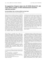
Co-localization of h-SH3 and NS1
Confocal microscopy was used to monitor the MDCK
cells transiently expressing h-SH3, NS1 (NS11, NS32, or
NS92), and co-expre ssing h-SH3 and NS1 (NS11, NS32,
or NS92). We found that h-SH3 mainly presented dif-
fused distribution within the nucleus and lesser within
the cytopl asm (Fig. 1A). And NS1 mainly presented dif-
fused distribution or some dot-like structures within the
nucleus (Fig. 1E). When co-expression, we found that h-
SH3 and NS1 mainly presented diffused distribution or
some dot-like structures within the nucleus (Fig. 1G and
1H), and h-SH3 located at the same position as NS1
which suggested the interaction between h-SH3 and
NS1 (Fig. 1I). Since NS1 from different influenza A
virus strains (NS11, NS32, or NS92) displayed the simi-
lar co-localized pattern with h-SH3 in MDCK cells, only
a representative figure was provided.
Differential suppressive effect of h-SH3 on the replication
kinetics of influenza A viruses
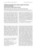
To examine if the replication of influenza A viruses
couldbeaffectedbyh-SH3,weexaminedviraltitersin
MDCK(SH3+) cells (MDCK cells transfected with SH3-
expressing plasmid) and control MDCK(SH3-) cells
(MDCK cells transfect ed with empty vecto r). Cells were
infected with a rela tively low dose of influenza A viruses
(MOI = 0.001) and virus yield was determined by plaque
assayinMDCKcells.Incomparisonwiththecontrol,
the MDCK(SH3+) showed variable degrees of reductio n
in viral titers at different time points (Fig. 2). The maxi-
mal reduction observed was about 8-fold with ST364,
about 6-fold wi th Qa199, about 4-fold with Ph2246, and
about 2-fold with ST 1233 and PR8. T hese findings indi-
cate differential sensitivities of influenza A virus sub-
types/strains to h-SH3.
Differential suppressive effect of h-SH3 on the
phosphorylation status of Akt
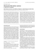
As the h-SH3 had differential suppressive effect on the
replication of influenza A viruses, we further examined
the phosphorylation status of Akt ( Akt-P) at Ser-473,
which is the marker commonly used to monitor the
activation state of PI3K [22], by measuring the ratio of
phosphorylated Akt (Akt-P)/total Akt (Akt-T) in virus-
infected MDCK(SH3+) and MDCK(SH3-) cells. Western
blot analysis showed that, compared to the uninfected
MDCK(SH3-) cells, the Akt-P level increased in PR8-,
ST364-, Ph2246-, or Qa199-infected MDCK(SH3-) cells
but not in ST1233-infected MDCK(SH3-) cells. More-
over, we observed the similar Akt-P and Akt-T levels in
MDCK(SH3+)andMDCK(SH3-)cells.Inaddition,we
found no change of Akt-P/Akt-T ratio in the ST1233-,
Ph2246-, or Qa199-infected MDCK(SH3+) cells, but sig-
nificant reduction of Akt-P/Akt-T ratio in the PR8- and
ST364-infected MDCK(SH3+) cells (Fig. 3). These data
suggested that in the absence of vi ral infection, h-SH3
had no effect on the phosphorylation of Akt and that
the suppressive effect of heterologous SH3 on the phos-
phorylation status of Akt was viral strain-specific.
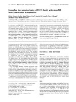
Differential roles of PI3K in the replication of ST364 and
PR8 viruses
Hav ing demonstrat ed the suppressive effe ct of h-SH3 on
the phosphorylation status of Akt in the ST364(H3N2)-
and PR8(H1N1)-infected cells, we further examined the
role of PI3K in the replication of these two viral strains
by treating them with the PI3K inhibitor, LY294002
(10 uM or 50 uM), followed by the plaque formation
assay. Significant reduction in the number of plaques was
observed in the ST364-infected cells, but there was no
obvious change with the PR8-infected cells. Treatment
with different concentrations (10 uM and 50 uM) of
LY294002 showed no obvious difference in replication.
Discussion
The binding of p85b subunit to the influenza A virus
NS1 protein results in the activation of PI3K/Akt path-
way, wh ich in turn can promote the vira l replic ation by
helping virus entry, viral RNA expression, nuclear
export of the RNPs, and preventing premature apoptos is
[19,23]. As NS1 interacts with p85b via direct binding
Figure 1 Confocal imaging of MDCK cells over- expressing SH3
and NS1. pSH3 (SH3-expressing plasmid based on a modified
pcDNA3 vector with the N-terminal Flag) and pNS1 (NS1-expressing
plasmid with the pEGFP-c1 backbone) were transiently transfected
into MDCK cells individually or together. 24 h post-transfection, cells
were stained with anti-Flag antibody and Cy3-labeled secondary
antibody. Detection of Cy3 channel seen as red (A, D, and G), GFP
channel seen as Green (B, E, and H), and Merged these two
channels (C, F, and I).
Zhang et al. Virology Journal 2010, 7:170
/>Page 2 of 6
to SH3, iSH2, or cSH2 domain of p85b, it can be specu-
lated that disturb this interaction by heterologously
expressed SH3, iSH2, or cSH2 may be a potential anti-
influenza strategy. Since iSH2 is also an interactive site
of p85b and p110 [16,17], overexpression of i SH2 may
interfere with the formation of PI3K in normal cells.
Additionally, given that the relative weak ability of cSH2
in mediating the interaction of p85b and NS1 [14 ], only
SH3 was thus chosen as a candidate antiviral agent in
this study. Our aim was to determine whether the SH3-
p85b site in the NS1 could be targeted by heterolo-
gously expressed SH3, thereby inhibiting influenza A
virus replication.
Subcellular localization in this study showed that in
MDCK cells transiently co-expressing NS11, NS32, or
NS92, the h-SH3 was colocalized with NS1 in the
nucleus (Fig. 1), implying the interaction of NS1 and
h-SH3. Combined with the results of Fig. 2, it can be
seen that in addition to its recognized interaction with
endogenous p85b subunit of PI3K via three binding
sites (SH3, iSH2, and cSH2) [14,16,17], NS1 can also
bind to heterologous SH3 and the interaction is strong
enough to result in suppression of viral replication,
though the degree of suppression was apparently differ-
ent among the viral strains.
As Akt-P is the major downstream product of acti-
vated PI3K, the Akt-P/Akt-T ratio was used to com-
pare the levels of virus-induced PI3K activation
affected by h-SH3 in cells infected with different influ-
enza A viruses. Remarkable elevation of Akt-P could
Figure 2 Replication kinetics of influenza A viruses in MDCK(SH3 +) and control MDCK(SH3-) cells. Cells were infected with influenza A
viruses at an MOI of 0.001 and incubated with serum-free medium containing 1 ug/ml TPCK-trypsin. The yield of virus in the culture
supernatant was titrated every 12 h, from 12 h post-infection, by plaque assay in MDCK cells. The time point where maximal reduction was
observed is indicated with asterisk (*).
Zhang et al. Virology Journal 2010, 7:170
/>Page 3 of 6
be seen in PR8-, ST364-, Ph2246-, or Qa199-infected
cells, but not in ST1233-infected cells. In addition, h-
SH3 could reduce the PI3K activation in PR8- or
ST364-infe cted cells, while no alteration was observed
in ST1233-, Ph2246-, or Qa199-infected cells (Fig. 3).
The reason for this is unclear. As iSH2 domain may be
more important than SH3 for NS1-p85b interaction
[16,17], it is reasonable to speculate that for some
influenza A virus strains, h-SH3 could not be abso-
lutely essential for the activation o f PI3K. Thus, even
through h-SH3 can disturb the NS1-p85b interaction,
its effect on the activation of PI3K and the ratio of
Akt-P/Akt-T may be negligible.
It has b een shown that the PI3K/Akt pathway acti-
vated by NS1 is beneficial for influenza A virus infection
[22], and influenza A virus titers are significantly
decreased when PI3K is inhibited [18]. Interestingly, h-
SH3’s effect on PI3K/Akt pathway was not well corre-
lated with the replication of some viruses in this study.
While h-SH3 could inhibit replication of every strain of
influenza A virus to varying degrees, no suppression of
PI3K (or PI3K/Akt pathway) was observed in ST1233-,
Ph2246-, or Qa199-infected MDCK c ells (Fig. 3 ). The
reason may involve the complicated relationship
between NS1 and cellular proteins. More than nine cel-
lular proteins were found directly interacted with NS1.
Among them, CPSF30 (30 k Da subunit of cleavage and
polyadenylation specificity factor) and p85b shared par-
tial overlapped binding sites (SH3 binding motif 1) in
NS1 [24]. Moreover, blocking the binding of endogen-
ous CPSF30 to NS1 protein can strikingly inhibit the
replication of influenza A virus [25]. Therefore, h-SH3
may also disrupt the interaction of NS1 and CPSF30
and then reduce the replication of some influenza A
viruses (such as ST1233, Ph2246, and Qa199) without
the reduction of Akt-P/Akt-T ratio.
Of note, the inhibitory effect of h-SH3 was stronger to
ST364 v irus than PR8 virus. This finding is not consis-
tent with the similar reduction in the Akt-P/Akt-T ratio
in PR8-infected cel ls and ST364-infected cells. One pos-
sible explanation is that influenza A virus varied in their
sensi tivity to PI3K/Akt pathway due to long-time evolu-
tion. And we found that compared to ST364 virus, PR8
virus was less susceptible to the treatment of LY29400 2
(PI3K inhibitor), which suggested that the activated
PI3K/A kt pathway is more important for ST364 re plica-
tion than PR8 replication (Fig. 4).
Conclusions
In conclusion, this study demonstrates that h-SH3 can
inhibit the replication of influenza A viruses (H1N1,
H3N2, and H9N2) at different degrees, and targeting
NS1-p85b interaction (PI3K/Akt pathway) or other
virus-host interaction ma y be an attractive strategy
against the infection of various influenza A virus sub-
types/strains.
Methods
Cell lines, viruses, plasmids, and reagents
Madin-Darby canine kidney (MDCK) cell lines were
routinely cultured in Dulbecco’s modified Eagle’smed-
ium (DMEM) supplemented with 10% fetal calf serum
and antibiotics (100 U penicillin and 100 ug/ml strepto-
mycin) at 37℃ in 5% CO
2
.
Figure 3 Western blot analysis of phosphorylated Akt level in MDCK cells. MDCK cells with or without heterologous SH3 were infected
with PR8, ST1233, ST364, Ph2246, or Qa199 at an MOI of 2 for 8 h. Akt-P (phosphorylated Akt at Serine 473) and Akt-T (total Akt) in cellular
lysate were detected using anti-Akt-P and anti-Akt-T antibodies, respectively, and peroxidase-conjugated secondary antibody.
Zhang et al. Virology Journal 2010, 7:170
/>Page 4 of 6
Influenza A virus strains A/PR/8/3 4(H1N1), A/ST/
1233/2006(H1N1), A/ST/364/2005(H3N2), A/Ph/ST/
2246/2006(H9N2), and A/Qa/ST/199/2006(H9N2),
abbreviated herein to PR8, ST1233, ST364, Ph2246, and
Qa199, respectively, were used in this study.
SH3 domain (9-79aa) of p85b subunit was cloned into
PNF vector (a modified pcDNA3 plasmid with an N-term-
inal Flag tag) at the BamHI and EcoRI sites to generate
pSH3. F ull-length NS1 s equence of H1N1, H3N2, or H 9N2
viruses was cloned into pEGFP-c1 at the BamHI site to
generate pNS11, pNS32, or pNS92 plasmids, respectively.
The constructs were verified by DNA sequencing.
Rabbit monoclonal anti-phospho-Akt (Ser473) anti-
body were purchased from C ell Signaling Technology
(Danvers, MA, USA), Rabbit monoclonal anti-Flag anti-
body from Sigma (St. Louis, MO, USA), Rabbit mono-
clonal anti-Akt antibody, Cy3-labeled goat anti-rabbit
antibody, peroxidase-conjugated g oat anti-rabbit a nti-
body, and LY294002 (PI3K inhibitor) from Beyotime
Biotechnology (Jiangsu, China), and Lipofectamine 2000
from Invitrogen (Carlsbad, CA, USA).
Co-localization analysis by confocal microscopy
MDCKcellculturesontheglasscoverslipsweretrans-
fected with indicated plasmids. 24 h post-transfection,
the cells were washed twice with phosphate-buffered sal-
ine (PBS), fixed for 10 min with 4% paraformaldehyde,
permeabilized with 0.2% Triton X-100 for 7 min, and
incubated for 30 min in PBS containing 3% Bovine
Serum Albumin (BSA). Cell s were subsequently incu-
bated at room temperature with anti-Flag antibody (1/
500) for 2 h and Cy3-labeled secondary antibody (1/300)
for 1 h. Images were captured with the Olympus confo-
cal microscopy.
Transient transfection, viral infection, and determination
of antiviral activity
MDCK cells were seeded onto six-well plates and trans-
fected with pSH3 or PNF empty vector at 60% conflu-
ence as previously described [26]. After 48 h of
transfection, MDCK cells were infected with influenza
A viruses at a multiplicity of infection (MOI) of 0.001
in serum-free DMEM containing 1 ug/ml TPCK-trypsi n
(Worthington, Freehold, NJ, USA) and incubated at 37°
C. An aliquot of the supernatant was harvested every 12
h and virus yield was titrated by plaque assay in MDCK
cells.
Western blot-based phosphorylation assay
MDCK cells transfected with indicated plasmids for 24 h
were infected with different influenza A virus strains at
an MOI of 2. At 8 h post infection the cells were lysed
with Laemmli sample buffer containing 5 mM of NaF
for 5 min in boiling water followed by brief sonication.
Protein concentration was determined using an RC-DC
kit (Bio-Rad, Hercules, CA, USA). Proteins (20 ug/lane)
wereseparatedin10%SDS-PAGEandtransferredonto
a nitrocellulose memb rane. The membrane was blocked
with Tris-buffered saline containing 0.1% Tween 20
(TBST) with 10% non-fat milk for 2 h and incubated
overnight at 4°C with anti-phospho-Akt (Ser473) (1/500)
or anti-Akt (1/700) antibody. The membrane was rinsed
extensively in TBST and incubated for 2 h with peroxi-
dase-conjugated secondary antibody (1/1000). Immuno-
blots were developed using the Enhanced
chemiluminescence (ECL) reagents (Pierce, Rockford, IL,
USA). Quantity One (Bio-Rad) software was used to
analyze images.
Determination of PI3K effect on the replication of ST364
and PR8 viruses
MDCK cells with 100% confluence were pretreated with
10 uM or 50 uM LY294002 (a specific PI3K inhibitor).
After 2 h, MDCK cells were infected with ST364 virus
or PR8 virus, and plaque assay was performed.
Figure 4 Diff erential roles of PI3K i n replication of ST364(A)
and PR8(B) viruses. MDCK cells treated with LY294002 (10 uM) for
2 h and MDCK cells treated with DMSO (control) were infected with
ST364(H3N2) or PR8(H1N1) viruses and were subjected to the
plaque assay.
Zhang et al. Virology Journal 2010, 7:170
/>Page 5 of 6
Acknowledgements
We thank Dr. William Ba-Thein for critical discussion and manuscript editing.
This study was supported by National Natural Science Foundation of China
(30771988, 30972766), Guangdong Natural Science Foundation
(8151503102000022, 9451503102003499), Specialized Research Fund for the
Doctoral Program of Higher Education (20094402110004), Outstanding
Young Scientists Foundation of Guangdong Province Education Department
(LYM08056), State Key Lab of Agriculture Microbiology Open Foundation
(AML200910), Shantou University Medical College Research Foundation, and
211 Project of Guangdong Province (Mechanism and Prevention of
Emerging Infectious Diseases).
Authors’ contributions
Conceived and designed the experiments: KSL, DGZ, WZL, GFW. Performed
the experiments: DGZ, WZL, GFW, CZ, XXC, YXX. Analyzed the data: DGZ,
WZL, KSL, GFW, YS, XXZ. Wrote the paper: DGZ, WZL, KSL. All authors read
and approved the final manuscript.
Competing interests
The authors declare that they have no competing interests.
Received: 12 April 2010 Accepted: 23 July 2010 Published: 23 July 2010
References
1. Nelson MI, Viboud C, Simonsen L, Bennett RT, Griesemer SB, St George K,
Taylor J, Spiro DJ, Sengamalay NA, Ghedin E, Taubenberger JK, Holmes EC:
Multiple reassortment events in the evolutionary history of H1N1
influenza A virus since 1918. PLoS Pathog 2008, 4:e1000012.
2. Peiris M, Yuen KY, Leung CW, Chan KH, Ip PL, Lai RW, Orr WK,
Shortridge KF: Human infection with influenza H9N2. Lancet 1999,
354:916-917.
3. Garcia-Sastre A: Inhibition of interferon-mediated antiviral responses by
influenza A viruses and other negative-strand RNA viruses. Virology 2001,
279:375-384.
4. Garcia-Sastre A, Egorov A, Matassov D, Brandt S, Levy DE, Durbin JE,
Palese P, Muster T: Influenza A virus lacking the NS1 gene replicates in
interferon-deficient systems. Virology 1998, 252:324-330.
5. Krug RM, Yuan W, Noah DL, Latham AG: Intracellular warfare between
human influenza viruses and human cells: the roles of the viral NS1
protein. Virology 2003, 309:181-189.
6. Hayman A, Comely S, Lackenby A, Murphy S, McCauley J, Goodbourn S,
Barclay W: Variation in the ability of human influenza A viruses to induce
and inhibit the IFN-beta pathway. Virology 2006, 347:52-64.
7. Hale BG, Jackson D, Chen YH, Lamb RA, Randall RE: Influenza A virus NS1
protein binds p85beta and activates phosphatidylinositol-3-kinase
signaling. Proc Natl Acad Sci USA 2006, 103:14194-14199.
8. Brazil DP, Yang ZZ, Hemmings BA: Advances in protein kinase B
signalling: AKTion on multiple fronts. Trends Biochem Sci 2004, 29:233-242.
9. Cantley LC: The phosphoinositide 3-kinase pathway. Science 2002,
296:1655-1657.
10. Cooray S: The pivotal role of phosphatidylinositol 3-kinase-Akt signal
transduction in virus survival. J Gen Virol 2004, 85:1065-1076.
11. J Yu, Zhang Y, McIlroy J, Rordorf-Nikolic T, Orr GA, Backer JM: Regulation of
the p85/p110 phosphatidylinositol 3’-kinase: stabilization and inhibition
of the p110alpha catalytic subunit by the p85 regulatory subunit. Mol
Cell Biol 1998, 18:1379-1387.
12. Cantrell DA: Phosphoinositide 3-kinase signalling pathways. J Cell Sci
2001, 114:1439-1445.
13. Vanhaesebroeck B, Ali K, Bilancio A, Geering B, Foukas LC: Signalling by
PI3K isoforms: insights from gene-targeted mice. Trends Biochem Sci 2005,
30:194-204.
14. Shin YK, Liu Q, Tikoo SK, Babiuk LA, Zhou Y: Influenza A virus NS1 protein
activates the phosphatidylinositol 3-kinase (PI3K)/Akt pathway by direct
interaction with the p85 subunit of PI3K. J Gen Virol 2007, 88:13-18.
15. Shin YK, Li Y, Liu Q, Anderson DH, Babiuk LA, Zhou Y: SH3 binding motif 1
in influenza A virus NS1 protein is essential for PI3K/Akt signaling
pathway activation. J Virol 2007, 81:12730-12739.
16. Y Li, Anderson DH, Liu Q, Zhou Y: Mechanism of influenza A virus NS1
protein interaction with the p85beta, but not the p85alpha, subunit of
phosphatidylinositol 3-kinase (PI3K) and up-regulation of PI3K activity. J
Biol Chem 2008, 283:23397-23409.
17. Hale BG, Kerry PS, Jackson D, Precious BL, Gray A, Killip MJ, Randall RE,
Russell RJ: Structural insights into phosphoinositide 3-kinase activation
by the influenza A virus NS1 protein. PNAS 2010, 107:1954-1959.
18. Ehrhardt C, Marjuki H, Wolff T, Nurnberg B, Planz O, Pleschka S, Ludwig S:
Bivalent role of the phosphatidylinositol-3-kinase (PI3K) during influenza
virus infection and host cell defence. Cell Microbiol 2006, 8:1336-1348.
19. Shin YK, Liu Q, Tikoo SK, Babiuk LA, Zhou Y: Effect of the
phosphatidylinositol 3-kinase/Akt pathway on influenza A virus
propagation. J Gen Virol 2007, 88:942-950.
20. Ehrhardt C, Wolff T, Pleschka S, Planz O, Beermann W, Bode JG,
Schmolke M, Ludwig S: Influenza A virus NS1 protein activates the PI3K/
Akt pathway to mediate antiapoptotic signaling responses. J Virol 2007,
81:3058-3067.
21. Zhirnov OP, Klenk HD: Control of apoptosis in influenza virus-infected
cells by up-regulation of Akt and p53 signaling. Apoptosis 2007,
12:1419-1432.
22. Ehrhardt C, Ludwig S: A new player in a deadly game: influenza viruses
and the PI3K/Akt signalling pathway. Cell Microbiol 2009, 11:863-871.
23. Franke TF, Hornik CP, Segev L, Shostak GA, Sugimoto C: PI3K/Akt and
apoptosis: size matters. Oncogene 2003, 22:8983-8998.
24. Hale BG, Randall RE, Ortin J, Jackson D: The multifunctional NS1 protein of
influenza A viruses. J Gen Virol 2008, 89:2359-2376.
25. Twu KY, Noah DL, Rao P, Kuo RL, Krug RM: The CPSF30 binding site on
the NS1A protein of influenza A virus is a potential antiviral target. J
Virol 2006, 80:3957-3965.
26. Li W, Wang G, Zhang H, Zhang D, Zeng J, Chen X, Xu Y, Li K: Differential
suppressive effect of promyelocytic leukemia protein on the replication
of different subtypes/strains of influenza A virus. Biochem Biophys Res
Commun 2009, 389:84-89.
doi:10.1186/1743-422X-7-170
Cite this article as: Zhang et al.: Heterologous SH3-p85b inhibits
influenza A virus replication. Virology Journal 2010 7:170.
Submit your next manuscript to BioMed Central
and take full advantage of:
• Convenient online submission
• Thorough peer review
• No space constraints or color figure charges
• Immediate publication on acceptance
• Inclusion in PubMed, CAS, Scopus and Google Scholar
• Research which is freely available for redistribution
Submit your manuscript at
www.biomedcentral.com/submit
Zhang et al. Virology Journal 2010, 7:170
/>Page 6 of 6