Báo cáo y học: " Herpes simplex virus UL56 interacts with and regulates the Nedd4-family ubiquitin ligase Itch" pps
Bạn đang xem bản rút gọn của tài liệu. Xem và tải ngay bản đầy đủ của tài liệu tại đây (3.34 MB, 11 trang )
RESEA R C H Open Access
Herpes simplex virus UL56 interacts with and
regulates the Nedd4-family ubiquitin ligase Itch
Yoko Ushijima, Chenhong Luo, Maki Kamakura, Fumi Goshima, Hiroshi Kimura, Yukihiro Nishiyama
*
Abstract
Background: Herpes simplex virus type 2 (HSV-2) is one of many viruses that exploits and modifies the cellular
ubiquitin system. HSV-2 expresses the tegument protein UL56 that has been implicated in cytoplasmic transport
and/or release of virions, and is a putative regulatory protein of Nedd4 ubiquitin ligase . In order to elucidate the
biological function of UL56, this study examined the interaction of UL56 with the Nedd4-family ubiquitin ligase Itch
and its role in the regulation of Itch. Additionally, we assessed the similarity between UL56 and regulatory proteins
of Itch and Nedd4, Nedd4-family-interactins proteins (Ndfip).
Results: UL56 interacted with Itch, independent of additional viral proteins, and mediated more striking
degradation of Itch, compared to Nedd4. Moreover, it was suggested that the lysosome pathway as well as the
proteasome pathway was involved in the degradation of Itch. Other HSV-2 proteins with PY motifs, such as VP5
and VP16, did not mediate the degradation of endogenous Itch. Ndfip1 and Ndfip2 were similar in subcellular
distribution patterns to UL56 and colocalized with UL56 in co-transfected cells.
Conclusions: We believe that this is the first report demonstrating the interaction of a HSV-specific protein and
Itch. Thus, UL56 could function as a regulatory protein of Itch. Th e mechanism, function and significance of
regulating Itch in HSV-2 infection remain unclear and warrant further investig ation.
Background
Viruses act as intracellular parasites, dep ending heavily
on functions provided by their host cells, and have
evolved diverse strategies to exploi t the biology and bio-
chemistry of hosts for their benefit [1]. The ubiquitin
system is one of the mechanisms exploited by many
viruses; it is involved in viral assembly and release, viral
transcriptional regulation, viral immune invasion, and
the suppress ion of apoptosis [2,3]. The ubiqui tin system
is a key regulatory mechanism fo r a diversity of cellular
processes including protein turnover, protein sorting
and trafficking, signal transduction, and cell-cycle con-
trol [4]. Ubiquitination is executed by a hierarchical cas-
cade of en zymes [5]. E3 ubiquitin ligases act as major
specificity determinants of the ubiquitin system by facili-
tating the transfer of ubiquitin to lysine residues of the
target proteins. The human genome encodes more than
600 putative E3 ligases [6], which generate the diversity
in the ubiquitin system. E3 ligases are classified into two
main groups: really in teresting novel genes (RING) and
homologous to E6AP carboxyl terminus (HECT) pro-
teins. The neuronal precursor cell-expressed develop-
mentally down-regulated 4 (Nedd4) family, comprised of
nine members, is one of the main HECT E3 protein
families.
Viruses encode their own E3 ligases, de-ubiquitinating
enzymes (DUBs) and adaptor/regulatory proteins to
modify the host ’ s ubiquitin system [2,3]. Herpes simplex
virus (HSV) is a large, enveloped, double-stranded-DNA
virus, which can cause various mild and life-thre atening
diseases, including herpes labialis, genital herpes, kerati-
tis, encephalitis, and neonatal herpes [7]. HSV encodes a
ubiquitin ligase (ICP0) [8,9] and a DUB (UL36) [10]. In
addition, we identified that the HSV type 2 (HSV-2)
tegument protein UL56 is a putative regulatory protein
of Nedd4 E3 ligase [11], specifically involved in protein
stability and subcellular localization. UL56 induces phos-
phorylation of Nedd4 and promotes the proteasome-
mediated degradation by increasing ubiquitination of
Nedd4, however UL56 itself is not ubiquitinated [11].
* Correspondence:
Department of Virology, Nagoya University Graduate School of Medicine, 65
Tsurumai-cho, Showa-ku, Nagoya 466-8550, Japan
Ushijima et al. Virology Journal 2010, 7:179
/>© 2010 Ushi jima et al; licensee BioMed Central Ltd. This is an Open Access article distributed under the terms of the Creative
Commons Attribution License ( which permits unrestricted use, distribution, and
reproduction in any medium, provided the original work is properly cited.
UL56 relocates Nedd4 primarily to the trans-Golgi net-
work (TGN) and partially to endosomes [12].
Approximately half of the 74 gene s encoded by HSV
are accessory genes that are not essential for viral repli-
cation in cell-culture system [7,13,14]. UL56 gene is an
accessory gene encoded by most members of the Alpha-
herpesvirinae family (References are listed in [12]).
Interestingly, UL56-deficient HSV-1 is substantially less
neuroinvasive in vivo [15,16], although little is known
about the molecular mechanisms of the attenuation.
Previously, we ha ve shown that UL56 deficiency reduces
the titer of extracellular HSV-2 [12]. These data suggest
that UL56 facilitates the cytoplasmic transport of virions
from the TGN to the plasma membrane and/or the
release of virions. In addition, we found that UL56 inter-
acts with two other proteins: KIF1A [17], the neuron-
specific kinesin; and HSV-2 UL11 [18], a tegument
protein that has dynamic membrane-trafficking proper-
ties [19] and plays a role in the envelopment and egress
of viral nucleocapsids [20]. These interactions also sup-
port the view that UL56 is involved in transports of
vesicles and virions, however the precise roles and func-
tions of UL56 remain elusive.
UL56 is a 235 amino acid (aa), carboxyl-terminal
anchored, type II membrane protein that is predicted to
be inserted into the viral envelope so that the amino-
terminal domain is located in the virion tegument [21].
In this topology, UL56 is predicted to have a 216 aa
cytoplasmic domain containing three PPXY (PY) motifs,
which are important for its interaction with Nedd4 E3
ligase (Fig. 1A).
In a previous study, Itch, a Nedd4-family ligase, was
identified as a UL56-interacting protein by a yeast t wo-
hybrid screen [11]. Itch is widely expressed in mamma-
lian tissues, and Itch-deficient mice develop a systemic
and progressive autoimmune disease that proves lethal
beginning at 6 months of age [22]. Itch is composed of
862 aa w ith a domain architecture similar to ot her
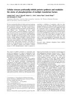
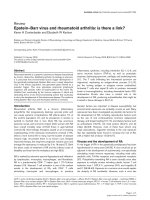
Figure 1 HSV infection causes a marked decrease of Itch in the presence of UL56. (A) Schematic representation of Itch and UL56. Itch (862
aa) contains a Ca
2+
/lipid binding C2 domain, four WW domains that interact with PY motifs, and a catalytic HECT domain. HSV UL56 (HSV-1, 234
aa; HSV-2, 235 aa) contains three PY motifs and a predicted transmembrane domain (TMD). (B) Infection with wild-type (HSV-1; F, HSV-2; 186)
and various mutant HSV (HSV-1; US3-deletion mutant R7041, UL13 deletion mutant R7356, g
1
34.5-deletion mutant R3616, HSV-2; US3-deletion
mutant L1BR1, UL56-reverted virus ΔUL56Zrev), but not infection with UL56-deficient HSV (HSV-1; HF10, HSV-2; ΔUL56Z), caused a marked
decrease of Itch. Vero cells were mock-infected or infected with wild-type or mutant viruses and harvested at 24 hpi. Nedd4 changed only in
cells infected with HSV-2 viruses except ΔUL56Z. WWP2, another Nedd4-family ubiquitin ligase, showed no remarkable change. (C-D) Itch
decreases as HSV-2 infection proceeds in Vero, HEp-2 (C), and HaCaT (C, D) cells. Wild-type (186) and ΔUL56Zrev infection caused a marked
decrease of Itch. In ΔUL56Z-infected cells, Itch was maintained at almost constant levels for up to 12 hpi. (D) VP5 and VP16 were detected
similarly in cells infected with all three viruses. a-tubulin or b-actin were used as loading controls.
Ushijima et al. Virology Journal 2010, 7:179
/>Page 2 of 11
Nedd4-family ligases: an amino-terminal C2 domain;
four protein-protein interacting WW domains, which
most commonly recognize PY motifs of binding pro-
teins; and a carboxyl terminal catalytic HECT domain
(Fig. 1A). Itch targets numerous proteins and has been
implicated in signal transduction, endocytosis, differen-
tiation, and transcription [23,24].
The catalytic activities of Nedd4-family ligases are in
part regulated by some PY-motif containing membrane
proteins such as Nedd4-family-interacting protein-1
(Ndfip1), -2 (Ndfip2), and Nedd4-b inding partner 1
(N4BP1) [ 25], although the mechanisms regulating the
catalytic activity of Nedd4-family ligases have not been
clearly defined. Ndfip proteins function as regulatory
proteins of multiple Nedd4-family ligases, including Itch
and Nedd4, by recruiting ligases to substrates and con-
trolling ligase activity [26].
In this study, to elucidate the biological function of
UL56 we studied the kinetics of Itch expression in HSV-
2-infected cells, and also assessed the similarity between
UL56 and Ndfip proteins.
Methods
Cells and viruses
Vero cells (African green monkey kidney cells) and
HEp2 cells (human laryngeal carcinoma cell line) were
obtained and maintained as previously described [12].
HaCaT cells (human keratinocyte cell line) [27] were
kindly provided by Dr. Norbert E Fusenig (German Can-
cer Research Center, Heidelberg, Germany). HaCaT cells
were maintained in Dulbecco’s modified Eagle’s medium
supplemented with 10% fetal calf serum, 100 U/ml peni-
cillin and 100 μg/ml streptomycin. Cell lines constitu-
tively expressing GFP-UL56 (Vero-GFP-UL56) or GFP
(Vero-GFP) were constructed as previously described
[28]. Briefly, Vero cells were transfected with pEGFP-
UL56 or pEGFP-N3 (Clontech, Mountain View, CA)
and selected with G418 (SIGMA, St. Louis, MO). The
expression of GFP-UL56 or GFP was verified with Wes-
tern blot analysis and Immunofluorescence confocal
microscopy . Vero-GFP-UL56 and Vero-GFP were main-
tained in Eagle’s minimum essential medium (MEM)
supplemented with 8% calf serum (CS), 100 U/ml peni-
cillin, 100 μg/ml streptomycin, and 350 μg/ml G418.
The wild-type HSV-2 strain (186) was used as the pro-
totype strain in this study. The generation of the UL56-
deletion mutant virus (ΔUL56Z) [18], the UL56-reverted
virus based on ΔUL56Z (ΔUL56Zrev) [11], and the
US3-deletion mutant virus (L1BR1) [29] was previo usly
described in detail. The HSV-1 wild type strain F, the
US3-deletion mutant (R7041), the UL13-deletion mutant
(R7356), and the g
1
34.5- deletion mutant (R3616) viruses
were generously provided by Dr . Bernard Roizman.
HSV-1 mutant HF10 [30], lacking the functional
expression of UL43, UL49.5, UL55 and UL56, and
latency-associated transcripts [31] was also used. Viruses
were propagated and the titers of viral stocks were
determined as previously described [12].
Antibodies and reagents
The following antibodies were used: polyclonal anti-
WWP2 (Abcam, Cambridge, UK), anti-Nedd4 (Milli-
pore, Billerica, MA), anti-GFP (MBL, Nagoya, Japan)
and anti-c-Myc (Santa Cruz Biotechnology, Santa Cruz,
CA); monoclonal anti-VP5 ( Abcam), anti-Itch (BD
Transduction Laboratories, Franklin Lakes, NJ), anti-b-
actin, anti-a-tubulin (SIGMA), and anti-c-Myc (Santa
Cruz Biotec hnology); horseradish peroxi dase-conjugated
goat anti-rabbit and anti- mouse IgG (Invitrogen), and
Alexa Fluor 488-conjugated goat anti-rabbit and 594-
conjugated goat anti-mouse IgG (Invitrogen). Protein G
affinity-purified normal mouse IgG was purchased from
Millipore. Polyclonal anti-UL56 [21] and anti-VP16 [32]
antisera were described previously. Reagents were pur-
chased from the following suppliers: cycloheximide
(CHX) and chloroquine (CQ), SIGMA; MG132, BIO-
MOL International (Plymouth Meeting, PA).
Expression vectors
Itch (GenBank: NM_031483), Ndfip1 (GenBank:
NM_030571) and Ndfip2 (GenBank: NM_019080)
cDNA were obtained from HEp-2 cells and cloned into
plasmids to generate pcDNA- Itch, pMyc-Itch, pNdfip1-
EGFP, and pNdfip2-EGFP. Total RNA was extracted
using ISOGEN (NIPPON GENE, Tokyo, Japan), and
then first-strand cDNA was synthes ized by po lymerase
chain reaction with reverse transcriptio n (RT-PCR)
using Transcriptor First Strand cDNA synthesis Kit
(Roche Applied Science, Ma nnheim, Germany) in accor-
dance with the manufacturer’s instructions. Fragments
of Itch, Ndfip1 or Ndfip2 cDNAs were amplified by PCR
withKODFX(TOYOBO,Osaka,Japan)andcloned
into pcDNA3.1(+) (Invitrogen), pCMV-Myc, or pEGFP-
N3 (Clontech). To generate pMyc-ICP0, HSV-2 ICP0
cDNA (GenBank: NC_001798) was reverse transcribed
andamplifiedfromtotalRNAfrom186-infectedVero
cells (multiplicity of infection (MOI) 3 PFU/ml, 6 h
post-infection) using the same procedures described
above, and then cloned into pCMV-Myc. To generate
pcDNA-VP5, the HSV-2 VP5 ORF (GenBank:
NC_001798) was amplified from HSV D NA which was
extracted from 186-infected Vero cells using QIAamp
DNA Blood Mini Kit (QIAgen, Hilden, Germany), and
cloned into pcDNA-3.1(+). pcDNA-UL56 [21] and
pcDNA-UL48 (pcDNA-VP16) [32] were generated as
described previously. The UL56 ORF was amplified by
PCR from pcDNA-UL56 and cloned into pEGFP-N3 to
generate pEGFP-UL56.
Ushijima et al. Virology Journal 2010, 7:179
/>Page 3 of 11
Transfection and infection
Cells plated in 35-mm dishes were transfected or
infected as previously described [12]. Bri efly, cells were
transfected with 1 μg of each plasmid using Lipofecta-
mine 2000 (Invitrogen), and in some experim ents,
further infected with HSV-2 at 48 h post-transfection.
Infections were routinely performed at an MOI of 3
PFU/cell (except where otherwise indicated).
Immunoblot assay
Cell lysates w ere extracted and analyzed as previously
described [11].
Co-immunoprecipitation assay
In assays on infected cells, Vero cells were pre-cultured
with CQ (100 μM) for 12 h, then infected with 186 and
harvested at 9 h post-infection. In assays on Vero-GFP-
UL56 or Vero-GFP, cells were cultured with CQ (100
μM) for 24 h and harvested. Harvested cells were ly sed
and clarified by centrifugation [11]. The lysates were
incubated for 1 h at 4°C with the Protein G Dynabeads
(Invitrogen) which were pre-incubated with anti-Itch
antibody or normal mouse IgG according to the manu-
facturer’s instructions. After washing with lysis buffer
(10 mM Tris-HCl, pH 7.4, 150 mM NaC l, 1% Nonident
P-40, 1 mM EDTA, 10 mM NaF, Protease Inhibitor
Cocktail [SIGMA]), the immunoprecipitated proteins
were eluted in 2x SDS sample buffer and subjected to
Western blot analysis.
Immunofluorescence confocal microscopy
Indirect immunofluorescence confocal microscopy was
performed as previously described [12]. In brief, cells
grown on cover slips were fixed in 4% paraformaldehyde
for 15 min, permeabilized with 0.1% Triton X-100 for 5
min, and incubated for 1 h at room temperature
sequentially with 20% normal g oat serum (DAKO,
Glostrup, Denmark), primary and secondary antibodies.
Confocal images were captured using the Zeiss LSM 510
system (Carl Zeiss, Oberkochen, Germany).
RNA interference
siRNAs for human Itch (ON-TARGETplus SMARTpool
L-007196-00, siItch), and a non-targeting control pool
of siRNA (ON-TARGETplu s Non-targeting Pool D-
001810-10, siCont) were obtained from Dharmacon
(Lafayette, CO). Vero cells were transfected using Lipo-
fectamine RNAiMAX (Invitrogen) according to the
manufacturer’s instructions. At 48 h post-transfection,
cells were used for further experiments.
Viral replication kinetics assay
Single-step and multi-step growth experiments were
performed using Vero cells as previ ously described [11].
Cells were t reated with siRNA for 48 h, and subse-
quently infected with the indicated viruses at an MOI of
3 (single-step) or 0.003 (multiple-step) PFU/cell.
Results
HSV infection causes a marked decrease of Itch in the
presence of UL56
Initially, we investigated the kinetics of Itch expression
after HSV infection. Itch was markedly decreased in
Vero cells infected with wild-type and various mutant
HSVthatexpressedUL56,butnotUL56-deficient
mutants (HSV-1, HF10; HSV-2, ΔUL56Z) at 24 h
post-infection (Fig. 1B). Itch showed no decrease in
cells infected with HF10, and remained at detectable
level in cells infected with ΔUL56Z. Nedd4 was
detected in two forms with different electrophoretic
mobilities and had decreased levels in cells infected
with HSV-2 viruses except ΔUL56Z, as previously
reported [11]. In contrast, WWP2, another Nedd4-
family ubiquitin ligase, which has be en also identified
as a UL56-intera cting protein by a yeast two-hybrid
screen[11],showednoremarkablechangeafterviral
infection. In time-course experiments, Itch decreased
markedly as the infection with wild-type (186) or
UL56-reverted (ΔUL56Zrev) viruses in multiple cell
lines: Vero, HEp-2 and HaCaT cells (Fig. 1C). In
ΔUL56Z-infected cells, Itch was maintained at almost
constant levels for up to 12 h post-infection whereas
slightly decreased at 24 h post-infection (Fig. 1C, D).
Thus, HSV infection causes a decrease of Itch, and
HSV-1 and -2 UL56 have a prominent role in the pro-
cess. In addition, the small decrease of Itch in cells
infected with ΔUL56Z suggests the presence of addi-
tional viral factors responsible for the decrease of Itch
in the course of HSV-2 infection.
UL56 causes the decrease of intrinsic Itch in the absence
of other viral proteins
We next investigated whether UL56 causes the decrease
of Itch without other HSV-2 proteins using stable UL56
transfected cells (Vero-GFP-UL56). Vero-GFP-UL56 was
similar in morphology and growth properties to GFP-
expressing Vero cells (Vero-GFP) that were used as a
control (data not shown). Itch was markedly decreased
in Vero-GFP-UL56 compared to control cells (Fig. 2A).
In contrast, WWP2 and Nedd4 showed relatively no
decrease in Vero-GFP-UL56. These data suggest that
UL56 specifically de creases Itch in the absence of any
other viral proteins. We have previously reported that
transient overexpression of UL56 caused the decrease of
exogenous Nedd4 but had no apparent effect on endo-
genous Nedd4 [11]. This discrep ancy may be due to the
relatively low transfection efficiency (approx. 20%) in the
previous study.
Ushijima et al. Virology Journal 2010, 7:179
/>Page 4 of 11
We further explored other possible HSV-2 proteins
involved in the decrease of It ch. HSV-1 and -2 genomes
encode four proteins with PY motif(s): VP5, a major
capsid protein, with one motif; VP16, a tegument pro-
tein which activates the transcription of immediately
early gene, with one motif; ICP0, a promiscuous transac-
tivator, with one motif [7]; and UL56, a tegument pro-
tein wit h three motifs. None of the three viral proteins
with one PY motif (VP5, VP16, ICP0) decreased endo-
genous Itch (data for VP5 is shown in Fig 2B), whereas
VP5 and VP16 caused the decrease of overexpressed
Itch in co-expressing cells (Fig. 2C). These results indi-
cated that VP5 and VP16 modulate the protein le vel of
overexpressed Itch in the absence of other viral proteins.
On the other hand, ove rexpression of Itch did not
affect the lev el of VP5, VP16, or ICP0 in co-expressing
cells (Fig. 2D). In infection experiments, both VP5 and
VP16 were detected at the same level in cells infected
with 186 and th ose infected with ΔUL56Z despite the
substantially different level of Itch expression (Fig. 1D).
These results suggest that VP5 and VP16 induce the
decrease of overexpressed Itch although Itch has no
apparent effect on VP5, VP16, or ICP0 levels.
UL56 promotes lysosome- and proteasome-mediated
degradation of Itch
To clarify the m echanism by which the protein levels of
Itch were decreased, we investigated the stability of Itch
in the presence of inhibitors. The treatment of unin-
fected cells with the protein synthesis inhibitor cyclo-
hexemide (CHX, 100 μg/ml) for 24 h did not alter the
level of Itch (Fig. 3A), indicating that Itch is very stable
in nature. In HSV-2-infected cells, the decrease of Itch
was greatly blocked by chloroquine (CQ, 100 μM), a
lysosome inhibitor, and only partially blocked b y
MG132 (10 μM), a proteasome inhibitor (Fig. 3B). Col-
lectively, these results suggest that Itch may be degraded
by the lysosome pathway and in part by the proteasome
pathway in HSV-2-infected cells. The decrease of Itch in
Vero-GFP-UL56 cells was also blocked by CQ and par-
tially by MG132 (Fig. 3C). Therefore, UL56 possibly
promotes the degradation of Itch via the lysosome and
proteasome in HSV-2-infected cells and UL56-expres-
sing cells. In Vero-GFP-UL56 cells, the amount of GFP-
UL56 was increased significantly by the addition of
MG132 and CQ, suggestingthatUL56isdegraded
together with Itch in this case.
HSV-2 UL56 interacts with Itch and changes the
subcellular localization of Itch
We next investigated whether UL56 and Itch interact.
Co-immunoprecipitation assay using anti-Itch antibody
revealed that UL56 was associated with Itch in cells
infected with wild-type HSV-2 (Fig. 4A) and cells stably
expressing UL56 (Fig. 4B). These results indicate that
UL56 interacts with Itch during HSV-2 infection and
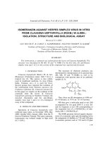
Figure 2 Effects of UL56 and other viral proteins with a PY motif on Itch. (A) Itch is mark edly decreased in cells stably expressing UL56
(Vero-GFP-UL56). Lysates from Vero, Vero-GFP-UL56, or Vero-GFP cells were analyzed for Itch and other Nedd4-family ubiquitin ligases. Itch was
markedly decreased in Vero-GFP-UL56 cells. (B) VP5 did not decrease endogenous Itch. Vero cells were transfected with plasmids encoding VP5
(pcDNA-VP5) or control plasmids (pcDNA3.1(+)). The levels of Itch did not change in cells transfected with pcDNA-VP5. (C) VP5 and VP16, but
not ICP0, caused the decrease of exogenous Itch. Vero cells were co-transfected with plasmids encoding Itch (pcDNA-Itch) and plasmids
encoding a viral protein (pMyc-ICP0, pcDNA-VP5, or pcDNA-VP16) or control plasmids (pCMV-Myc or pcDNA3.1(+)). The levels of Itch decreased
in cells transfected with pcDNA-VP5 or pcDNA-VP16. (D) Overexpression of Itch has no effect on the protein levels of VP5, VP16, or ICP0. Vero
cells were co-transfected with plasmids encoding a viral protein (pMyc-ICP0, pcDNA-VP5 or pcDNA-VP16) and either pcDNA-Itch or control
plasmids (pcDNA-3.1(+)). The levels of viral proteins did not change with the overexpression of Itch. b-actin was used as a loading control.
Ushijima et al. Virology Journal 2010, 7:179
/>Page 5 of 11
that no other viral proteins are required for the interac-
tion. In infected cells, VP16 was also detected at a much
lower level whereas VP5 was not detected (Fig. 4A).
Confocal immunofluorescence analysis revealed the
colocalization of UL56 and Itch in H SV-2-infected cells
and transiently UL56-expressing cells. M yc-tagged Itch
(Myc-Itch) was mainly distributed throughout the cyto-
plasm with partial vesicular distribution in uninfected
cells (Fig. 4C, top left panel). The Myc-Itch showed
reduced signal intensity and altered subcellular distribu-
tion after 6 hpi, concomitant with UL56 detection (Fig.
4C, middle and bottom panels). Myc-Itch accumulated
in the perinu clear region with punctate distribution and
colocalized wit h UL56 at 6 hpi and 9 hpi. These results
support the view that Itch interacts with UL56, and
decreases during HSV-2 infection. Co-expression with
UL56 also reduced signal intensity and altered the distri-
bution of Myc-Itch (Fig. 4D); Myc-Itch showed the clear
vesicular distribution and colocalized with UL56. These
results highlight that UL56 interacts directly with Itch
and causes Itch to decrease even without other viral
proteins.
siRNA knockdown of Itch has no apparent effect on the
growth of either wild-type or UL56-deficient HSV-2
ToassesstheroleofItchinHSV-2replication,the
effect of Itch knockdown on the efficiency of viral
growth in Vero cells was measured. Itch protein levels
were efficiently and specifically down-regulated by Itch
siRNA (siItch) (Fig. 5A). Wild-type viruses sh owed simi-
lar growth kinetics in siCont-treated cells and s iItch-
treated cells both in multiple- (MOI 0.003, Fig. 5B) and
single- (MOI 3 , data not shown) growth experiments.
ΔUL56Z also showed similar growth kinetics in siCont-
and siItch-treated cells (Fig.5B).Additionally,amajor
capsid protein V P5 and tegument proteins, VP16 and
UL56, showed similar expression patterns in siCont-
and siItch-treated cells (Fig. 5C). Thus, the knockdown
of Itch did not influence the replication of wild-type and
UL56-deficient HSV-2 in Vero cells.
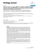
Figure 3 Lysosome inhibitor and proteasome inhibitor block the decrease of Itch. (A) Itch expression is stable in the absence of UL56.
Vero cells were cultured for 24 h with or without cycloheximide (CHX, 100 μg/ml) and the lysates were analyzed for Itch expression. Cyclin D3
was used as a control. (B) Vero cells were mock-treated or treated for 12 h with either MG132 (10 μM) or chloroquine (CQ, 100 μM) and infected
with wild-type HSV-2. Itch was detected in both cells treated with MG132 and those with CQ at 12 hpi, but only in those with CQ at 24 hpi. (C)
The decrease of Itch is blocked by a lysosome inhibitor and partially by a proteasome inhibitor in cells stably expressing UL56. Vero, Vero-GFP-
UL56, or Vero-GFP cells were mock-treated or treated with either MG132 (10 μM) or CQ (100 μM) for 24 h. b-actin was used as a loading control.
Ushijima et al. Virology Journal 2010, 7:179
/>Page 6 of 11
HSV-2 UL56 colocalizes with Ndfip proteins
Ndfip1 and Ndfip2 are small membrane proteins with
multiple PY motifs (Fig. 6A) that regulate Nedd4 family
ligases including Itch and Nedd4 by directly controlling
ligase activity and relocating ligases [26]. To provide
evidence of similarity between UL56 and Ndfip proteins,
we investigated whether UL56 and Ndfip proteins colo-
calize in H SV-2-infect ed cells a nd cells co-expressing
UL56andaNdfipprotein.EGFP-tagged-Ndfip1
(Ndfip1-EGFP) (Fig. 6B) and -Ndfip2 (Ndfip2-EGFP)
(Fig. 6C) showed vesicular distribution with accumula-
tion to the perinuclear space, consistent with results of
other studies [33,34]. When co-expressed with UL56,
Ndfip1-EGFP and Ndifip2-EGFP did not change their
subcellular distribution and they largely colocalized with
UL56. In HSV-2 infected cells, Ndfip proteins altered
their distribution and formed perinuclear clumps
(Fig. 6D, E). UL56 showed a similar distribution pattern
and accumulated in the perinuclear space, but only par-
tially colocalized with Ndfip proteins.
Discussion
This study demonstrates that HSV-2 UL56 interacts
with t he Nedd4-family ubiquitin ligase Itch, and more-
over, targets Itch for degradation primarily via the
lysosome pathway and partially via the ubiquitin-protea-
some pathway in the course of HSV-2 infection. UL56
interacted with Itch and induced the degradation of Itch
independent of any other viral proteins. To the best of
our knowledge, this is the first report demonstrating
that an HSV protein interacts with Itch. In addition,
UL56 and Ndfip proteins, regulatory proteins of Nedd4
and Itch, showed similar subcellular distribution and
colocalized.
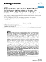
Figure 4 UL56 interacts with Itch and changes the subcellular localization of Itch. Co-immunoprecipitation assay on HSV-2-infected cells
(A) or stably UL56-expressing cells (B). (A)Vero cells were treated with chloroquine (CQ) for 12 h and subsequently mock-infected or infected
with wild-type HSV-2 (186). Whole cell lysates (WCL) were immunoprecipitated (IP) with an anti-Itch antibody at 9 hpi. (B) WCL from Vero-GFP or
Vero-GFP-UL56 cells treated with CQ for 24 h and immunoprecipitated with an anti-Itch antibody. UL56 was detected in the Itch-
immunoprecipitates in HSV-2-infected cells (A) and stably UL56-expressing cells (B). b-actin was used as a loading control. Confocal
immunofluorescence analysis of the subcellular localizations of Myc-Itch and UL56 in HSV-2-infected cells (C) or co-expressing cells (D). (C) HEp-2
cells were transfected with plasmids encoding Myc-Itch and subsequently infected with wild-type HSV-2. The Myc-Itch (red) showed the altered
subcellular distribution with the reduced signal intensity after 6 hpi, when UL56 (green) became detectable. Myc-Itch colocalized with UL56 in
the vesicular pattern. (D) HEp-2 cells were transfected with plasmids encoding UL56 (pcDNA-UL56) and/or pCMV-Myc-Itch. In co-expressing cells
(bottom panels), Myc-Itch changed its distribution and colocalized with UL56 in the vesicular pattern. The Myc signal was reduced in co-
expressing cells. Scale bars, 10 μm.
Ushijima et al. Virology Journal 2010, 7:179
/>Page 7 of 11
Itch differed from Nedd4, another UL56-interacting
E3 ligase, in the following aspects: Itch decreased in
both HSV-1-infected cells and HSV-2-infected cells,
whereas Nedd4 decreased only in HSV-2-infected cells;
endogenous Itch was degraded much more efficiently
than Nedd4 in UL56-expressing cells; Itch was degraded
primarily by the lysosome pathway wherea s Nedd4 was
degraded by the protea some pathway [11]; and Itch was
co-immunoprecipitated with a major tegument protein
VP16 whereas Nedd4 was not [11]. UL56 cause d more
strikingchangesinItchthaninNedd4asawhole.The
distinct effects of UL56 on Itch and Nedd4 support the
view that each of Nedd4-family ligases is regulated in
the specific way in spite of sharing many common prop-
erties [35]. HSV-1 UL56 (234 aa) and HSV-2 UL56 (235
aa) share three PY motifs and one C-terminal trans-
membrane domain, and exhibit 62.6% identity on the
amino acid level. UL56 itself and/or other viral proteins
could account for the different effect of HSV-1 and
HSV-2 on Nedd4 and Itch.
To our knowledge, UL56 is the first example of a pro-
tein which induces Itch to degrade except Itch i tself.
Itch is regulated by multiple mechanisms: phosphoryla-
tion mediated by Jun amino-terminal kinase [36] and
Figure 5 siRNA knockdown of Itch has no apparent effect on the viral growth. Vero cells were mock-transfected (RNA [-]) or t ransfected
with either negative control siRNAs (siCont) or siRNAs specific to Itch (siItch) for 48 h (A), and subsequently infected with indicated viruses (B, C).
(A) siItch efficiently and specifically down-regulates protein levels of Itch, whereas levels of Nedd4 and b-actin were constant. (B) A multi-step
growth curve in siRNA-treated cells (MOI 0.003 PFU/ml). Both wild-type (186) and ΔUL56Z viruses showed similar growth kinetics in siCont-
treated cells and siItch-treated cells. (C) Expression of Itch and viral proteins with PY motifs (VP5, VP16, and UL56) by immunoblot. There was no
difference in viral protein level between siCont- and siItch-treated cells. b-actin was used as a loading control.
Ushijima et al. Virology Journal 2010, 7:179
/>Page 8 of 11
Fyn [37]; conformational change and relocation induced
by adaptor/regulatory proteins (Ndfip-1 [38] and -2 [26],
and N4BP1 [25]); and modulation o f the level of Itch
ubiquitination mediated by Itch itself [36], FAM/USP9X
[39], and Akt1 [40]. Little has been done to clarify how
Itch degradation is controlled because of its high s tabi-
lity, and moreover, the limited results obtained so far
are contr oversial. One report showed that autoubiquiti-
nated Itch is degraded by the proteasome [39], however
others showed Itch is very stable even in polyubiquiti-
nated state, and the level of ubiquitination has no dis-
cernible impact on Itch stability [40,41]. UL56 originally
induced Itch to degrade via primarily the lysosome and
partially the proteasome pathways. From the view point
of the degradation of ubiquitinated proteins, this result
concurs with the report which showed ubiquitinated
proteins can undergo lysosomal degradation [42]. In
addition, a proteasome inhibitor blocked the degradation
at12hpibutnotat24hpi,whereasalysosomeinhibi-
tor blocked both at 12 and 24 hpi. These data suggest
that the degradation pathway c ould change during the
course of HSV-2 infection.
Of the three viral proteins with PY motif(s) other than
UL56, only VP16 was detected in the Itch-immunopreci-
pitates, albeit at a much lower level than UL56. VP16 is
a te gument protein which activates viral transcription of
immediate early genes after infection and plays an
essential role during assembly in the late ph ase of infec-
tion [43]. VP16 interacts with multiple envelo pe- and
tegument- proteins including UL36 (VP1/2), and
appears to function in linking the outer tegument/glyco-
protein and capsid/inner tegument complexes [44,45].
UL36 is a large inner tegument protein with the deubi-
quitinating activity [10], and required for the addition of
VP16 to the viral capsid [46]. It is noteworthy that VP16
associates with Itch, a cellular E3 ligase, and also with
UL36, a viral DUB.
We explored the possibility that additional viral pro-
teins mediated the decrease of Itch, since there was a
small decrease in cells infected with UL56-deficient
HSV-2. Two of three HSV-2 proteins with one PY motif
other than UL56, VP5 and VP16 caused the decrease of
overexpressed Itch i n co-expressing cells. In contrast,
ICP0, a viral component with promiscuous transactivity
and ubiquiti n ligase activity, did not influence Itch
expression. These re sults indicate that only specific viral
proteins with PY motifs are capable of inducing Itch
degradation. In addition, transient expressions of VP5
and VP16 caused no decrease of endogenous Itch,
whereas stable expression of UL56 caused the striking
decrease of endogenous Itch. These results support the
notion that UL56 plays a prime role, and VP5 and VP16
can play secondary roles in the decrease of Itch during
HSV-2 infection. The reason why UL56-deficient HSV-1
did not cause the decrease of Itch remains unknown.
The experiments with inhibitors provided insights into
the mechanism of UL56 degradation. Treatment with a
lysosome inhibitor caused the increase of UL56 in cells
stably expressing UL56. This result suggests that UL56 is
Figure 6 UL56 co-localizes with Ndfip1 and Ndfip2.(A)
Schematic representation of UL56 and Ndfip proteins. HSV-2 UL56
(235 aa) contains three PY motifs and one predicted
transmembrane domain (TMD). Ndfip1 (221 aa) and Ndfip2 (336 aa)
contain two and three PY motifs respectively, and three TMD. (B-C)
HEp-2 cells were transfected with plasmids encoding either Ndfip1-
EGFP (pNdfip1-EGFP) (B) or Ndfip2-EGFP (pNdfip2-EGFP) (C) alone
(left panels), or in combination with plasmids encoding UL56 (right
panels). UL56 colocalized with Ndfip1-EGFP and Ndfip2-EGFP in
co-expressing cells. (D-E) HEp-2 cells were transfected with pNdfip1-
EGFP (D) or pNdfip2-EGFP (E), and subsequently infected with
wild-type HSV-2. UL56 partially colocalized with Ndfip1-EGFP and
Ndfip2-EGFP in infected cells. Scale bars, 10 μm.
Ushijima et al. Virology Journal 2010, 7:179
/>Page 9 of 11
also degraded via the lysosome pathway in UL56-expres-
sing cells. Interestingly, treatment with a proteasome inhi-
bitor also caused the increase of UL56, although the effect
was minimal, suggesting that t he proteasome pathway is
also involved in UL56 degradation. Given that UL56 is
lysine-free and not ubiquitinated [11], some additional fac-
tors may be involved in the degradation of UL56. In this
study, we used only one lysosome-inhibitor and one-pro-
teasome-inhibitor, and did not analyze ubiquitinated sub-
strates or free ubiquitin. Further investigation is n eeded
concerning the turnover of UL56.
We also investigated whether Itch can change the pro-
tein levels of VP5, VP16, and ICP0 because they contain
a PY motif and also lysine residues, which are targets of
ubiquitination. Contrary to expectations , overexpression
of Itch did not affect the level of VP5, VP16, or ICP0,
and knockdown of Itch did not change the expression
patterns of VP5 and VP16 during the course of infec-
tion. Thus, the levels of these three viral proteins do not
appear to be regulated by Itch.
siRNA knockdown of Itch has no apparent effect on
the growth of either w ild-type or UL56-deficient HSV-2
in Vero cells. Itch is reported to be involved in viral repli-
cation and pathogenicity in Epstein-Barr virus (EBV),
which belongs to Gammaherpesvirinae family, and Molo-
ney murine leukemia virus (MoMLV). Itch interacts with
latent membrane protein (LMP) 2A of EBV and down-
regulates LMP2A activity in B-cell signaling [47,48], and
rescues a release-deficient MoMLV independent of PY
motif of the Gag protein [49]. More investigations are
needed to elucidate how the decrease of Itc h is involved
in the replication and pathogenicity of HSV-2.
UL56 and Ndfip proteins share some common fea-
tures: small membrane proteins (Ndfip1, 221 aa; Ndfip2,
336 aa; and UL56, 234 aa); contain multiple PY motifs
in the cytoplasmic domains (Ndfip1, two; Ndfip2 a nd
UL56, three); interaction with Nedd4 and Itch via PY
motifs; and relocate Nedd4 and Itch for degradation.
This study revealed that UL56 and Ndfip proteins a re
similar in their subcellular localiz ation. In co-expressing
cells, UL56 and Ndfip proteins colocalized to the vesi-
cles. UL56 localized primarily the TGN and early endo-
somes [12], while Ndfip proteins localized to the TGN,
early endosomes, and late endosomes/multi vesicular
bodies [33,34]. The partial colocalization of UL56 and
either Ndfip-1 or -2 in infecte d cells suggests that UL56
and Ndfip proteins behave similarly during the course of
HSV infection. It is interesting that the viral p rotein
UL56 shares so many properties with cellular regulatory
proteins of Itch and Nedd4.
Conclusions
This study demonstrates that HSV-2 UL56 interacts
withaNedd4-familyubiquitin ligase Itch, and
moreover, relocates Itch and induces Itch to degrade
in the course of HSV-2 infection. UL56 caused more
striking changes in Itch than in N edd4 as a whole. I n
addition, UL56 shared multiple common properties
with Ndfip proteins. In light of these results, we pro-
pose that UL56 functions as a regulatory protein of
Itch. The mechan ism, function and significa nce of reg-
ulating Itch in HSV-2 infection remain unclear and
warrant further investigation.
Acknowledgements
We would like to thank Bernard Roizman for HSV-1 F, R7041, R7356, and
R3616; and Norbert E Fusenig for HaCaT cells. We would also like to thank
Akane Ohta, Yoshifumi Muto, and Seiko Iwata for technical suggestions and
discussions, and Hiromi Noma for technical assistance. This study was
supported by grant-in-aid for scientific research on priority areas (18073007
to YN) and grant-in-aid for Japan Society for the Promotion of Science (JSPS)
fellows (20•7388 to YU) from the Ministry of Education, Culture, Sports,
Science and Technology of Japan. YU was supported by Research
Fellowships for Young Scientists from JSPS.
Authors’ contributions
YU and YN designed the research, YU, CL, and MK performed the
experimental work, YU conducted the data analysis and drafted the
manuscript, and FG, HK, and YN participated in the data analysis and review
of the manuscript. All authors read and approved the final manuscript.
Competing interests
The authors declare that they have no competing interests.
Received: 16 March 2010 Accepted: 3 August 2010
Published: 3 August 2010
References
1. Ball L: Virus Replication Strategies. Fields Virology Philadelphia: Lippincott
Williams & WilkinKnipe DM, Howley PM , 5 2007, 1:119-139.
2. Isaacson MK, Ploegh HL: Ubiquitination, ubiquitin-like modifiers, and
deubiquitination in viral infection. Cell Host Microbe 2009, 5:559-570.
3. Randow F, Lehner PJ: Viral avoidance and exploitation of the ubiquitin
system. Nat Cell Biol 2009, 11:527-534.
4. Weissman AM: Themes and variations on ubiquitylation. Nat Rev Mol Cell
Biol 2001, 2:169-178.
5. Pickart CM: Mechanisms underlying ubiquitination. Annu Rev Biochem
2001, 70:503-533.
6. Li W, Bengtson MH, Ulbrich A, Matsuda A, Reddy VA, Orth A, Chanda SK,
Batalov S, Joazeiro CA: Genome-wide and functional annotation of
human E3 ubiquitin ligases identifies MULAN, a mitochondrial E3 that
regulates the organelle’s dynamics and signaling. PLoS One 2008, 3:
e1487.
7. Roizman B, DM Knipe, RJ Whitely: Herpes Simplex Viruses. Fields Virology
Philadelphia: Lippincott Williams & WilkinKnipe DM, Howley PM , 5 2007,
2:2502-2601.
8. Boutell C, Sadis S, Everett RD: Herpes simplex virus type 1 immediate-
early protein ICP0 and is isolated RING finger domain act as ubiquitin E3
ligases in vitro. J Virol 2002, 76:841-850.
9. Van Sant C, Hagglund R, Lopez P, Roizman B: The infected cell protein 0
of herpes simplex virus 1 dynamically interacts with proteasomes, binds
and activates the cdc34 E2 ubiquitin-conjugating enzyme, and
possesses in vitro E3 ubiquitin ligase activity. Proc Natl Acad Sci USA 2001,
98:8815-8820.
10. Kattenhorn LM, Korbel GA, Kessler BM, Spooner E, Ploegh HL: A
deubiquitinating enzyme encoded by HSV-1 belongs to a family of
cysteine proteases that is conserved across the family Herpesviridae. Mol
Cell 2005, 19:547-557.
11. Ushijima Y, Koshizuka T, Goshima F, Kimura H, Nishiyama Y: Herpes simplex
virus type 2 UL56 interacts with the ubiquitin ligase Nedd4 and
increases its ubiquitination. J Virol 2008, 82:5220-5233.
Ushijima et al. Virology Journal 2010, 7:179
/>Page 10 of 11
12. Ushijima Y, Goshima F, Kimura H, Nishiyama Y: Herpes simplex virus type
2 tegument protein UL56 relocalizes ubiquitin ligase Nedd4 and has a
role in transport and/or release of virions. Virol J 2009, 6:168.
13. Dolan A, Jamieson FE, Cunningham C, Barnett BC, McGeoch DJ: The
genome sequence of herpes simplex virus type 2. J Virol 1998,
72:2010-2021.
14. McGeoch DJ, Dalrymple MA, Davison AJ, Dolan A, Frame MC, McNab D,
Perry LJ, Scott JE, Taylor P: The complete DNA sequence of the long
unique region in the genome of herpes simplex virus type 1. J Gen Virol
1988, 69(Pt 7):1531-1574.
15. Berkowitz C, Moyal M, Rosen-Wolff A, Darai G, Becker Y: Herpes simplex
virus type 1 (HSV-1) UL56 gene is involved in viral intraperitoneal
pathogenicity to immunocompetent mice. Arch Virol 1994, 134:73-83.
16. Rosen-Wolff A, Darai G: Identification and mapping of the UL56 gene
transcript of herpes simplex virus type 1. Virus Res 1991, 19:115-126.
17. Koshizuka T, Kawaguchi Y, Nishiyama Y: Herpes simplex virus type 2
membrane protein UL56 associates with the kinesin motor protein
KIF1A. J Gen Virol 2005, 86:527-533.
18. Koshizuka T, Kawaguchi Y, Goshima F, Mori I, Nishiyama Y: Association of
two membrane proteins encoded by herpes simplex virus type 2, UL11
and UL56. Virus Genes 2006, 32:153-163.
19. Loomis JS, Bowzard JB, Courtney RJ, Wills JW: Intracellular trafficking of
the UL11 tegument protein of herpes simplex virus type 1. J Virol 2001,
75:12209-12219.
20. Baines JD, Roizman B: The UL11 gene of herpes simplex virus 1 encodes
a function that facilitates nucleocapsid envelopment and egress from
cells. J Virol 1992, 66:5168-5174.
21. Koshizuka T, Goshima F, Takakuwa H, Nozawa N, Daikoku T, Koiwai O,
Nishiyama Y: Identification and characterization of the UL56 gene
product of herpes simplex virus type 2. J Virol 2002, 76:6718-6728.
22. Perry WL, Hustad CM, Swing DA, O’Sullivan TN, Jenkins NA, Copeland NG:
The itchy locus encodes a novel ubiquitin protein ligase that is
disrupted in a18 H mice. Nat Genet 1998, 18:143-146.
23. Matesic LE, Copeland NG, Jenkins NA: Itchy mice: the identification of a
new pathway for the development of autoimmunity. Curr Top Microbiol
Immunol 2008, 321:185-200.
24. Melino G, Gallagher E, Aqeilan RI, Knight R, Peschiaroli A, Rossi M, Scialpi F,
Malatesta M, Zocchi L, Browne G, et al: Itch: a HECT-type E3 ligase
regulating immunity, skin and cancer. Cell Death Differ 2008,
15:1103-1112.
25. Oberst A, Malatesta M, Aqeilan RI, Rossi M, Salomoni P, Murillas R, Sharma P,
Kuehn MR, Oren M, Croce CM, et al: The Nedd4-binding partner 1
(N4BP1) protein is an inhibitor of the E3 ligase Itch. Proc Natl Acad Sci
USA 2007, 104:11280-11285.
26. Mund T, Pelham HR: Control of the activity of WW-HECT domain E3
ubiquitin ligases by NDFIP proteins. EMBO Rep 2009, 10:501-507.
27. Boukamp P, Petrussevska RT, Breitkreutz D, Hornung J, Markham A,
Fusenig NE: Normal keratinization in a spontaneously immortalized
aneuploid human keratinocyte cell line. J Cell Biol 1988, 106:761-771.
28. Yamauchi Y, Wada K, Goshima F, Daikoku T, Ohtsuka K, Nishiyama Y:
Herpes simplex virus type 2 UL14 gene product has heat shock protein
(HSP)-like functions. J Cell Sci 2002, 115:2517-2527.
29. Nishiyama Y, Yamada Y, Kurachi R, Daikoku T: Construction of a US3 lacZ
insertion mutant of herpes simplex virus type 2 and characterization of
its phenotype in vitro and in vivo. Virology 1992, 190:256-268.
30. Nishiyama Y, Kimura H, Daikoku T: Complementary lethal invasion of the
central nervous system by nonneuroinvasive herpes simplex virus types
1 and 2. J Virol 1991, 65:4520-4524.
31. Ushijima Y, Luo C, Goshima F, Yamauchi Y, Kimura H, Nishiyama Y:
Determination and analysis of the DNA sequence of highly attenuated
herpes simplex virus type 1 mutant HF10, a potential oncolytic virus.
Microbes Infect 2007, 9:142-149.
32. Kato K, Daikoku T, Goshima F, Kume H, Yamaki K, Nishiyama Y: Synthesis,
subcellular localization and VP16 interaction of the herpes simplex virus
type 2 UL46 gene product. Arch Virol 2000, 145:2149-2162.
33. Harvey KF, Shearwin-Whyatt LM, Fotia A, Parton RG, Kumar S: N4WBP5, a
potential target for ubiquitination by the Nedd4 family of proteins, is a
novel Golgi-associated protein. J Biol Chem 2002, 277:9307-9317.
34. Shearwin-Whyatt LM, Brown DL, Wylie FG, Stow JL, Kumar S: N4WBP5A
(Ndfip2), a Nedd4-interacting protein, localizes to multivesicular bodies
and the Golgi, and has a potential role in protein trafficking. J Cell Sci
2004, 117:3679-3689.
35. Rotin D, Kumar S: Physiological functions of the HECT family of ubiquitin
ligases. Nat Rev Mol Cell Biol 2009, 10:398-409.
36. Gao M, Labuda T, Xia Y, Gallagher E, Fang D, Liu YC, Karin M: Jun turnover
is controlled through JNK-dependent phosphorylation of the E3 ligase
Itch. Science 2004, 306:271-275.
37. Yang C, Zhou W, Jeon MS, Demydenko D, Harada Y, Zhou H, Liu YC:
Negative regulation of the E3 ubiquitin ligase itch via Fyn-mediated
tyrosine phosphorylation. Mol Cell 2006, 21:135-141.
38. Oliver PM, Cao X, Worthen GS, Shi P, Briones N, MacLeod M, White J,
Kirby P, Kappler J, Marrack P, Yang B: Ndfip1 protein promotes the
function of itch ubiquitin ligase to prevent T cell activation and T helper
2 cell-mediated inflammation. Immunity
2006, 25:929-940.
39. Mouchantaf R, Azakir BA, McPherson PS, Millard SM, Wood SA, Angers A:
The ubiquitin ligase itch is auto-ubiquitylated in vivo and in vitro but is
protected from degradation by interacting with the deubiquitylating
enzyme FAM/USP9X. J Biol Chem 2006, 281:38738-38747.
40. Panner A, Crane CA, Weng C, Feletti A, Parsa AT, Pieper RO: A novel PTEN-
dependent link to ubiquitination controls FLIPS stability and TRAIL
sensitivity in glioblastoma multiforme. Cancer Res 2009, 69:7911-7916.
41. Scialpi F, Malatesta M, Peschiaroli A, Rossi M, Melino G, Bernassola F: Itch
self-polyubiquitylation occurs through lysine-63 linkages. Biochem
Pharmacol 2008, 76:1515-1521.
42. Chastagner P, Israel A, Brou C: Itch/AIP4 mediates Deltex degradation
through the formation of K29-linked polyubiquitin chains. EMBO Rep
2006, 7:1147-1153.
43. Mossman KL, Sherburne R, Lavery C, Duncan J, Smiley JR: Evidence that
herpes simplex virus VP16 is required for viral egress downstream of the
initial envelopment event. J Virol 2000, 74:6287-6299.
44. Lee JH, Vittone V, Diefenbach E, Cunningham AL, Diefenbach RJ:
Identification of structural protein-protein interactions of herpes simplex
virus type 1. Virology 2008, 378:347-354.
45. Vittone V, Diefenbach E, Triffett D, Douglas MW, Cunningham AL,
Diefenbach RJ: Determination of interactions between tegument proteins
of herpes simplex virus type 1. J Virol 2005, 79:9566-9571.
46. Ko DH, Cunningham AL, Diefenbach RJ: The major determinant for
addition of tegument protein pUL48 (VP16) to capsids in herpes simplex
virus type 1 is the presence of the major tegument protein pUL36 (VP1/
2). J Virol 2010, 84:1397-1405.
47. Winberg G, Matskova L, Chen F, Plant P, Rotin D, Gish G, Ingham R,
Ernberg I, Pawson T: Latent membrane protein 2A of Epstein-Barr virus
binds WW domain E3 protein-ubiquitin ligases that ubiquitinate B-cell
tyrosine kinases. Mol Cell Biol 2000, 20:8526-8535.
48. Ikeda M, Ikeda A, Longnecker R: Lysine-independent ubiquitination of
Epstein-Barr virus LMP2A. Virology 2002, 300:153-159.
49. Jadwin JA, Rudd V, Sette P, Challa S, Bouamr F: Late domain-independent
rescue of a release-deficient Moloney murine leukemia virus by the
ubiquitin ligase itch. J Virol 2010, 84:704-715.
doi:10.1186/1743-422X-7-179
Cite this article as: Ushijima et al.: Herpes simplex virus UL56 interacts
with and regulates the Nedd4-family ubiquitin ligase Itch. Virology
Journal 2010 7:179.
Submit your next manuscript to BioMed Central
and take full advantage of:
• Convenient online submission
• Thorough peer review
• No space constraints or color figure charges
• Immediate publication on acceptance
• Inclusion in PubMed, CAS, Scopus and Google Scholar
• Research which is freely available for redistribution
Submit your manuscript at
www.biomedcentral.com/submit
Ushijima et al. Virology Journal 2010, 7:179
/>Page 11 of 11