Báo cáo y học: " Genetic diversity and evolution of human metapneumovirus fusion protein over twenty years" ppt
Bạn đang xem bản rút gọn của tài liệu. Xem và tải ngay bản đầy đủ của tài liệu tại đây (458.29 KB, 10 trang )
BioMed Central
Page 1 of 10
(page number not for citation purposes)
Virology Journal
Open Access
Research
Genetic diversity and evolution of human metapneumovirus fusion
protein over twenty years
Chin-Fen Yang
†4
, Chiaoyin K Wang
†4
, Sharon J Tollefson
1
,
Rohith Piyaratna
1
, Linda D Lintao
4
, Marla Chu
4
, Alexis Liem
4
, Mary Mark
4
,
Richard R Spaete
4
, James E Crowe Jr
1,2,3
and John V Williams*
1,2,3
Address:
1
Department of Pediatrics, Vanderbilt University School of Medicine, Nashville, TN, USA,
2
Monroe Carell Jr Children's Hospital at
Vanderbilt, Nashville, TN, USA,
3
Department of Microbiology and Immunology, Vanderbilt University School of Medicine, Nashville, TN, USA
and
4
MedImmune Vaccines, Inc, Mountain View, CA, USA
Email: Chin-Fen Yang - ; Chiaoyin K Wang - ;
Sharon J Tollefson - ; Rohith Piyaratna - ;
Linda D Lintao - ; Marla Chu - ; Alexis Liem - ;
Mary Mark - ; Richard R Spaete - ; James E Crowe - ;
John V Williams* -
* Corresponding author †Equal contributors
Abstract
Background: Human metapneumovirus (HMPV) is an important cause of acute respiratory illness
in children. We examined the diversity and molecular evolution of HMPV using 85 full-length F
(fusion) gene sequences collected over a 20-year period.
Results: The F gene sequences fell into two major groups, each with two subgroups, which
exhibited a mean of 96% identity by predicted amino acid sequences. Amino acid identity within
and between subgroups was higher than nucleotide identity, suggesting structural or functional
constraints on F protein diversity. There was minimal progressive drift over time, and the genetic
lineages were stable over the 20-year period. Several canonical amino acid differences
discriminated between major subgroups, and polymorphic variations tended to cluster in discrete
regions. The estimated rate of mutation was 7.12 × 10
-4
substitutions/site/year and the estimated
time to most recent common HMPV ancestor was 97 years (95% likelihood range 66-194 years).
Analysis suggested that HMPV diverged from avian metapneumovirus type C (AMPV-C) 269 years
ago (95% likelihood range 106-382 years).
Conclusion: HMPV F protein remains conserved over decades. HMPV appears to have diverged
from AMPV-C fairly recently.
Background
Human metapneumovirus (HMPV) is a recently
described respiratory virus in the order Mononegavirales,
family Paramyxoviridae, subfamily Pneumovirinae, genus
Metapneumovirus [1]. HMPV is a leading cause of lower
respiratory infection (LRI) in infants and children world-
wide [2-13]. HMPV is also associated with severe disease
in immunocompromised hosts or persons with underly-
ing conditions [14-20]. Most reports of HMPV molecular
epidemiology have included only a few seasons, and the
Published: 9 September 2009
Virology Journal 2009, 6:138 doi:10.1186/1743-422X-6-138
Received: 27 July 2009
Accepted: 9 September 2009
This article is available from: />© 2009 Yang et al; licensee BioMed Central Ltd.
This is an Open Access article distributed under the terms of the Creative Commons Attribution License ( />),
which permits unrestricted use, distribution, and reproduction in any medium, provided the original work is properly cited.
Virology Journal 2009, 6:138 />Page 2 of 10
(page number not for citation purposes)
genetic variability of HMPV over decades has not been
determined. Candidate vaccines for HMPV are under
development [21-25], and the fusion (F) protein is the
major antigenic determinant of protection [22,24,26-28]
Therefore, it is critical to understand the potential for
immune escape through virus evolution over time, and
the likelihood that immunity against a particular F pro-
tein included in a vaccine candidate will be broadly pro-
tective.
The virus most closely related genetically to HMPV is
avian metapneumovirus type C (AMPV-C) [1]. AMPV is
an emerging pathogen of poultry that was identified in
1979. Subtypes AMPV-A and AMPV-B circulate in Europe
and Africa, while AMPV-C was discovered in Minnesota
and has been detected in the US and Korea [29,30]. Pro-
ductive experimental infection of poultry with HMPV has
not been successful, and serological studies have failed to
detect evidence of human infection by AMPV [1]. Recent
data suggest that F protein is responsible for this species
restriction [31]. Thus, HMPV infection of humans may
arise from a relatively recent trans-species transmission
from AMPV-C.
We analyzed full-length F gene sequences from 68 isolates
of HMPV collected over a 20-year period from otherwise
healthy children with respiratory disease and 17 pub-
lished full-length F gene sequences from other regions of
the world. Our data show that HMPV F is highly con-
served geographically over several decades. Distinct
amino acid changes were present between different
genetic lineages, but these amino acids were conserved
within lineages. Variations that were present clustered in
discrete regions, suggesting antigenic sites possibly driven
by selective immune pressure. However, HMPV F gene
sequences did not display progressive drift over time,
unlike influenza viruses. The mutation rate of HMPV was
similar to that of other RNA viruses, and the time to most
recent common ancestor suggested recent divergence
from AMPV-C.
Results
Comparison of sequence identity between subgroups
Full-length F gene sequences were obtained for 68 Tennes-
see strains of HMPV and assigned to one of the four pro-
posed lineages (A1, A2, B1, or B2) based on phylogenetic
analysis, discussed further below [32]. Of the 68 strains
sequenced, 34 (50%) were of the B2 lineage, 18 (26%)
A2, 7 (10%) B1 and 9 (13%) A1 lineage. Sequences
obtained in this study were compared to 17 published
full-length HMPV F gene sequences. The overall mean
nucleotide identity between all 85 isolates was 89%, with
a minimum identity of 83.7% (Table 1). The identity
within major groups was higher, mean 96% (minimum
93.9%) between A1 and A2, and mean 97% (minimum
93.5%) between B1 and B2. The B2 lineage diverged more
from the A lineages than the B1 lineage. B2 mean identity
with A1 and A2 was 86.7% and 89.7%, respectively, while
B1 identity with A1 and A2 was 91.3% and 94.7%, respec-
tively. Mean nucleotide identity was >97% within all
minor lineages, although the minimum identity for the
B2 isolates was the lowest at 93.5%, showing more diver-
sity within this lineage.
Amino acid identity was more conserved than nucleotide
identity between and within all groups, with overall min-
imum identity of 93.7% and mean identity 96.3%. Amino
acid identity within major groups was 98.7% for A1 and
A2, and 99.3% for B1 and B2. The minimum amino acid
identity between all lineages was approximately 94%; the
greater divergence of the B2 lineage at the nucleotide level
was not represented in the amino acid sequence.
Table 1: Comparison of nucleotide and amino acid identity of full-length human metapneumovirus F genes within or between
subgroups.
Group Number of sequences Minimum % nt identity Mean %
nt identity
Minimum % aa identity Mean %
aa identity
A1 13 97.5 98.2 99.3 99.6
A2 23 97.2 98.7 98.9 99.6
All A1+A2 36 93.9 96 98 98.7
B1 11 97.6 98.5 98.7 99.3
B2 38 93.5 97.5 99.4 99.9
All B1+B2 49 93.5 97 98.7 99.3
All A1+B1 24 84 91.3 93.7 97
All A1+B2 51 83.7 86.7 94.2 95.7
All A2+B1 34 84 94.7 93.9 98.1
All A2+B2 61 84.1 89.7 94.6 96.7
All 85 83.7 89 93.7 96.3
nt = nucleotide; aa = amino acid.
Virology Journal 2009, 6:138 />Page 3 of 10
(page number not for citation purposes)
Distinct and conserved amino acid changes between
lineages
There were a number of amino acid residues distinct to
each group or subgroup (Table 2). The greatest number of
divergent and subgroup-specific residues was identified in
the F1 domain, between the two heptad repeat (HR)
regions. At several positions all subgroups had either
arginine or lysine but maintained a basic residue: 82, 348,
450, 479 and 518; only position 82 has been shown to be
cleaved during infection [33,34]. Many subgroup-specific
residues were similar biochemically between groups.
Some variations, however, were unexpected, such as the
presence of a proline at position 404 only in B subgroup
viruses. Fourteen cysteine residues were conserved among
all isolates except one Japanese sequence (JPS03.178)
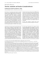
with a reported C292W variation [35]. Three potential N-
glycosylation sites were conserved in all sequences: N58,
N172 and N350 (Figure 1).
There were a number of single amino acid variations
present in only one or a few sequences; these amino acids
are listed in Table 3 and shown graphically in Figure 1.
Many of these variant amino acids were biochemically
quite dissimilar, though the biological significance of this
finding is not clear. Interestingly, the highest variability
was in the region between amino acids 260 to 300, anal-
ogous to the major antibody antigenic site A of the related
RSV F protein [36] (Figure 1). Some of the variations in
this region, such as E294G, were present in viruses of both
the A1 and A2 subgroups. Viruses of the A2 lineage had
the greatest number of such variations in the region
between amino acids 230 to 300, but not elsewhere in the
protein.
Phylogenetic diversity and evolution over time
We performed phylogenetic and evolutionary analysis of
the aligned full-length F sequences with six different mod-
els using the BEAST program suite [37]. The phylogenetic
Table 2: Comparison of distinct amino acid variations in the indicated functional domains of F protein between groups or subgroups of
unique human metapneumovirus strains.
Functional domain AA residues in
domain
No. of AA AA Position A
(n = 36)
B
(n = 49)
A1
(n = 13)
A2
(n = 23)
B1
(n = 11)
B2
(n = 38)
Signal peptide 1-22 2 6 V M/V V V M/V M/V
9FI
F2 subunit 23-102 2 61 A/S T/(S*) A A/S T T/(S*)
82 RKKK
Fusion peptide 103-125 1 122 V I
Heptad repeat A 131-172 4 135 T N
139 N G
143 K K/T Q/(K*) K/T
167 D E
F1 subunit 173-453 10 175 R S
185 A/D A A D A A
233 N Y
286 V I
296 K/R N/D K K/R N/(D*) D
312 Q K
348 K R
404 N/S P N N/S P P
449 V/I I V V/I I I
450 K R/K
Heptad repeat B 454-486 3 466 S/N S S N/S S S
479 R K/(R*) R R K/(R*) K
482 S/(N*) N/(S*) S S/(N*) N/(S*) N
Transmembrane 490-514 6 498 I I/V I I V I
503 S L
504 T/S T/A T S T T/A
507 L S
510 V/I I V V/I I I
511 F I
Cytoplasmic tail 515-539 4 518 K/(R*) R/(K*) K K/(R*) R/(K*) R
528 S N
533 N G
539 N/(S*) S/N S S
Amino acids (AA) in bold type did not vary within the group or subgroup.
*Amino acids found in only one isolate within the group or subgroup.
Virology Journal 2009, 6:138 />Page 4 of 10
(page number not for citation purposes)
Schematic representation of putative structure and mutation map of human metapneumovirus F proteinFigure 1
Schematic representation of putative structure and mutation map of human metapneumovirus F protein. SS =
signal sequence; FP = fusion peptide; HRA = heptad repeat A; HRB = heptad repeat B; TM = transmembrane domain; and CT
= cytoplasmic tail. Arrow indicates cleavage site; arrowheads indicate putative N-glycosylation sites. Amino acid variations are
indicated by asterisks, with the number of asterisks representing the number of distinct strains in which the variation was
found.
100 200 300 400 539500
SS
FP
HRA HRB
TM
CT
** ***
*
*
*******
*
*
*
****
*
*
** ***
*
**
*
*
**
*
*
**
*
*
*
*
*
*
*
** *
*
*
*
**
*
*
****
F1F2
Table 3: Distinct amino acid variations detected in the indicated domains of F protein in human metapneumovirus strains.
Domain AA positions of domain Subgroup
A1 A2 B1 B2
Signal peptide 1-22 K20Q S21N S21N
F2 subunit 23-102 D72E (3)
E93V E96G
Cleavage site 102-103 S101P (2)* S101P
Fusion peptide 103-125 T114A A115T
Heptad repeat A 131-172 K172R
F1 subunit 173-453 K179Q R179K (2)
S232P F196Y
I248F G239E
L249P (2) G261E
M270T
V271I D280G
C292G I285T
C292W
E294G (4) E294G (2)
K296R (3)
N298S
Y310N
A314T E323K
I352V N358K
H368N R396W (2)
N404S (2)
T419I K438R
Heptad repeat B 454-486 D475E (2)
Transmembrane 490-514 I492T I492V I492V
I514T L507P
Cytoplasmic tail 515-539 K519R P520Q P525L P520T
* Numbers in parentheses indicate the number of distinct strains with the variation.
Virology Journal 2009, 6:138 />Page 5 of 10
(page number not for citation purposes)
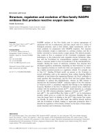
tree representing the sequence relationships by nucleotide
substitutions identified four genetic subgroups (Figure 2),
consistent with previous analyses [32]. The four distinct
subgroups remained stable over time, and viruses within
these lineages were closely related genetically, despite
being isolated at time points separated by as many as
twenty years. Thus, the clustering did not correlate closely
with chronological origin of the sequences. For example,
one subcluster within the B2 lineage contained nearly
identical sequences from Tennessee in 1989, 1990, 1991,
1992, 1993, 1994, 1995, 1996, 1998, 1999 and 2001, as
well as Netherlands in 1994 and Canada in 1998 and
2000 (Figure 2). Similar clustering of chronologically and
geographically disparate sequences was present within
each subgroup. In the A1 subgroup, Tennessee sequences
from 1994, 1996, and 2003 were closely related to Cana-
dian sequences from 1999 and 2000 and a Japanese
sequence from 2003. To examine further the evolution of
HMPV F gene sequences over time, we aligned sequences
within each subgroup in chronological order (see Addi-
tional files 1, 2, 3 and 4). A few nucleotide changes per-
sisted in later chronological viruses and thus represented
progressive evolution at those sites. However, the majority
of the nucleotide changes from year to year were not pre-
served and often reverted in subsequent isolates, showing
a lack of major drift over time.
Analysis of multiple sequences collected over time
allowed a molecular clock calculation of viral nucleotide
changes. The mutation rate of HMPV F was 7.12 × 10
-4
substitutions/site/year (95% HPD 4.23 × 10
-4
, 1.01 × 10
-
3
). The estimated time to most recent common ancestor
(tMRCA) of all HMPV strains was 97 years (95% HPD 66-
194) (Figure 2). The estimated time of divergence of the A
subgroup into A1 and A2 was 51 years (95% HPD 38-92)
and between B1 and B2 subgroups 40 years (95% HPD
38-97). Similar analysis using the limited number of
available AMPV full-length F sequences (n = 24, including
16 AMPV-C F sequences collected between 1998-2007)
suggested a tMRCA between AMPV-C and HMPV of 269
years (95% HPD 106-382) (Figure 2). However, very few
full-length AMPV type C F sequences were available, and
most were obtained within the last few years. The effect of
these limitations is reflected in the wide 95% HPD inter-
vals and thus the estimates for divergence of HMPV from
AMPV-C must be considered with some caution.
Discussion
We analyzed 85 full-length HMPV F gene sequences
obtained over a twenty-year period from Tennessee, Can-
ada, Japan, and the Netherlands. Our data confirm that
there are four distinct genetic lineages of HMPV, provi-
sionally designated as A1, A2, B1 and B2 [32]. These data
further show that these genetic subgroups are stable over
time in circulating viruses in a population of children with
respiratory illnesses. Thus, HMPV does not appear to
exhibit progressive genetic evolution, unlike influenza
virus that exhibits rapid genetic drift associated with anti-
genic variation resulting in immune escape. In this
respect, HMPV appears to be similar to other paramyxovi-
ruses. RNA viruses mutate frequently due to the infidelity
and lack of proofreading ability of RNA-dependent RNA
polymerases [38]. Our data confirm that the HMPV
polymerase also allows frequent errors resulting in the cir-
culation of field strains with nucleotide variations at a
similarly high rate. The rate of mutations we identified in
HMPV F (7.12 × 10
-4
substitutions/site/year) was interme-
diate between the lower rate of measles virus H gene
mutation (9 × 10
-5
substitutions/site/year) and the higher
rate of influenza A virus HA (1.8 × 10
-3
substitutions/site/
year)[39]. Nonetheless, while paramyxoviruses including
RSV and measles exhibit mutations and genotype varia-
tion over time [40,41], these nucleotide mutations do not
result in progressive antigenic "drift" over time with loss
of neutralizing epitopes [36,42,43]. This finding is in con-
trast to the data from studies of the influenza virus hemag-
glutinin protein, which progressively evolves both
genetically and antigenically, necessitating annual vaccine
updates [44-47]. The reason for the lack of directional
antigenic drift in paramyxoviruses is not clear. There
could be functional constraints on paramyxovirus fusion
proteins to prevent such drastic amino acid changes. The
fact that nucleotide diversity is greater than amino acid
diversity among HMPV F sequences supports this hypoth-
esis. In contrast to paramyxoviruses, the analogous influ-
enza virus hemagglutinin and human immunodeficiency
virus gp120 fusion proteins are capable of substantial
mutation to escape neutralizing antibodies without loss
of function. Alternatively, the nature of immune pressure
on fusion protein sequences by human antibodies could
differ between paramyxoviruses and orthomyxoviruses.
Experimental live wild-type virus challenge of previously
infected adults with a single lot of virus can achieve pro-
ductive infection in a repetitive fashion within months of
previous infection with the same virus [48]. The mecha-
nism of the functional constraints on paramyxovirus
fusion protein diversity warrants further investigation.
The finding that HMPV F gene sequences do not evolve
rapidly in a progressive fashion is important for the devel-
opment of monoclonal antibodies (mAbs) and vaccines.
The HMPV F protein is the major determinant of protec-
tion in animal models [21,22,24,26,28]. Studies with a
limited number of virus strains in these models suggest a
degree of cross-protective efficacy mediated by prior infec-
tion with viruses of differing subgroups [26,49]. The high
degree of conservation of F protein over time suggests that
interventions such as mAbs or vaccines likely will not
need to be continuously updated.
Virology Journal 2009, 6:138 />Page 6 of 10
(page number not for citation purposes)
Maximum clade credibility tree of HMPV and AMPV F nucleotide diversity by tMRCAFigure 2
Maximum clade credibility tree of HMPV and AMPV F nucleotide diversity by tMRCA. Phylogenetic analysis of 85
full-length HMPV F nucleotide sequences from Canada (CAN), Japan (JPS or JPY), Tennessee (TN), or the Netherlands (NL)
and 16 AMPV F sequences. The first two digits of the HMPV sequence names indicate the year of the isolate. The names of the
AMPV sequences indicate geographic origin (US = United States; UK = United Kingdom; MN = Minnesota) and year. The pos-
terior probability of divergence is indicated at each node. Mean TMRCA nodes on the MCC tree differ slightly from those
reported in text, although all are contained with the same 95% HPD values. Scale bar represents time in years. Tree was con-
structed as described in Methods.
A1
B1
A2
B2
HMPV-C
1
0.73
0.99
1
0.99
1
1
1
200019001800
Virology Journal 2009, 6:138 />Page 7 of 10
(page number not for citation purposes)
We identified a number of group and subgroup-specific
amino acid residues, some in putative functional
domains. The biological importance of these variations is
not clear, since definitive evidence of pathogenic differ-
ences between HMPV strains has not been described. A
previous analysis of 84 partial HMPV F sequences did not
identify subgroup-specific amino acid differences
between A1 and A2 viruses; however, a 441-nt gene seg-
ment was analyzed and most of the viruses were of recent
derivation [32]. The subgroup-specific amino acid
changes in F genes also were conserved over time, raising
the question of whether these residues possess critical bio-
logical features for virus infection or transmission. Some
of these variant amino acids were found in regions pre-
sumed to be essential, such as the heptad repeat (HR)
regions. Synthetic HR peptides mediate potent in vitro
inhibition of HMPV infection [50,51] and the HR are pre-
dicted to form a six-membered helical bundle [50], sug-
gesting that HMPV F is a Class I viral fusion protein. We
have cultivated multiple strains of all four subgroups that
exhibit similar growth kinetics and syncytial formation in
vitro, and similar levels of replication in vivo in rodents
(data not shown), suggesting that the fusion function of
all these strains is intact despite amino acid variations.
The HMPV F protein, like other Class I viral fusion pro-
teins, requires cleavage for activation and most strains
require exogenous trypsin for in vitro growth. Schickli et
al described a cleavage site mutation S101P that arose in
two strains of HMPV during cell passage and was associ-
ated with trypsin-independent viral growth in vitro [34].
The variant viruses did not differ from wild type in repli-
cation in Syrian hamsters [34]. We identified an S101P
variation in three distinct viruses in this study from 1989,
1994, and 1999. The F gene sequences in the current study
were amplified directly from specimens collected from
children with URI, and thus these viruses are natural vari-
ants. One of these had an associated E93V variation that
also was observed by Schickli et al. None of these three
viruses in our study was associated with more severe clin-
ical disease (data not shown).
Human and rodent F-specific mAbs have been described
with neutralizing activity in vitro and protective effects in
vivo, and several overlapping antigenic sites have been
identified using these mAbs [52,53]. However, the precise
location of these epitopes on the protein has not been
defined. We find it intriguing that the greatest concentra-
tion of amino acid variations among these 85 field iso-
lates lies in a region found between residues 260 to 300,
which is roughly analogous to the major antibody anti-
genic site A in the human RSV F protein [36,42]. The pre-
cise definition of neutralizing epitopes, especially
conserved epitopes for broadly neutralizing antibodies, is
critical for the development of prophylactic mAbs.
Phylogenetic and evolutionary analysis of multiple full-
length HMPV and AMPV F sequences obtained over
twenty years showed that HMPV may have diverged from
AMPV-C nearly 300 years ago, and the divergence of the
four HMPV genotypes likely occurred within the last hun-
dred years. de Graaf et al recently reported estimated
tMRCA values of ~120 years for the four HMPV genotypes
and 200 years for HMPV divergence from AMPV-C [54].
These estimates were based on analysis of 76 HMPV G
sequences, 107 partial HMPV F sequences, 12 partial
AMPV-C F sequences, 21 HMPV N sequences, and 15
AMPV-C N sequences from isolates collected over approx-
imately 12 years. Thus, the number of genes included was
greater, but the spread in years was less and most
sequences were from recent isolates. Despite these differ-
ences, we estimated remarkably similar rates of divergence
for both major and minor subgroups. Padhi et al analyzed
published HMPV G sequences and estimated a tMRCA of
only 25-50 years; however, the majority of viruses in that
study were isolated between 2001 and 2003 [55]. Analysis
of complete genome sequences from HMPV strains
obtained over many years would provide the most robust
estimates of genetic diversity and evolution.
Our phylogenetic and evolutionary analysis suggest that
HMPV may have diverged fairly recently from AMPV,
although the power of this analysis was limited by the
small number of available AMPV F gene sequences. Suc-
cessful productive infection of chickens and turkeys with
HMPV has not been reported [1], although inflammation,
HMPV RNA and antigen could be detected in turkey
poults inoculated with a large inoculum of HMPV [56].
HMPV and AMPV contain analogous open reading frames
in the same order that are distinct from those of the Pneu-
movirus genus, and metapneumoviruses lack the NS1 and
NS2 genes of pneumoviruses [57]. This finding suggests
that HMPV diverged from AMPV-C. Other viruses includ-
ing influenza and HIV are thought to have originated in
animal reservoirs but are now established primary human
pathogens; HMPV may have arisen as a human pathogen
by similar zoonotic transfer.
Methods
HMPV isolates
Virus sequences were derived from specimens collected
over a twenty-year period from 1982-2002 in the Vander-
bilt Vaccine Clinic, as previously described [2,3]. Nasal
wash specimens were collected from children <5 years of
age with acute respiratory tract illness. We extracted RNA
from these samples and used quantitative real-time RT-
PCR to test for HMPV by detection of nucleoprotein gene
sequences [2]. Specimens that tested positive for HMPV
were subjected to nested RT-PCR for the F gene as
described below. Viral nomenclature used in this study
uses a letter code representing the geographic site of isola-
Virology Journal 2009, 6:138 />Page 8 of 10
(page number not for citation purposes)
tion (e.g., "TN" represents Tennessee) followed by the year
of isolation, month in which the virus was isolated and
isolate number.
RNA extraction, RT-PCR and sequencing of F genes
RNA was extracted from 220 μl of nasal wash sample on a
Qiagen BioRobot 9604 Workstation using the QIAamp
Viral RNA kit (Qiagen), as described [2]. Amplification of
the entire F open reading frame (ORF) was carried out by
RT-PCR followed by nested PCR. The primers used to
amplify the F regions were FF1 (5'-ATGTCTGTACTTC-
CCAAA-3') and FR (5'-CCCGYACTTCATATTTGCA-3') for
RT-PCR, and FF2 (5'-AATATGCAAGACTTGGAGCC-3'
and 5'-AGGATCTGCAAGAGCTGGAG-3') and FR (5'-
CCCGYACTTCATATTTGCA-3') for nested PCR. The Ther-
moscript/Platinum Taq Polymerase Kit (Invitrogen) was
used in a 50 μL RT-PCR reaction with 10 μL of diluted
RNA as template. The RT-PCR was carried out at 50°C for
50 min and 95°C for 3 min, followed by 5 cycles of 94°C
for 30 sec, 50°C for 1 min, and 68°C for 3 min, and addi-
tional 30 cycles of 94°C for 30 sec, 55°C for 1 min, and
68°C for 3 min. For nested PCR, 2 μL of RT-PCR product
was added to a 50 μL reaction using Platinum PCR Super-
mix (Invitrogen). The reaction was incubated at 95°C for
3 min followed by 5 cycles of 94°C for 30 sec, 50°C for
30 sec, and 68°C for 2 min, and additional 30 cycles of
94°C for 30 sec, 55°C for 30 sec, and 68°C for 2 min. For
all reactions a final extension at 68°C for 7 min was
included. The resulting products were about 1.9 kb for the
F ORF and flanking sequences. The majority of PCR prod-
ucts generated after RT-PCR and nested PCR were specific
and migrated as a single band of the expected size (data
not shown). Agarose gel purification of the desired PCR
products was performed when multiple products were
generated.
Sequencing reactions were carried out using ABI PRISM
BigDye Terminator Cycle Sequencing Ready Reaction Kit
(Applied Biosystems). Eight sequencing primers were
used for each fragment to ensure a two-fold coverage of
the open reading frame. Sequencing primers are available
upon request. The products were processed by capillary
electrophoresis using ABI 3730 DNA Analyzer (Applied
Biosystems), and analyzed using DNA Sequencing Analy-
sis (Applied Biosystems) and Sequencher (Gene Codes
Corp.).
Sequence alignment and phylogenetic analysis
Final sequences were edited and aligned using the Clus-
talW algorithm in MacVector version 10.0 (Accelrys) and
MEGA version 3.1 [58]. Published AMPV and HMPV F
sequences were obtained from GenBank (Accession num-
bers AY145287
-AY145301, AY304360-AY304362,
AYAY622381
, EF051124, EF081369, EF199771-
EF199772
, EF589610, AF176593, AF187153-AF187154,
AF298642
-AF298650, AF368170, AF085228, AJ400728,
AJ400730
, DQ175630-DQ175634, DQ207607, D00850,
EU658938
, Y14290-Y14294). Sequences identified in this
study have been submitted to GenBank under accession
numbers EU857542
-EU857610. Pairwise sequence align-
ment, multiple sequence alignment, and percent nucle-
otide identity calculations were performed using
MacVector version 9.0. Inference of phylogeny and overall
rates of evolutionary change (nucleotide substitutions per
site per year) and the time to most recent common ances-
tor (tMRCA) were estimated using the Bayesian Markov
chain Monte Carlo (MCMC) approach available in the
BEAST package />[37]. Because
the sequences analyzed were very closely related and
exhibited few multiple substitutions at single nucleotide
sites, we used the simple HKY85 model of nucleotide sub-
stitution in each case, as more complex models some-
times failed to converge (data not shown). Data sets were
analyzed under demographic models of constant popula-
tion size, exponential population growth, and expansion
population growth, using strict or relaxed (uncorrelated
logarithmic) molecular clocks. Comparison of the output
of each model showed that the relaxed clock, exponential
population growth model gave the best estimation based
on 95% highest posterior density (HPD)(not shown). All
runs were visually examined to ensure convergence and
Estimated Sample Size of >200. MCMC chains were run
for 30 million steps with a burn-in rate of 10%, and two
separate runs were combined using the Log Combiner
program />[37], with uncertainty
in parameter estimates reported as the 95% HPD. Output
sets of trees were combined using LogCombiner and ana-
lyzed with the TreeAnnotator program to produce a Max-
imum Clade Credibility tree with a posterior probability
limit of >50%. Final tree was produced with FigTree [37].
Competing interests
Chin-Fen Yang, Chiaoyin K. Wang, Linda Lintao, Marla
Chu, Alekis Liem, Mary Mark and Richard R. Spaete were
employees of MedImmune at the time of this study. James
E. Crowe, Jr. has served as a consultant for Anaptys,
Immunobiosciences, Mapp, MedImmune, and Novartis
and has had research support from MedImmune, Mapp,
Alnylam, and sanofi Pasteur. John V. Williams has served
as a consultant for MedImmune and Novartis.
Authors' contributions
CFY, CKW, LDL, MC, AL, MM, RRS, and RP performed RT-
PCR, cloning and sequencing of HMPV isolates. SJT culti-
vated HMPV isolates and performed RT-PCR, cloning and
sequencing. JVW and JEC conceived the study, partici-
pated in its design and coordination, and helped to draft
the manuscript. JVW performed sequence alignment and
phylogenetic analysis. All authors read and approved the
final manuscript.
Virology Journal 2009, 6:138 />Page 9 of 10
(page number not for citation purposes)
Additional material
Acknowledgements
Financial support:
Supported by NIH R03 AI 054790 and R21 AI 082417 to JVW, a MedIm-
mune research grant to JEC, and a Burroughs Wellcome Fund Clinical Sci-
entist Award in Translation Research to JEC. The Vanderbilt Vaccine Clinic
was supported in part by NIH Respiratory Pathogens Research Unit N01
AI65298, NIH GCRC award RR00095 and NIH CTSA grant 1 UL1-
RR024975.
References
1. Hoogen BG van den, de Jong JC, Groen J, Kuiken T, de Groot R,
Fouchier RA, Osterhaus AD: A newly discovered human pneu-
movirus isolated from young children with respiratory tract
disease. Nat Med 2001, 7:719-724.
2. Williams JV, Wang CK, Yang CF, Tollefson SJ, House FS, Heck JM,
Chu M, Brown JB, Lintao LD, Quinto JD, et al.: The role of human
metapneumovirus in upper respiratory tract infections in
children: a 20-year experience. J Infect Dis 2006, 193:387-395.
3. Williams JV, Harris PA, Tollefson SJ, Halburnt-Rush LL, Pingsterhaus
JM, Edwards KM, Wright PF, Crowe JE Jr: Human metapneumov-
irus and lower respiratory tract disease in otherwise healthy
infants and children. N Engl J Med 2004, 350:443-450.
4. Hoogen BG van den, van Doornum GJ, Fockens JC, Cornelissen JJ,
Beyer WE, de Groot R, Osterhaus AD, Fouchier RA: Prevalence
and clinical symptoms of human metapneumovirus infection
in hospitalized patients. J Infect Dis 2003, 188:1571-1577.
5. Peiris JS, Tang WH, Chan KH, Khong PL, Guan Y, Lau YL, Chiu SS:
Children with respiratory disease associated with metapneu-
movirus in Hong Kong. Emerg Infect Dis 2003, 9:628-633.
6. Mullins JA, Erdman DD, Weinberg GA, Edwards K, Hall CB, Walker
FJ, Iwane M, Anderson LJ: Human metapneumovirus infection
among children hospitalized with acute respiratory illness.
Emerg Infect Dis 2004, 10:700-705.
7. McAdam AJ, Hasenbein ME, Feldman HA, Cole SE, Offermann JT,
Riley AM, Lieu TA: Human metapneumovirus in children
tested at a tertiary-care hospital. J Infect Dis 2004, 190:20-26.
8. Mackay IM, Bialasiewicz S, Jacob KC, McQueen E, Arden KE, Nissen
MD, Sloots TP: Genetic diversity of human metapneumovirus
over 4 consecutive years in Australia. J Infect Dis 2006,
193:1630-1633.
9. Foulongne V, Guyon G, Rodiere M, Segondy M: Human metapneu-
movirus infection in young children hospitalized with respi-
ratory tract disease. Pediatr Infect Dis J 2006, 25:354-359.
10. Esper F, Martinello RA, Boucher D, Weibel C, Ferguson D, Landry
ML, Kahn JS: A 1-year experience with human metapneumov-
irus in children aged <5 years. J Infect Dis
2004, 189:1388-1396.
11. Ebihara T, Endo R, Kikuta H, Ishiguro N, Ishiko H, Hara M, Takahashi
Y, Kobayashi K: Human metapneumovirus infection in Japa-
nese children. J Clin Microbiol 2004, 42:126-132.
12. Dollner H, Risnes K, Radtke A, Nordbo SA: Outbreak of human
metapneumovirus infection in norwegian children. Pediatr
Infect Dis J 2004, 23:436-440.
13. Boivin G, De Serres G, Cote S, Gilca R, Abed Y, Rochette L, Bergeron
MG, Dery P: Human metapneumovirus infections in hospital-
ized children. Emerg Infect Dis 2003, 9:634-640.
14. Williams JV, Martino R, Rabella N, Otegui M, Parody R, Heck JM,
Crowe JE Jr: A prospective study comparing human metap-
neumovirus with other respiratory viruses in adults with
hematologic malignancies and respiratory tract infections. J
Infect Dis 2005, 192:1061-1065.
15. Williams JV, Crowe JE Jr, Enriquez R, Minton P, Peebles RS Jr, Hamil-
ton RG, Higgins S, Griffin M, Hartert TV: Human metapneumov-
irus infection plays an etiologic role in acute asthma
exacerbations requiring hospitalization in adults. J Infect Dis
2005, 192:1149-1153.
16. Vicente D, Montes M, Cilla G, Perez-Trallero E: Human metapneu-
movirus and chronic obstructive pulmonary disease. Emerg
Infect Dis 2004, 10:1338-1339.
17. Pelletier G, Dery P, Abed Y, Boivin G: Respiratory tract reinfec-
tions by the new human Metapneumovirus in an immuno-
compromised child. Emerg Infect Dis 2002, 8:976-978.
18. Madhi SA, Ludewick H, Abed Y, Klugman KP, Boivin G: Human
metapneumovirus-associated lower respiratory tract infec-
tions among hospitalized human immunodeficiency virus
type 1 (HIV-1)-infected and HIV-1-uninfected African
infants. Clin Infect Dis 2003, 37:1705-1710.
19. Larcher C, Geltner C, Fischer H, Nachbaur D, Muller LC, Huemer
HP: Human metapneumovirus infection in lung transplant
recipients: clinical presentation and epidemiology. J Heart
Lung Transplant 2005, 24:
1891-1901.
20. Englund JA, Boeckh M, Kuypers J, Nichols WG, Hackman RC, Mor-
row RA, Fredricks DN, Corey L: Brief communication: fatal
human metapneumovirus infection in stem-cell transplant
recipients. Ann Intern Med 2006, 144:344-349.
21. Buchholz UJ, Nagashima K, Murphy BR, Collins PL: Live vaccines for
human metapneumovirus designed by reverse genetics.
Expert Rev Vaccines 2006, 5:695-706.
22. Cseke G, Wright DW, Tollefson SJ, Johnson JE, Crowe JE Jr, Williams
JV: Human metapneumovirus fusion protein vaccines that
are immunogenic and protective in cotton rats. J Virol 2007,
81:698-707.
23. Herfst S, de Graaf M, Schickli JH, Tang RS, Kaur J, Yang CF, Spaete RR,
Haller AA, Hoogen BG van den, Osterhaus AD, Fouchier RA: Recov-
ery of human metapneumovirus genetic lineages a and B
from cloned cDNA. J Virol 2004, 78:8264-8270.
24. Tang RS, Mahmood K, Macphail M, Guzzetta JM, Haller AA, Liu H,
Kaur J, Lawlor HA, Stillman EA, Schickli JH, et al.: A host-range
restricted parainfluenza virus type 3 (PIV3) expressing the
human metapneumovirus (hMPV) fusion protein elicits pro-
tective immunity in African green monkeys. Vaccine 2005,
23:1657-1667.
25. Mok H, Tollefson SJ, Podsiad AB, Shepherd BE, Polosukhin VV, John-
ston RE, Williams JV, Crowe JE Jr: An alphavirus replicon-based
human metapneumovirus vaccine is immunogenic and pro-
tective in mice and cotton rats. J Virol 2008, 82:11410-11418.
26. Herfst S, de Graaf M, Schrauwen EJ, Ulbrandt ND, Barnes AS, Senthil
K, Osterhaus AD, Fouchier RA, Hoogen BG van den: Immunization
Additional file 1
Supplemental Figure 1. Nucleotide sequence alignment of full-length F
genes from subgroup A1 HMPV isolates, listed in chronological order.
Click here for file
[ />422X-6-138-S1.pdf]
Additional file 2
Supplemental Figure 2. Nucleotide sequence alignment of full-length F
genes from subgroup A2 HMPV isolates, listed in chronological order.
Click here for file
[ />422X-6-138-S2.pdf]
Additional file 3
Supplemental Figure 3. Nucleotide sequence alignment of full-length F
genes from subgroup B1 HMPV isolates, listed in chronological order.
Click here for file
[ />422X-6-138-S3.pdf]
Additional file 4
Supplemental Figure 4. Nucleotide sequence alignment of full-length F
genes from subgroup B2 HMPV isolates, listed in chronological order.
Click here for file
[ />422X-6-138-S4.pdf]
Publish with BioMed Central and every
scientist can read your work free of charge
"BioMed Central will be the most significant development for
disseminating the results of biomedical research in our lifetime."
Sir Paul Nurse, Cancer Research UK
Your research papers will be:
available free of charge to the entire biomedical community
peer reviewed and published immediately upon acceptance
cited in PubMed and archived on PubMed Central
yours — you keep the copyright
Submit your manuscript here:
/>BioMedcentral
Virology Journal 2009, 6:138 />Page 10 of 10
(page number not for citation purposes)
of Syrian golden hamsters with F subunit vaccine of human
metapneumovirus induces protection against challenge with
homologous or heterologous strains. J Gen Virol 2007,
88:2702-2709.
27. Skiadopoulos MH, Biacchesi S, Buchholz UJ, Amaro-Carambot E, Sur-
man SR, Collins PL, Murphy BR: Individual contributions of the
human metapneumovirus F, G, and SH surface glycopro-
teins to the induction of neutralizing antibodies and protec-
tive immunity. Virology 2006, 345:492-501.
28. Mok H, Tollefson SJ, Podsiad AB, Shepherd BE, Polosukhin VV, John-
ston RE, Williams JV, Crowe JE Jr: An Alphavirus Replicon-Based
Human Metapneumovirus Vaccine is Immunogenic and Pro-
tective in Mice and Cotton Rats. J Virol 2008, 82(22):11410-8.
29. Njenga MK, Lwamba HM, Seal BS: Metapneumoviruses in birds
and humans. Virus Res 2003, 91:163-169.
30. Toquin D, de Boisseson C, Beven V, Senne DA, Eterradossi N: Sub-
group C avian metapneumovirus (MPV) and the recently iso-
lated human MPV exhibit a common organization but have
extensive sequence divergence in their putative SH and G
genes. J Gen Virol 2003, 84:2169-2178.
31. de Graaf M, Schrauwen EJ, Herfst S, van Amerongen G, Osterhaus
AD, Fouchier RA: Fusion protein is the main determinant of
metapneumovirus host tropism. J Gen Virol 2009, 90:1408-1416.
32. Hoogen BG van den, Herfst S, Sprong L, Cane PA, Forleo-Neto E, de
Swart RL, Osterhaus AD, Fouchier RA: Antigenic and genetic var-
iability of human metapneumoviruses. Emerg Infect Dis 2004,
10:658-666.
33. Schowalter RM, Smith SE, Dutch RE: Characterization of human
metapneumovirus F protein-promoted membrane fusion:
critical roles for proteolytic processing and low pH. J Virol
2006, 80:10931-10941.
34. Schickli JH, Kaur J, Ulbrandt N, Spaete RR, Tang RS: An S101P sub-
stitution in the putative cleavage motif of the human metap-
neumovirus fusion protein is a major determinant for
trypsin-independent growth in vero cells and does not alter
tissue tropism in hamsters. J Virol 2005, 79:10678-10689.
35. Ishiguro N, Ebihara T, Endo R, Ma X, Kikuta H, Ishiko H, Kobayashi
K: High genetic diversity of the attachment (G) protein of
human metapneumovirus. J Clin Microbiol 2004, 42:3406-3414.
36. Beeler JA, van Wyke Coelingh K:
Neutralization epitopes of the
F glycoprotein of respiratory syncytial virus: effect of muta-
tion upon fusion function. J Virol 1989, 63:2941-2950.
37. Drummond AJ, Rambaut A: BEAST: Bayesian evolutionary anal-
ysis by sampling trees. BMC Evol Biol 2007, 7:214.
38. Drake JW: Rates of spontaneous mutation among RNA
viruses. Proc Natl Acad Sci USA 1993, 90:4171-4175.
39. Jenkins GM, Rambaut A, Pybus OG, Holmes EC: Rates of molecu-
lar evolution in RNA viruses: a quantitative phylogenetic
analysis. J Mol Evol 2002, 54:156-165.
40. Rima BK, Earle JA, Baczko K, ter Meulen V, Liebert UG, Carstens C,
Carabana J, Caballero M, Celma ML, Fernandez-Munoz R: Sequence
divergence of measles virus haemagglutinin during natural
evolution and adaptation to cell culture. J Gen Virol 1997, 78(Pt
1):97-106.
41. Garcia O, Martin M, Dopazo J, Arbiza J, Frabasile S, Russi J, Hortal M,
Perez-Brena P, Martinez I, Garcia-Barreno B, et al.: Evolutionary
pattern of human respiratory syncytial virus (subgroup A):
cocirculating lineages and correlation of genetic and anti-
genic changes in the G glycoprotein. J Virol 1994, 68:5448-5459.
42. Crowe JE, Firestone CY, Crim R, Beeler JA, Coelingh KL, Barbas CF,
Burton DR, Chanock RM, Murphy BR: Monoclonal antibody-
resistant mutants selected with a respiratory syncytial virus-
neutralizing human antibody fab fragment (Fab 19) define a
unique epitope on the fusion (F) glycoprotein. Virology 1998,
252:373-375.
43. Tamin A, Rota PA, Wang ZD, Heath JL, Anderson LJ, Bellini WJ:
Antigenic analysis of current wild type and vaccine strains of
measles virus. J Infect Dis 1994, 170:795-801.
44. Cox NJ, Black RA, Kendal AP: Pathways of evolution of influenza
A (H1N1) viruses from 1977 to 1986 as determined by oligo-
nucleotide mapping and sequencing studies. J Gen Virol 1989,
70(Pt 2):299-313.
45. Russell CA, Jones TC, Barr IG, Cox NJ, Garten RJ, Gregory V, Gust
ID, Hampson AW, Hay AJ, Hurt AC, et al.: The global circulation
of seasonal influenza A (H3N2) viruses. Science
2008,
320:340-346.
46. Six HR, Webster RG, Kendal AP, Glezen WP, Griffis C, Couch RB:
Antigenic analysis of H1N1 viruses isolated in the Houston
metropolitan area during four successive seasons. Infect
Immun 1983, 42:453-458.
47. Smith DJ, Lapedes AS, de Jong JC, Bestebroer TM, Rimmelzwaan GF,
Osterhaus AD, Fouchier RA: Mapping the antigenic and genetic
evolution of influenza virus. Science 2004, 305:371-376.
48. Hall CB, Walsh EE, Long CE, Schnabel KC: Immunity to and fre-
quency of reinfection with respiratory syncytial virus. J Infect
Dis 1991, 163:693-698.
49. Skiadopoulos MH, Biacchesi S, Buchholz UJ, Riggs JM, Surman SR,
Amaro-Carambot E, McAuliffe JM, Elkins WR, St Claire M, Collins PL,
Murphy BR: The two major human metapneumovirus genetic
lineages are highly related antigenically, and the fusion (F)
protein is a major contributor to this antigenic relatedness.
J Virol 2004, 78:6927-6937.
50. Miller SA, Tollefson S, Crowe JE Jr, Williams JV, Wright DW: Exam-
ination of a fusogenic hexameric core from human metap-
neumovirus and identification of a potent synthetic peptide
inhibitor from the heptad repeat 1 region. J Virol 2007,
81:141-149.
51. Deffrasnes C, Hamelin ME, Prince GA, Boivin G: Identification and
evaluation of a highly effective fusion inhibitor for human
metapneumovirus. Antimicrob Agents Chemother 2008, 52:279-287.
52. Williams JV, Chen Z, Cseke G, Wright DW, Keefer CJ, Tollefson SJ,
Hessell A, Podsiad A, Shepherd BE, Sanna PP, et al.: A recombinant
human monoclonal antibody to human metapneumovirus
fusion protein that neutralizes virus in vitro and is effective
therapeutically in vivo. J Virol 2007, 81:8315-8324.
53. Ulbrandt ND, Ji H, Patel NK, Riggs JM, Brewah YA, Ready S, Donacki
NE, Folliot K, Barnes AS, Senthil K, et al.: Isolation and character-
ization of monoclonal antibodies which neutralize human
metapneumovirus in vitro and in vivo. J Virol 2006,
80:7799-7806.
54. de Graaf M, Osterhaus AD, Fouchier RA, Holmes EC: Evolutionary
dynamics of human and avian metapneumoviruses. J Gen Virol
2008, 89:2933-2942.
55. Padhi A, Verghese B: Positive natural selection in the evolution
of human metapneumovirus attachment glycoprotein. Virus
Res 2008, 131:121-131.
56. Velayudhan BT, Nagaraja KV, Thachil AJ, Shaw DP, Gray GC, Halvor-
son DA: Human metapneumovirus in turkey poults. Emerg
Infect Dis 2006, 12:1853-1859.
57. Hoogen BG van den, Bestebroer TM, Osterhaus AD, Fouchier RA:
Analysis of the genomic sequence of a human metapneumo-
virus. Virology 2002, 295:119-132.
58. Kumar S, Tamura K, Nei M: MEGA3: Integrated software for
Molecular Evolutionary Genetics Analysis and sequence
alignment. Brief Bioinform 2004, 5:150-163.