Báo cáo khoa học: "Characterisation of a GII-4 norovirus variant-specific surface-exposed site involved in antibody binding" docx
Bạn đang xem bản rút gọn của tài liệu. Xem và tải ngay bản đầy đủ của tài liệu tại đây (1008.29 KB, 11 trang )
BioMed Central
Page 1 of 11
(page number not for citation purposes)
Virology Journal
Open Access
Research
Characterisation of a GII-4 norovirus variant-specific
surface-exposed site involved in antibody binding
David J Allen*
1,2
, Rob Noad
2,4
, Dhan Samuel
3
, Jim J Gray
1
, Polly Roy
2
and
Miren Iturriza-Gómara
1
Address:
1
Enteric Virus Unit, Virus Reference Department, Centre for Infections, Health Protection Agency, Colindale, London, NW9 5EQ,
2
Pathogen Molecular Biology Unit, Department of Infectious & Tropical Diseases, London School of Hygiene & Tropical Medicine, Keppel Street,
London, WC1 5HT,
3
Serology Development Unit, Virus Reference Department, Centre for Infections, Health Protection Agency, Colindale,
London, NW9 5EQ and
4
Department of Pathology and Infectious Diseases, Royal Veterinary College, Hawkshead Lane, Hatfield, AL9 7TA, UK
Email: David J Allen* - ; Rob Noad - ; Dhan Samuel - ;
Jim J Gray - ; Polly Roy - ; Miren Iturriza-Gómara -
* Corresponding author
Abstract
Background: The human noroviruses are a highly diverse group of viruses with a single-stranded
RNA genome encoding a single major structural protein (VP1), which has a hypervariable domain
(P2 domain) as the most exposed part of the virion. The noroviruses are classified on the basis of
nucleotide sequence diversity in the VP1-encoding ORF2 gene, which divides the majority of human
noroviruses into two genogroups (GI and GII). GII-4 noroviruses are the major aetiological agent
of outbreaks of gastroenteritis around the world. During a winter season the diversity among the
GII-4 noroviruses has been shown to fluctuate, driving the appearance of new virus variants in the
population. We have previously shown that sequence data and in silico modelling experiments
suggest there are two surface-exposed sites (site A and site B) in the hypervariable P2 domain. We
predict these sites may form a functional variant-specific epitope that evolves under selective
pressure from the host immune response and gives rise to antibody escape mutants.
Results: In this paper, we describe the construction of recombinant baculoviruses to express VLPs
representing one pre-epidemic and one epidemic variant of GII-4 noroviruses, and the production
of monoclonal antibodies against them. We use these novel reagents to provide evidence that site
A and site B form a conformational, variant-specific, surface-exposed site on the GII-4 norovirus
capsid that is involved in antibody binding.
Conclusion: As predicted by our earlier study, significant amino acid changes at site A and site B
give rise to GII-4 norovirus epidemic variants that are antibody escape mutants.
Background
The ability of RNA viruses to maintain plasticity as well as
functionality in their genome has been well documented
as a survival mechanism, allowing RNA viruses to adapt to
changes in their environment, maintaining fitness in the
viral population [1]. Mutation in vivo can have a number
of effects including increasing the virulence of a virus [2]
or acquisition of antiviral resistance [3,4]. An important
consequence of the accumulation of point mutations in
viral structural proteins is the rise of antibody escape
Published: 25 September 2009
Virology Journal 2009, 6:150 doi:10.1186/1743-422X-6-150
Received: 25 August 2009
Accepted: 25 September 2009
This article is available from: />© 2009 Allen et al; licensee BioMed Central Ltd.
This is an Open Access article distributed under the terms of the Creative Commons Attribution License ( />),
which permits unrestricted use, distribution, and reproduction in any medium, provided the original work is properly cited.
Virology Journal 2009, 6:150 />Page 2 of 11
(page number not for citation purposes)
mutants [5-7]. RNA viruses generate this diversity in their
genome via the lack of fidelity of the viral RNA-dependent
RNA polymerase (RdRp), and the mutants with most
increased fitness are selected from the progeny by envi-
ronmental factors such as the host immune response.
Norovirus is a genus in the Caliciviridae family, that
includes pathogens of humans and animals [8]. Human
noroviruses are a highly diverse group of viruses with a
single-stranded RNA genome made up of three open read-
ing frames (ORFs), [9]. Noroviruses are classified on the
basis of nucleotide sequence diversity in the ORF2 gene,
which divides the majority of human noroviruses into
two genogroups (GI and GII) and approximately 19
genetic clusters within them [10]. The genogroup II-geno-
type 4 (GII-4) noroviruses have been the dominant circu-
lating strain since the early 1990s [11], and in 2002 a
variant GII-4 norovirus emerged that caused unusually
high numbers of outbreaks of gastroenteritis in the sum-
mer of 2002, and epidemic gastroenteritis around the
world in the winter of 2002/2003 [12]. This variant pos-
sessed a 3 nucleotide (nt) insertion in the hypervariable
P2 domain of the VP1 protein at position 6265. This epi-
demiological pattern was repeated in 2006 when another
novel GII-4 norovirus variant emerged, however, no inser-
tions or deletions were observed in the genome of this
virus (J Gray, personal communication).
Noroviruses are the major aetiological agent of outbreaks
of gastroenteritis in the community and in semi-closed
settings around the world. During a winter season (Sep-
tember-March), the diversity among the GII-4 noroviruses
has been shown to fluctuate, driving the appearance of
new virus variants in the population [13]. Studies of the
genetic diversity of these viruses have shown that new GII-
4 variants appear periodically in the population following
evolution of the viruses along neutral networks, and that
accumulation of mutations in the hypervariable P2
domain results in antibody escape mutant viruses which
go on to cause epidemic gastroenteritis [14-16].
Computer modelling experiments have previously sug-
gested that there are two 3-amino acid motifs (site A and
site B) in the hypervariable P2 domain that define the
appearance of epidemiologically significant GII-4 variant
norovirus strains [14]. Based on these observations, we
predicted that these two motifs may be a functional vari-
ant-specific epitope that evolves under selective pressure
from the host immune response and give rise to antibody
escape mutants.
Due to the lack of a tissue culture system [17] and suitable
animal models in which to study noroviruses, we synthe-
sised recombinant virus-like particles (VLPs) using a bac-
ulovirus expression system based on previously described
methods [18,19]. These VLPs were used to generate mon-
oclonal antibodies (mAbs) in order to test the functional-
ity of site A and site B. We use these novel reagents to
provide evidence that site A and site B form a conforma-
tional, variant-specific, surface-exposed site on the GII-4
norovirus capsid that is involved in antibody binding, and
that as predicted, significant amino acid changes at site A
and site B give rise to GII-4 norovirus epidemic variants
that represent antibody escape mutants.
Results
Construction of Recombinant Baculoviruses Expressing
GII-4 Norovirus VLPs
Analysis of sequence data for the strains used in this study
showed >88% nucleotide identity in the ORF2 gene
between strains GII-4v0, GII-4v2 and the reference strain
Lordsdale virus (LV), and >90% nucleotide identity
between GII-4v0 and GII-4v2 sequences (data not
shown). Deduced amino acid sequences for VP1 revealed
>95% similarity between the GII-4v0 virus and LV (data
not shown). Detailed comparison of coding and non-cod-
ing nucleotide sequence substitutions showed that the
majority of non-synonymous substitutions occurred in
the P2 domain (data not shown)
Wild-type VLPs were purified from recombinant baculovi-
rus-infected Sf9 cells and were analysed by EM (Figure
1(d)). Negative staining of preparations showed intact
VLPs had formed for both strains, and no significant mor-
phological variation was observed within the VLP prepa-
rations (Figure 1(d)). Particles were 30-35 nm in
diameter, and the surface structure of the particles could
be visualised at high magnification (Figure 1(d)). The
VLPs were morphologically indistinguishable from native
norovirus virions found in clinical specimens, but >10-
fold more VLPs were observed per viewing field than are
typically observed in clinical specimens.
The two wild-type expressing plasmid constructs pRN-
GII4v0 and pRN-GII4v2 were modified by PCR site
directed mutagenesis so that the ORF2 encoded a VP1
protein that was identical to either the GII-4v0 or GII-4v2
parental protein except at either of the putative epitope
positions 296-298 (site A) or positions 393-395 (site B),
where the protein would be of the heterologous (non-
parental) strain (Figure 2). Successful mutagenesis at the
target site without alteration of the remaining norovirus
insert was confirmed through sequence analysis (data not
shown).
All four hybrid VLP-expressing recombinant baculovi-
ruses efficiently expressed VP1 (data not shown). Further,
EM analysis of GII4v0/A
2
B
0
, GII4v0/A
0
B
2
and GII4v2/
A
2
B
0
VLPs formed by mutant recombinant VP1 proteins
were morphologically indistinguishable from wild-type
VLPs (data not shown). However, the hybrid construct
pRN-GII4v2/A
0
B
2
did not form VLPs, despite expressing
Virology Journal 2009, 6:150 />Page 3 of 11
(page number not for citation purposes)
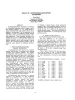
GII-4 Norovirus Major Sturctural ProteinFigure 1
GII-4 Norovirus Major Sturctural Protein. (a) Schematic representing the norovirus VP1 protein, highlighting the hyper-
variable P2 domain and putative epitopes Site A and Site B (as described in Allen et al., 2008). (b) Table showing amino acid var-
iation at Site A and Site B in the period 1999-2006, as previously described in (Allen et al., 2008). Strains used in the work
described here are highlighted. (c) Model of the norovirus VP1 P domain showing the location of Site A and Site B in the three-
dimensional protein (structure from Cao et al., 2007). (d) Electron micrograph showing GII-4v2 VLPs purified from Sf9 cells,
the morphology of which is representative for all VLPs described here. Magnification is 105 000× and VLPs are stained with
1.5% phosphotungstic acid. Scale bar is 100 nm.
Virology Journal 2009, 6:150 />Page 4 of 11
(page number not for citation purposes)
VP1 (data not shown), therefore the hybrid VLP GII-4v2/
A
0
B
2
was not available for subsequent work.
In addition to the hybrid VLPs described above, a reverse
mutant construct, pRN-GII4v0/A
0
B
0R
, was also generated
by back mutation of the hybrid construct pRN-GII4v0/
A
0
B
2
by site-directed mutagenesis (Figure 2). This reverted
the amino acid sequence at the site B from STA (GII-4v2)
back to N~N (GII-4v0). This plasmid was sequenced to
confirm the mutation at the target site had taken place,
and that no changes had taken place in the rest of the
insert (data not shown). The plasmid was used as
described to produce recombinant baculoviruses and pro-
duce the VLP GII-4v0/A
0
B
0R
.
Production and Characterisation of Variant Specific Anti-
GII-4 Norovirus Monoclonal Antibodies
Mice were inoculated with either GII-4v0 VLPs or GII-4v2
VLPs, and five monoclonal antibodies were fully charac-
terised for their isotype, titre and binding specificity
(Table 1). Test bleeds from both GII-4v0 and GII-4v2
inoculated mice prior to fusion showed that only a low
level of cross-reactivity with the heterologous antigen by
ELISA (data not shown).
Urea treatment of the homologous antigen significantly
reduced recognition of the epitope by all of the mono-
clonal antibodies (Table 1), and urea treatment of the het-
Schematic representation of norovirus protein coding region of pRN16 constructs expressing wild-type and hybrid VLPsFigure 2
Schematic representation of norovirus protein coding region of pRN16 constructs expressing wild-type and
hybrid VLPs. Following construction of plasmids pRN-GII4v0 and pRN-GII4v2 expressing wild-type GII-4v0 and GII-4v2 VLPs,
respectively, these plasmid constructs were modified by site directed mutagenesis at either putative epitope site A (nt 886-891,
aa 296-298), or putative epitope site B (nt 1176-1182, aa 383-395) to generate plasmid constructs that expressed hybrid VLPs.
The schematic shows a representation of the region of the plasmid encoding norovirus structural proteins (3'UTR and remain-
der of plasmid not shown for clarity). PCR mutagenesis was used to generate plasmid constructs that encoded an ORF2 iden-
tical to either GII-4v0 or GII-4v2, except at either site A or site B, which was modified to be as equivalent to that position in
the heterologous variant. The resulting expressed VP1 protein was a hybrid of the two variants, and so the VLP formed from
the hybrid VP1 was antigenically hybrid. Plasmid construct names are given on the left, whilst the hybrid VLPs are represented
with VLP names next to them on the right. All GII-4v0 derived regions are shown in solid black, all GII-4v2 derived regions are
shown in hatched lines.
Virology Journal 2009, 6:150 />Page 5 of 11
(page number not for citation purposes)
erologous antigen did not confer recognition (data not
shown).
Identification of a Variant-Specific Surface-Exposed Site
Involved In Antibody Binding
Each of the three anti-GII-4v0 mAbs displayed a slightly
different binding pattern, although the data suggested
that all three mAbs recognised an epitope formed or influ-
enced by both site A and site B (Figure 3(a)).
More than 75% reduction in binding of mAbGII4v0.5 to
its homologous antigen (GII-4v0) was observed following
blocking with the homologous antigen, whereas no
reduction in binding was observed following incubation
with the heterologous antigen (GII-4v2). Following
replacement of GII-4v0 site A with the heterologous GII-
4v2 site A, VLP GII-4v0/A
2
B
0
failed to block any of the
binding of mAbGII4v0.5 to its homologous antigen, dem-
onstrating that site A is essential for mAb recognition of
the antigen. When the GII-4v0 site B was replaced with the
GII-4v2 site B, VLP GII-4v0/A
0
B
2
, reduced binding to the
GII-4v0 antigen by ~19% indicating that when the
homologous site A was intact, partial mAb recognition
occurred, but without the corresponding homologous site
B, mAb recognition was impaired. Similarly, substitution
of the heterologous site B in the GII-4v2 with the GII-4v0
site B (VLP GII-4v2/A
2
B
0
) was not sufficient for recogni-
tion of this hybrid VLP by mAbGII4v0.5. Importantly,
restoring site B to the VLP GII-4v0/A
0
B
2
was sufficient to
restore wild-type levels of binding, to >70% reduction.
A 25% reduction in binding was observed following
blocking of mAbGII4v0.8 with the homologous GII-4v0
VLP, compared to only 5% reduction in binding follow-
ing blocking with the heterologous GII-4v2 VLP. Blocking
with any one of the three hybrid VLPs resulted in ≤10%
reduction in binding; the highest level of reduction fol-
lowing blocking among these VLPs was with the GII-4v2/
A
2
B
0
VLP which displays site B from the homologous (GII-
4v0) antigen, thereby indicating that mAbGII4v0.8 was
able to partially recognise site B, but that complete recog-
nition required the homologous site A to be displayed
concurrently. Blocking with the reverse mutant VLP GII-4/
A
0
B
0R
restored binding reduction to 27%, comparable to
blocking levels by wild-type GII-4v0. This further supports
the observation that both homologous site A and site B
must be displayed simultaneously on the virus surface for
mAb recognition.
Approximately 68% of mAbGII4v0.10 binding was
reduced following blocking by GII4v0 VLP, and only one
tenth of this reduction in binding observed following
blocking with GII-4v2 VLP. Blocking with any one of the
three hybrid VLPs resulted in ≤10% reduction in binding;
again the highest level of reduction following blocking
with a hybrid VLP was with the GII-4v2/A
2
B
0
VLP which
displays site B from the homologous (GII-4v0) antigen,
suggesting a role for site B in mAb recognition. Blocking
with the reverse mutant VLP GII-4/A
0
B
0R
restored binding
reduction to levels comparable to blocking levels by wild-
type GII-4v0, of approximately 70% reduction. This con-
firmed that both homologous site A and site B must be
present for mAb recognition of the antigen.
Both anti-GII-4v2 mAbs behaved the same in competition
immunoassays (Figure 3(b)). Blocking of both mAbs with
the homologous GII-4v2 antigen resulted in >85% reduc-
tion in binding in both mAbs, and blocking with the het-
erologous GII-4v0 antigen resulted in <2.5% reduction in
both mAbs. When blocking was performed using one of
the two GII-4v0-derived hybrid VLPs (GII-4v0/A
2
B
0
, or,
GII-4v0/A
0
B
2
), or the reverse mutant VLP, <7.5% reduc-
tion in binding was observed in both anti-GII-4v2 mAbs.
In contrast, blocking with the hybrid VLP GII-4v2/A
2
B
0
(which is all GII-4v2 except at site B) produced reduction
in binding equivalent to that observed with the GII-4v2
Table 1: Monoclonal antibody characterisation.
Monoclonal Antibody Homologous Antigen Isotype Titre
1
% Reduction in Binding to Urea-Treated Homologous Antigen
mAbGII4v0.5 GII-4v0 IgG1 100 71.8%
mAbGII4v0.8 GII-4v0 IgG2a 10 000 80.9%
mAbGII4v0.10 GII-4v0 IgG2b 10 000 79.8%
mAbGII4v2.5 GII-4v2 IgG1 10 000 44.0%
mAbGII4v2.6 GII-4v2 IgG1 10 000 51.2%
1
Titres were determined by serial dilution between 1:10 and 1:10 000 000 in an EIA. Titres were taken as the reciprocal of the last dilution to give
an optical density >0.5 at 450 nm.
Three anti GII-4v0 mAbs (mAbGII4v0.5, mAbGII4v0.8, mAbGII4v0.10) and two anti-GII-4v2 mAbs (mAbGII4v2.5, mAbGII4v2.6) were
characterised for reactivity, isotype and titre. The ability of the five mAbs to recognise their respective homologous antigen that had been
chemically denatured was also assessed by ELISA.
Virology Journal 2009, 6:150 />Page 6 of 11
(page number not for citation purposes)
Average percent reduction in binding of (a) anti-GII-4v0 and (b) anti-GII-4v2 mAbs to wild-type and mutant VLPs in a cross adsorption ELISAFigure 3
Average percent reduction in binding of (a) anti-GII-4v0 and (b) anti-GII-4v2 mAbs to wild-type and mutant
VLPs in a cross adsorption ELISA. As described in the Materials and Methods, each mAb was pre-incubated in a blocking
step with the antigen indicated on the x-axis, before being transferred to a microtitre plate coated with antigen homologous to
the mAbs being tested. Percent reduction in binding was then calculated using a PBS control. Cross absorption assays were
repeated 3 times independently and the average data is presented here with bars showing the standard error of the mean. Car-
toons representing the antigenic structure of the antigen (as described in Figure 2) are shown above the bars (with corre-
sponding labels below the bars). All mAbs were used at 1:10 000 dilution, except mAbGII4v0.5, which was used at 1:1000.
Virology Journal 2009, 6:150 />Page 7 of 11
(page number not for citation purposes)
antigen of >80% reduction. These data indicated that both
the anti-GII-4v2 mAbs recognised an epitope that is vari-
ant specific, but is not formed of either site A or site B,
alone or in combination.
Discussion
Efforts to identify sites on the norovirus capsid involved in
antibody binding have been hampered by the lack of a cell
culture system for human noroviruses [17], and therefore
epitope mapping studies using infectious virus have not
been possible. Here we have used VLPs synthesised in the
baculovirus expression system (BES) as a surrogate for
infectious virus in a mutagenesis study to identify sites on
the GII-4 norovirus capsid important in antibody recogni-
tion. Previous work has shown that when VLPs expressed
in the BES were compared with VLPs expressed in a mam-
malian recombinant protein expression system, no dis-
cernable differences in the biochemistry or structure of the
two differently expressed VLPs were observed [20].
The data presented here show the expression of high
yields of VLPs representing two norovirus strains, one
from each of the previously identified neutral networks:
(i) pre-2002 epidemic, and (ii) 2002 epidemic-200 [14].
These VLPs were used to immunize mice to produce mon-
oclonal antibodies against these strains.
Both the anti-GII-4v0 and anti-GII-4v2 polyclonal anti-
body responses were generally specific for the homolo-
gous antigen, but a low level of cross-reactivity was
observed (data not shown). Cross-reactivity is expected in
polyclonal serum because the different antibodies present
recognise a range of different epitopes, and have different
affinities; therefore polyclonal antibodies will, at least in
part, recognise a heterologous antigen. However, follow-
ing sub-cloning by limiting dilution, cross-reactivity was
lost as mAbs were isolated. This confirmed the specificity
of these antibodies for a single GII-4 norovirus variant
strain through recognition of an epitope that was unique
to that variant norovirus and offered no cross-reactivity
between other GII-4 norovirus variants.
The absence of any cross-reactivity in the EIA between the
mAbs and their heterologous antigen also showed that the
mAbs were not recognising epitopes from baculovirus
proteins or from Sf9 cell-derived proteins. Both antigen
preparations were made in the same protein expression
system and purified in the same way. Therefore any bacu-
lovirus or cell-derived proteins that co-purified with the
VLPs were present in both the GII-4v0 and the GII-4v2
VLP preparations used in the immunization of the mice
and in the preparation used as antigen in the EIA. Thus
any mAb reacting to a baculovirus or cell-derived protein
would react equally with both antigen preparations. This
is not the case, with all mAbs displaying specificity for the
homologous antigen preparation, thereby demonstrating
that all five mAbs were raised against norovirus proteins
and not baculovirus or insect cell proteins.
Urea treatment of both GII-4v0 and GII-4v2 VLPs revealed
the three anti-GII-4v0 mAbs recognised a conformational
epitope, whereas the anti-GII-4v2 mAbs recognised a par-
tially conformational epitope (Table 1). Treatment of a
macromolecular protein with a chaotropic agent such as 8
M urea will denature the three-dimensional structure of
the protein by disrupting the non-covalent intra-molecu-
lar interactions such as hydrogen bonding and van der
Waals forces. If the mAbs recognised a linear epitope,
binding would remain unaffected following chaotropic
treatment. However, the epitope recognised by the mAbs
must be conformational, as the level of mAb binding to
the antigen was reduced following denaturing treatment
of the antigen. Demonstrating that the mAbs recognised
conformational epitopes was important because site A
and site B identified by sequence analysis [14] were
shown to be surface exposed loop structures on the virus
surface separated by 100 amino acid residues in the linear
protein, but in close proximity in the three-dimensional
protein (Figure 1(a) &1(c)). Therefore, it was expected
that any antibodies raised against these sites would, at
least in part, recognise the conformation of the surface
structure at these positions, which is why it was important
that VLPs were used as the immunogen rather than linear
VP1 protein. This was corroborated through the failure to
detect a VP1 band in western blots (data not shown).
Site-directed mutagenesis was used to modify the norovi-
rus ORF2 gene in the plasmids pRN-GII4v0 and pRN-
GII4v2 at putative epitopes site A (aa296-298) or site B
(aa393-395). The aim was to generate both GII-4v0 and
GII-4v2 hybrid VLPs which displayed either an heterolo-
gous site A or site B. It was predicted that the changes engi-
neered at site A or site B would differently affect the ability
of mAbs to recognise the antigen, and so demonstrate the
roles of site A and site B as surface-exposed sites involved
in antibody binding.
The three hybrid VLPs that were isolated were found to be
morphologically indistinguishable from wild-type VLPs
as determined by EM, demonstrating that the mutagenesis
had no adverse effect on the structural integrity of the VLP.
The exception was the hybrid VLP expressed from the
recombinant baculovirus BAC-GIIv2/A
0
B
2
, which despite
expressing VP1 to high levels, did not form VLPs. As no
coding errors were observed in the ORF2 gene, and a high
level of protein expression was observed by SDS-PAGE,
the lack of VLP formation was not due to truncation of the
protein, failure of the baculovirus and expression vector to
undergo recombination, or failure of the recombinant
baculovirus to express the protein. Therefore, it seems
most likely that the mutations engineered in the P2
domain were structurally unfavourable and that they
Virology Journal 2009, 6:150 />Page 8 of 11
(page number not for citation purposes)
either perturb the correct conformation of the protein or
interfered with the subunit-subunit interactions, thus pre-
cluding particle formation.
It was predicted that the mAbs raised against the GII-4v0
and the GII-4v2 antigens would recognise a site formed of
both site A and site B, or would recognise a site formed of
one of these sites alone. This was tested in a cross absorp-
tion EIA using wild-type VLPs, hybrid VLPs, the reverse
mutant VLP and the five mAbs.
All three anti-GII-4v0 mAbs recognised an antigenic
region formed or influenced directly by both site A and
site B, as replacement of either of these sites abolished rec-
ognition of the GII-4v0 antigen by the mAbs. This obser-
vation was supported by the data from the reverse mutant
VLP. The GII-4v0/A
0
B
2
VLP failed to block binding of the
anti-GII-4v0 mAbs to the GII-4v0 antigen, but reverse
mutation of site B in this antigen back to GII-4v0 concur-
rently restored the ability of the antigen to block mAb
binding. The conclusion that the anti-GII-4v0 mAbs
require both site A and site B for antibody binding reflects
predictions made using bioinformatics data [14]. It was
noted that epidemiologically significant variant strains
appeared in the population following a cluster transition
event in which biochemically significant amino acid sub-
stitutions (or insertions/deletions) were observed at site A
and site B concurrently which itself suggested that both
site A and site B are required for defining epidemiologi-
cally important strains and allowing GII-4 noroviruses to
evade immunity existing in the population.
The back mutation of the hybrid GII-4v0/A
0
B
2
at site B cre-
ated the VLP GII-4v0/A
0
B
0R
that had twice undergone site-
directed mutagenesis at site B, so that it was structurally
and antigenically identical to the wild-type GII-4v0 anti-
gen. This experiment confirmed: (i) that the site-directed
mutagenesis process had no effect on the integrity of the
antigenic properties of the particle, other than those cre-
ated by the targeted mutation, and, (ii) that recognition of
an unrecognised hybrid antigen by a mAb could be
restored by replacement of the mutated site, thus demon-
strating that the mutated site was necessary for antibody
recognition of the antigen.
The anti-GII-4v2 mAbs recognised only the GII-4v2 VLP
and the GII-4v2/A
2
B
0
VLP, therefore demonstrating that
these mAbs recognise either an epitope that is dependent
on site A being in the structural context of the GII-4v2
antigen, but is independent of site B, or, an epitope that is
formed of neither site A nor site B. The former is difficult
to evaluate because the VLP GII-4v2/A
0
B
2
was not availa-
ble, but the latter could be investigated by construction of
a GII-4v2 hybrid VLP with site A and site B from GII-4v0,
which if recognised by the mAbs, would demonstrate that
the mAbs bind a site that is neither site A nor site B. Con-
versely, if such a VLP was not recognised, this would dem-
onstrate that site A and site B were important for antibody
binding, and more detailed mutagenesis studies where
individual amino acid residues in the GII-4v2 VLP were
mutated would aid in revealing the residues critical for
antibody recognition. Whether the five mAbs described
here recognise the same or different epitopes remains to
be tested in blocking EIAs.
In this study, we have used several well characterised
experimental systems in conjunction with in silico models,
to identify sites on the GII-4 norovirus capsid that are
important in antibody recognition. The use of antigeni-
cally hybrid VLPs to study capsid-antibody interactions
was used as a surrogate for infectious virus because there
is no cell culture system available for these viruses, and
our approach of systematic mutation of VLPs led to the
identification of two 3aa sites on the surface of the capsid
required for antibody binding. Whether the regions iden-
tified in this work represent neutralisisng epitopes
remains to be investigated, but these investigations
remain hampered by the lack of a replicative in vitro sys-
tem or suitable animal model. It would also be interesting
to determine whether the mAbs described here could
interfere with the ability of VLPs to interact with histo-
blood group antigens (HBGAs) when used in a VLP-
HBGA binding assay [21].
Methods
Clinical Samples
Two faecal specimens were selected from outbreaks that
had been characterised by PCR as being caused by a GII-4
norovirus at the Enteric Virus Unit, Centre for Infections,
Health Protection Agency, London, UK. The two viruses
were: (i) a GII-4 norovirus circulating before the 2002 epi-
demic, classified as a variant 0 (GII-4v0) virus; and, (ii) a
GII-4 norovirus circulating after the 2002 epidemic, and
classified as a variant 2 (GII-4v2) virus. Samples were pre-
pared as 10% suspensions in balanced salt solution
(Medium 199, Sigma, Dorset, UK) prior to nucleic acid
extraction.
Nucleic Acid Extraction & Reverse Transcription
Total nucleic acid was extracted from a 250 μl aliquot of
the 10% faecal suspension using a guanadinium isothio-
cyanate/silica method as previously described [22].
Extracted nucleic acid was incubated at 42°C for 60 min-
utes with 50 pmol of poly(T)-TVN primer in Tris-HCl
buffer, pH8.3, 5 mM MgCl
2
, 1 mM each dNTP, and 200 U
SuperScript
®
III reverse transcriptase (Invitrogen, Paisley,
UK).
PCR and Amplicon Purification
The genes ORF2 and ORF3 encoding the major structural
protein VP1 and the minor structural protein VP2, respec-
tively, and the 3' untranslated region (3'UTR), were
Virology Journal 2009, 6:150 />Page 9 of 11
(page number not for citation purposes)
amplified by PCR using primers ORF1/2-F1 [14] and
TVN-linker. The resulting amplicon
3'ORF1+ORF2+ORF3+3'UTR, was either 2513 bp or 2516
bp in length, depending on the strain. Reactions were per-
formed using High Fidelity PCR System (Roche Diagnos-
tics Ltd, Burgess Hill, UK). PCR amplified amplicons were
purified either from solution using Montage
®
PCR Filter
Units (Millipore, Watford, UK), or from agarose gels using
Geneclean
®
Spin Kit (Qbiogene, Cambridge, UK). Both
were used as according to manufacturers' instructions.
Amplicon Sequencing and Sequence Analysis
Sequencing PCR was performed using 10 pmol of primer
and 100 fmol template DNA. All sequencing was per-
formed using GenomeLab™ DTCS - Quick Start Kit (Beck-
man Coulter, High Wycombe, UK) according to the
manufacturer's instructions, and a CEQ8000 automated
sequencer (Beckman Coulter).
Nucleotide sequence contigs were generated from trace
data using the Assembler tool in BioNumerics v3.5
(Applied Maths, Kortrijk, Belgium). Multiple alignment
and phylogenetic analysis was performed using appropri-
ate algorithms in BioNumerics v3.5 (Applied Maths).
Amino acid sequence data was deduced from nucleotide
data and analysed using BioEdit [23], and also using
BioNumerics v3.5 (Applied Maths).
Cloning
Each of the purified 3'ORF1+ORF2+ORF3+3'UTR ampli-
cons was cloned into the vector pCR2.1-TOPO
®
(Invitro-
gen) according to the manufacturer's instruction. The
3'ORF1+ORF2+ORF3+3'UTR amplicon was then modi-
fied by PCR using primers deigned to: (i) remove the par-
tial 3'ORF1 sequence at the 5' end of the amplicon; (ii)
include two restriction enzyme sites at each end of the
amplicons in order to allow for directional cloning into
vector pRN16; and, (iii) modify the translation initiation
context of ORF2 to match that of the baculovirus polyhe-
drin gene (PH). The GII-4v0 amplicon was modified as
follows: 5'-A-StuI-SacI-PH-ORF2-ORF3-3'UTR-XbaI-StuI-
A-3'. The GII-4v2 amplicon was modified as follows: 5'-A-
StuI-KpnI-PH-ORF2-ORF3-3'UTR-XbaI-StuI-A-3'. The vec-
tor pRN16 contains a region of the Autographa californica
nuclear polyhedrosis virus (AcMNPV) around the polyhe-
drin gene (ORF7 (735)) which overlaps the essential
ORF8 (1629) gene. pRN16 was produced by ligating the
BstXI/HindIII polyhedrin promoter-polylinker fragment
from pAcCL29.1 [24] into BstXI/HindIII cut pBacPAK8
(BD Clontech). After digestion of both inserts and vector
pRN16 with the appropriate restriction enzymes ligation
was performed using T4 DNA ligase (Fermentas, York) to
produce the plasmids pRN-GII4v0 and pRN-GII4v2.
Positive clones were grown overnight in a 50 ml LB broth
culture containing 50 μg/ml ampicillin, and the plasmid
isolated using a plasmid preparation kit (Plasmid Midi
Kit, QIAGEN, West Sussex, or SNAP Midi-Prep Kit, Invit-
rogen) according to the manufacturer's instructions.
Site-Directed Mutagenesis
Wild-type sequences for GII-4v0 and GII-4v2 VLPs were
mutated in the P2 domain at site previously identified as
forming a putative epitope [14] (Figure 1(a)-(c)). Plas-
mids pRN-GII4v0 and pRN-GII4v2 were mutated in a site
specific mutagenic PCR reaction at either a 9 nt site at
positions 886-894 (site A), or a 6 or 9 nt site (depending
on the strain) at positions 1176-1182 (site B) from the
GII-4v0 sequence to the GII-4v2 sequence, or vice versa
(Figure 2). For this, the GeneTailor Site-Directed Muta-
genesis System (Invitrogen) was used according to manu-
facturer's instruction, using a touchdown PCR method to
mutate and amplify the plasmids. Mutated plasmids were
transformed into DH5αT1
R
E. coli cells (Invitrogen) and
purified using SNAP Midi Prep Kit (Invitrogen) according
to manufacturer's instruction.
Purified plasmids were used to generate recombinant bac-
uloviruses as has been previously described [19]. Follow-
ing recombination, a clonal population of recombinant
baculoviruses was obtained by plaque purification. The
resulting recombinant baculoviruses expressed either GII-
4v0 VLPs (BAC-GIIv0) or GII-4v2 VLPs (BAC-GIIv2).
Plaque purified viruses were used to seed stock cultures of
each virus, and these stocks were titred by plaque assay.
Generation of VLPs
Suspension cultures of Sf9 cells were infected with either
BAC-GII4v0 or BAC-GII4v2 at a moi of 2-3 and incubated
at 28°C for 48-72 hours. Virus-like particles were purified
from the intracellular phase by treatment with phosphate
buffer containing 1% IGEPAL (Sigma Aldrich) and
sequential centrifugation steps for clarification, and
finally through 15%-60% sucrose cushions to concentrate
the VLPs. Fractions were collected and analysed by SDS-
PAGE on a 12% polyacrylamide gel (NuPAGE kit (Invitro-
gen), according to manufacturer's instruction) and elec-
tron microscopy (EM).
Monoclonal Antibody (mAb) Production
BALB/c mice were inoculated subcutaneously with 100 μg
of either wild-type GII-4v0 or wild-type GII-4v2 VLPs in
Freunds incomplete adjuvant in order to produce mAbs.
After boosting the mice fortnightly on a further four occa-
sions, the spleen cells were harvested and fused with
mouse myeloma cells (NSI) by standard procedures [25].
Hybridoma Cloning, Screening and Selection
The fused cells were dispensed into 96 well tissue culture
plates and cultured in RPMI1640+GlutaMAX media (Inv-
itrogen), supplemented with 2% hypoxanthine-thymi-
dine (HT) (Invitrogen), 1% oxaloacetate-pyruvate-insulin
Virology Journal 2009, 6:150 />Page 10 of 11
(page number not for citation purposes)
(OPI) (Sigma) and 1% antibiotic-antimycotic (AbAm)
(Invitrogen). Ten to 14 days post fusion, supernatants
from the fusions were tested for antibodies to GII-4v0 and
GII-4v2 by EIA as described below. Hybridomas secreting
norovirus variant-specific antibodies were then cloned
twice by limiting dilution.
Enzyme-Linked Immunoassay (EIA)
Microtiter plates (Greiner Bio-One, Stonehouse) were
coated with either GII-4v0 or GII-4v2 VLPs at a concentra-
tion of 1 μg/ml diluted in PBS + 0.08% azide at 4°C. A
100 μl aliquot of test supernatants were diluted between 1
in 100 and 1 in 10000000 in PBST, and detection was per-
formed using a rabbit anti-Mouse IgG-HRP conjugate
antibody (Dako, Cambridgeshire) at 1 in 4000 dilution in
conjugate diluent (Microimmune) and TMB Substrate
(Europa Bioproducts, Cambridge).
Isotyping of Monoclonal Antibodies
A 100 μl sample of culture supernatant from each hybrid-
oma was added to coated microtiter plates and antibody
isotype determined using a goat anti-mouse IgG1a, IgG2a,
IgG2b, IgG2c, IgG3 or IgM (Jackson Laboratories, Maine,
USA) antibody, diluted 1 in 2000 in conjugate diluent
(Microimmune). Detection was performed using rabbit
anti-goat HRP-conjugate diluted 1 in 20000 in conjugate
diluent (Microimmune) containing mouse serum (Sigma-
Aldrich, Dorset, UK) and TMB Substrate (Europa Bioprod-
ucts).
Reactivity of Monoclonal Antibodies with Denatured
Antigen
The EIA was performed as described above, but before the
addition of the mAb to the plate, the VLP antigen bound
to the plate surface was treated with either 8 M urea in PBS
or PBS for 1 hour at room temperature. Wells were then
washed 3 times with PBST and the EIA performed as
described above.
Competitive Immunoassay
In the competitive immunoassay, monoclonal antibodies
were diluted in PBS 1 in 1000 - 1 in 10000 and pre-incu-
bated with either the homologous or heterologous wild-
type VLP at a concentration of 1 μg/ml, one of the antigen-
ically hybrid VLPs at 1 μg/ml, or PBST as a control. Pre-
incubated monoclonal antibodies were then added to
microtiter plates coated with 1 μg/ml of the homologous
antigen (as described above) to which the monoclonal
antibody was raised. The monoclonal antibody was then
allowed to attach, and detected with an anti-mouse HRP
conjugate antibody in an EIA as described above. Results
are shown as per cent reduction in binding of mAb to
homologous antigen (OD
test
) compared to level of bind-
ing in PBST control (OD
PBST
): % reduction in binding =
([OD
PBST
- OD
test
]/OD
PBST
) × 100.
Competing interests
The authors declare that they have no competing interests.
Authors' contributions
DJA participated in the design of the experiments, con-
ducted the experiments, and drafted the manuscript. RN
participated in the design of the experiments, provided
reagents and expertise for production of the VLPs, and
editing of the manuscript. DS participated in the design of
the experiments, provided reagents and expertise for pro-
duction of the monoclonal antibodies, and editing of the
manuscript. JJG participated in the design and coordina-
tion of the study, analysis of the data and editing of the
manuscript. PR provided reagents, participated in the
coordination of the study and editing of the manuscript.
MIG participated in the design and coordination of the
study, analysis of the data and drafting and editing of the
manuscript. All authors read and approved the final man-
uscript.
Acknowledgements
The authors are grateful to Ian Jones (University of Reading) for the gift of
BAC10:KO1629 and pAcCL29.1. This work was supported by the Euro-
pean Commission, DG Research Quality of Life Program, under the 6
th
Framework (EVENT;SP22-CT-2004-502571).
References
1. Domingo E, Holland JJ: RNA virus mutations and fitness for sur-
vival. Annu Rev Microbiol 1997, 51:151-178.
2. Cann AJ, Stanway G, Hughes PJ, Minor PD, Evans DM, Schild GC,
Almond JW: Reversion to neurovirulence of the live-attenu-
ated Sabin type 3 oral poliovirus vaccine. Nucleic Acids Res 1984,
12:7787-7792.
3. Hay AJ, Zambon MC, Wolstenholme AJ, Skehel JJ, Smith MH: Molec-
ular basis of resistance of influenza A viruses to amantadine.
J Antimicrob Chemother 1986, 18(Suppl B):19-29.
4. Shulman N, Winters M: A review of HIV-1 resistance to the
nucleoside and nucleotide inhibitors. Curr Drug Targets Infect
Disord 2003, 3:273-281.
5. Lambkin R, McLain L, Jones SE, Aldridge SL, Dimmock NJ: Neutrali-
zation escape mutants of type A influenza virus are readily
selected by antisera from mice immunized with whole virus:
a possible mechanism for antigenic drift. J Gen Virol 1994,
75(Pt 12):3493-3502.
6. Palombo EA, Bugg HC, Masendycz PJ, Bishop RF: Sequence of the
VP7 gene of an atypical human rotavirus: evidence for
genetic and antigenic drift. DNA Seq 1997, 7:307-311.
7. Speller SA, Sangar DV, Clarke BE, Rowlands DJ: The nature and
spatial distribution of amino acid substitutions conferring
resistance to neutralizing monoclonal antibodies in human
rhinovirus type 2. J Gen Virol 1993, 74(Pt 2):193-200.
8. Mayo MA: A summary of taxonomic changes recently
approved by ICTV. Archives of Virology 2002, 147:1655-1663.
9. Clarke IN, Lambden PR: The molecular biology of caliciviruses.
J Gen Virol 1997, 78(Pt 2):291-301.
10. Green KY: Caliciviridae: The Noroviruses. In Fields Virology Fifth
edition. Edited by: Knipe DM, Howley PM. Wolters Kluwer Health/
Lippincott Williams and Wilkins, Philadelphia; 2007:949-979.
11. Hale A, Mattick K, Lewis D, Estes M, Jiang X, Green J, Eglin R, Brown
D: Distinct epidemiological patterns of Norwalk-like virus
infection. J Med Virol 2000, 62:99-103.
12. Lopman B, Vennema H, Kohli E, Pothier P, Sanchez A, Negredo A,
Buesa J, Schreier E, Reacher M, Brown D, et al.: Increase in viral
gastroenteritis outbreaks in Europe and epidemic spread of
new norovirus variant. Lancet 2004, 363:682-688.
Publish with BioMed Central and every
scientist can read your work free of charge
"BioMed Central will be the most significant development for
disseminating the results of biomedical research in our lifetime."
Sir Paul Nurse, Cancer Research UK
Your research papers will be:
available free of charge to the entire biomedical community
peer reviewed and published immediately upon acceptance
cited in PubMed and archived on PubMed Central
yours — you keep the copyright
Submit your manuscript here:
/>BioMedcentral
Virology Journal 2009, 6:150 />Page 11 of 11
(page number not for citation purposes)
13. Gallimore CI, Iturriza-Gomara M, Xerry J, Adigwe J, Gray JJ: Inter-
seasonal diversity of norovirus genotypes: emergence and
selection of virus variants. Arch Virol 2007, 152:1295-1303.
14. Allen DJ, Gray JJ, Gallimore CI, Xerry J, Iturriza-Gómara M: Analysis
of Amino Acid Variation in the P2 Domain of the GII-4 Noro-
virus VP1 Protein Reveals Putative Variant-Specific
Epitopes. PLoS ONE 2008, 3:e1485.
15. Lindesmith LC, Donaldson EF, Lobue AD, Cannon JL, Zheng DP, Vinje
J, Baric RS: Mechanisms of GII.4 Norovirus Persistence in
Human Populations. PLoS Med 2008, 5:e31.
16. Siebenga JJ, Vennema H, Renckens B, de Bruin E, Veer B van der,
Siezen RJ, Koopmans M: Epochal Evolution of GGII.4 Norovirus
Capsid Proteins from 1995 to 2006. J Virol 2007.
17. Duizer E, Schwab KJ, Neill FH, Atmar RL, Koopmans MP, Estes MK:
Laboratory efforts to cultivate noroviruses. J Gen Virol 2004,
85:79-87.
18. Jiang X, Wang M, Graham DY, Estes MK: Expression, self-assem-
bly, and antigenicity of the Norwalk virus capsid protein. J
Virol 1992, 66:6527-6532.
19. Kitts PA, Possee RD: A method for producing recombinant
baculovirus expression vectors at high frequency. Biotech-
niques 1993, 14:810-817.
20. Baric RS, Yount B, Lindesmith L, Harrington PR, Greene SR, Tseng
FC, Davis N, Johnston RE, Klapper DG, Moe CL: Expression and
self-assembly of norwalk virus capsid protein from venezue-
lan equine encephalitis virus replicons. J Virol 2002,
76:3023-3030.
21. Lindesmith L, Moe C, Marionneau S, Ruvoen N, Jiang X, Lindblad L,
Stewart P, LePendu J, Baric R: Human susceptibility and resist-
ance to Norwalk virus infection. Nat Med 2003, 9:548-553.
22. Boom R, Sol CJ, Salimans MM, Jansen CL, Wertheim-van Dillen PM,
Noordaa J van der: Rapid and simple method for purification of
nucleic acids. J Clin Microbiol 1990, 28:495-503.
23. Hall TA:
BioEdit: a user friendly biological sequence align-
ment editor and analysis program for Windows 95/98/NT.
Nucleic Acids Symp Ser 1999, 41:95-98.
24. Livingstone C, Jones I: Baculovirus expression vectors with sin-
gle strand capability. Nucleic Acids Res 1989, 17:2366.
25. Kohler G, Milstein C: Continuous cultures of fused cells secret-
ing antibody of predefined specificity. Nature 1975,
256:495-497.