Báo cáo khoa học: " Characterization of a VHS virus genotype III isolated from rainbow trout (Oncorhychus mykiss) at a marine site on the west coast of Norway" doc
Bạn đang xem bản rút gọn của tài liệu. Xem và tải ngay bản đầy đủ của tài liệu tại đây (1.22 MB, 15 trang )
RESEARC H Open Access
Characterization of a VHS virus genotype III
isolated from rainbow trout (Oncorhychus mykiss)
at a marine site on the west coast of Norway
Henrik Duesund, Stian Nylund, Kuninori Watanabe, Karl F Ottem, Are Nylund
*
Abstract
Background: Norwegian production of rainbow trout (Oncorhynchus mykiss) has been without any outbreaks of
VHS for many years until the disease emerged in a farm in western Norway in November 200 7. The fish were, in
addition to VHS virus, positive for gill chlamydia-like bacteria, Flavobacterium psychrophilum, and a microsporidian. A
new VHS virus genotype III was isolated from the fish in RTgill-W1 cells and the complete coding region (11,065
nucleotides) was sequenced. This virus was also used in a challenge experiment to see if it could cause any
mortality in rainbow trout in sea water.
Results: This is the first time a nearly complete sequence of a genotype III virus isolate has been presented. The
organization of the genes is the same as in the other VHS virus genotypes studied (GI and GIV). Between the ORFs
are nontranslated regions that contain highly conserved sequence s encompassing the polyadenylation signal for
one gene, and the putative transcription initiation site of the next gene. The intergenic regions vary in length from
74 nt to 128 nt. The nucleotide sequence is more similar to genotype I isolates compared to isolates from
genotype II and IV. Analyses of the sequences of the N and G protein genes show that this new isolate is distinct
from other VHS virus isolates and groups closely together with isolates from genotype III. In a challenge
experiment, using intraperitoneal (ip) injection of the isolate, co-habitation with infected fish, and bath challenge,
mortalities slightly above 40% were obtained. There was no significant difference in mortality between the bath
challenged group and the ip injected group, while the mortality in the co-habitation group was as low as 30%.
Conclusions: All VHS virus isolates in genotype III are from marine fish in the North East Atlantic. Unlike the other
known genotype III isolates, which are of low virulence, this new isolate is moderately virulent. It was not possible
to detect any changes in the virus genome that could explain the higher virulence. A major problem for the study
of virulence factors is the lack of information about other genotype III isolates.
Background
Viral haemorrhagic septicaemia virus (VHSV) is an
enveloped, single stranded, negative-strand RNA virus
belonging to the genus Novirhabdovirus, family Rhabdo-
viridae [1]. The VHS virus genome consists of a pproxi-
mately 11 k nucleotides and six genes encoding
nucleocapsid- (N), phospho- (P), matrix- (M), glyco-
(G), non-structural- (Nv) and RNA polymerase (L) pro-
tein. Based on phylogenetic analysis of the N, P, G and
Nv protein genes the VHS virus isolates have been
divided into four different genotypes; VHS virus
genotypes I, II, III and IV [2-7]. The third VHS virus
genotype (III) represents isolates from marine fish spe-
cies in Kattegat, Skagerrak and the North Sea [8] and a
member of this genotype wa s in the autumn of 2007
associated with about 10% mortality in a rainbow trout
farm in western Norway [9,10]. VHS virus genotype III
has been found in eel (Anguilla anguilla), cod (Gadus
morhua), herring (Clupea harengus), sprat (Sprattus
sprattus), haddock (Melanogrammus aeglefinus), Norway
pout (Trisopterus esmarkii), poor cod (Trisopterus minu-
tus), blue whiting (Micromesistius poutassou), withing
(Merlangius merlangus), turbot (Scophthalmus maxi-
mus), greenland halibut (Reinhardtius hippoglossoides)
and lesser argentine (Argentina sphyraena)[cf
* Correspondence:
Department of Biology, University of Bergen, Thormohlensgt 55, 5020
Bergen, Norway
Duesund et al. Virology Journal 2010, 7:19
/>© 2010 Duesund et al; licensee BioMed Central Lt d. This is an Open Acces s article distributed under the terms of the Creative
Commons Attribution License ( which permits unrestricted use, distribution, and
reproduction in any medium, provided the or iginal work is properly cited.
[3,5,8,11]]. The outbreak of VHS in Norway is the first
time an isolate belonging to genotype III is found in
rainbow trout.
According to existing literature challenge of rainbow
trout with the VHS virus, genotype III, should not result
in any significant mortality [12]. However, it is to be
expected that viruses which enter into farmed popula-
tions of fish may show some virulence and possibly
cause mortality. It has been shown that VHS virus
belonging to genotype III may cause mortality when
challenging turbot [13-15] and halibut [16], which sug-
gests that the susceptibility of the host species is also
important for the expected mortality. This has also been
observed for other viruses isolated from fish [17,18]. A
challenge experiment on rainbow trout fingerlings (10.1
gram s) in fresh water, using a VHS virus isolate type III
from the same outbreak as the isolate used in this study,
has already been carried out resulting in mortality after
immersion and injections of 70% and 100%, respectively
[9]. However, this genotype III VHS virus is a purely
marine virus a nd experiment on fi ngerlings in fresh
water may not be representative for the susceptibility of
larger rainbow trout in sea water, and the resulting mor-
tality in sea water cannot be predicted based on this
challenge experiment.
TheaimofthepresentstudyistoseeifthisVHS
virus genotype III with the first completely characterized
coding region may cause any mortality when challenging
rainbow trout in full sea water. The genome of this iso-
late will be compared with partly sequenced VHS virus,
genotype III, and completely sequenced coding regions
and intergenic regions of other VHS virus, genotypes I
and IV. Such a comparison may also give clues as to
which changes in the genome may influence t he viru-
lence or ability to cause mortality in rainbow trout
populations.
Results
Genome of isolate FA28.11.07
ThefirstgenomeofaVHSvirusingenotypeIII,strain
FA28.11.07, containing all protein coding sequences
(CDS) and intergenic regions (ITRs), has been
sequenced (accession no: EU481506). The sequence is
11,065 nucleotides (nt) long and co ntains six open read-
ing frames (ORF) in the order 3’-N-P-M-G-NV-L-5’.
This arrangement is identical to what has been found
for other, fully sequenced, VHS virus isolates in geno-
type I [19,20] and IV (accession no: AB490792). Between
the ORFs are nontranslated regions that include highly
conserved sequences encompassing the polyadenylation
signal for one gene, and the putative transcription initia-
tion site of the next gene. An alignment of the con-
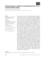
served nontranslated sequences is shown in Figure 1.
Theintergenicregionsvaryinlength,from74nt
between the G and NV ORFs, to 128 nt between the
NVandLORFs.Thisisconsistentwithwhathasbeen
found for VHS virus isolates in other genotypes except
that the length of the intergenic sequences in genotype
IV isolates are slightly different. The polyadenylation
signal is also present after the ORF of the L protein (21
nt downstream), but the sequence (aga ttg aaa aaa a) is
slightly different from that found in the intergenic
regions.
Characteristics of the different protein coding genes
and their deduced ORFs are listed in table 1. When
comparing the amino acid identity of the deduced pro-
teins of isolate FA28.11.07 (genotype III) to that of fully
or partially sequenced VHS virus isolates, all proteins
share a higher identity to the genotype I isolates than
isolates from genotypes II and IV (table 2). The only
genes sequenced from other genotype III isolates are
those coding for the G and NV proteins and these sh ow
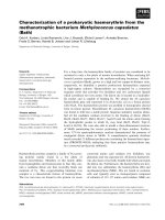
the highest identity to FA28.11.07. The short ORF (366
nt), located between the G and L protein, encodes the
NV protein which is the most variable protein based on
amino acid (aa) sequences. The variation is found
throughout the aa sequence, but the latter 14 aa are dis-
tinctly different in the three genotypes I, III and IV (Fig-
ure 2). Most of the variation in the nucleoprotein (N) of
VHS virus isolates, comparing genotypes I, III and IV, is
found between aa 37 - 132 and among the last 37 aa in
the peptide, avoi ding the con served RNA binding
domain suggested to be in the middle region of the pro-
tein. The aa variation in the P protein, among VHS
virus isolates, is mainly in the first third of the protein,
while there is little variation in the M protein.
Variation in aa sequence of the G protein is found
throughout the length of the protein, but with little var-
iation in the putative transmembrane regions. The
Figure 1 Comparison of the conserved sequence p arts of the
intergenic regions within the VHS virus isolate, FA28.11.07,
genome. The sequences between the genes N and P (N-P), P and
M (P-M), M and G (M-G), G and NV (G-NV), NV and L (NV-L) are
listed in message sense along with the consensus sequence. The
sequences consist of a polyadenylation signal and a putative
transcription initiation site, respectively.
Duesund et al. Virology Journal 2010, 7:19
/>Page 2 of 15
TMpred program />TMPRED_form.html was used for prediction of trans-
membrane regions a nd orientation in the G protein of
FA28.11.09. The most strongly supported model sug-
gested transmembrane regions between aa 1 - 18 and aa
462 - 483 where the latter has the highest support.
The last ORF (1984 aa) in the VHS virus genome is
the large (L) protein that encodes the RNA-dependent
RNA polymerase. The L protein from FA28.11.07 is
highly similar to the other L protein genes sequenced
from other VHS virus isolates, with the exception of
one isolate of genotype I, isolate FR-07-71 (Accession
nos: AJ233396 and AJ009814) from France. The con-
served domains III and IV containing the four major
motifs A, B, C and D localized between aa 566 and 790
[cf [21,20]] are also present in i solate FS28.11.09. A
putative ATP biding site , with sequence GEGVRG - (20
aa) - K in position aa 1223 to aa 1249, is present in
FA28.11.07 and the ge notype I isolates with the excep-
tion of isolate FR-07-71 from France. The ATP binding
site in FR-07-71 and isolates in genotype IV are
GEGVRR - (20 aa) - K and GEGIRG - (20 aa) - K,
respectively.
Ten amino acid residues that may play a role in the
determination of virulence in genotype I isolates have
been identified [19] (table 3). Compared to the genotype
I isolates, the VHS virus isolate FA28. 11.09 genotype III
from rainbow trout in Norway, share 2 and 6 amino
acids with the avirulent and virulent strains, respectively.
No information is available about the N, P and L
proteins from other genotype III isolates. It has also
been suggested that two regions, related to fusion activ-
ity, within the G protein may play a role in deter mina-
tion of virulence [22]. The VHS virus isolate,
FA28.11.09, from rainbow trout in Norway shares six
out of seven amino acids, believed to be important for
determination o f virulence, with the highly virulent FR-
07-71 strain (table 4). However, so do all other VHS
virus genotype III isolates (see accession numbers in
table 2).
Phylogeny
Analyses of the relationship of the VHS virus isolate
FA28.11.07, based on nucleotides of the complete o pen
reading frame (ORF) of the N (1215 nt) and G (1524 nt)
proteins, show that this isolate belong to genotype III
(Figures 3 and 4). The closest relatives, based on the
ORF of the G protein, are VHS virus isolates from
Atlantic cod, Norway pout, and haddock collected in
the North sea and herring an dturbotcollectedinSka-
gerrak and Ireland, respectively. The genotype, GIII,
constitutes a sister group to GI in both phylogenies.
ThenucleotidesequenceoftheGproteinfrom
FA28.11.07 is identical to that published (Accession no:
EU547740) by Dale et al [9]. The VHS virus from her-
ring (CH15.02.08), collected in the mouth of Storfj or-
den, belongs to genogroup Ib. The rainbow trout isolate
from Norway (FA28.11.07) is the only fully sequenced
member of the GIII. The phylogeny based on the
nucleotide sequence s of the ORF of the N protein
shows stronger support values compared to a similar
analysis using the G gene, however, this could be a
result of the choice of isolates and the number of iso-
lates included in the two phylogenies.
Challenge experiment
The rainbow trout used in the challenge experiment,
came from fresh water and were put directly in full sea
water, where they suffered some mortality (8.8%) during
the acclimatization period. Most of the mortality seemed
to be due to poor smoltification, but bacteria (Vibrio
spp. and Aliivibrio spp.) were isolated from a few fish
(Accession nos: EU862328, EU862329, EU862330,
EU862331, EU862332, EU862333, EU862334) and IPN
virus was present in all of the fish. The dominating bac-
teria were Vibrio splendidus-like. The mortality stopped
one week bef ore the start of the experiment. However,
the fish were still positive for IPN virus at the time of
challenge, i.e. they were carriers of the virus (Ct values
above 30). The fish remained positive for IPN virus
throughout the experimental period and a few fish were
also positive for Vibrio spp and Aliivibrio spp. All fish
tested before the start of the experiment were negative
for VHS virus.
The mortalities in the different groups varied from
2.9% in the bath control group, BK (N = 68), and up
Figure 2 The CO e nd (last 18 amino acids) of the deduced NV
protein sequence from VHS virus isolates belonging to
genotypes I, III and IV.
Table 1 VHSV isolate FA28.11.07 genome transcription
units and deduced protein products.
mRNA features (nt) Deduced protein features
(aa) Calculated
Gene Length 5’UTR ORF 3’UTR Length Mr pI
NP 1368 113 1212 43 404 44.1 5.2
P 761 58 666 37 222 24.5 8.5
Matrix 742 83 603 56 201 22.3 9.3
G 1610 35 1521 54 507 57.0 6.5
NV 423 23 366 34 122 13.6 5.4
L 6086 97 5952 37 1984 224.4 7.6
Duesund et al. Virology Journal 2010, 7:19
/>Page 3 of 15
48.4% in the i.p. challenged group, V (N = 31) (Figure
5). The mortality in the bath challenged group (BV, N =
68) was 44.1% while the mortality among the co-habi-
tants (KV, N = 30) was 30%. The total mortality in the
tank challenged by homogenate from rainbow trout
(group H, N = 61) was 41%. The group that was chal-
lenged with homogenate from VHS virus positive her-
ring (group CH) suffered 6.3% mortality.
Not all fish that died in the different groups were posi-
tive for presence of VHS virus. If the fish that were nega-
tive for presence of VHS virus are removed from the
mortalities the pattern of mortality remains, however,
moreorlessthesame(Figure6).Themortalitiesasso-
ciated with presence of VHS virus in the V and BV were
41.9% and 44.1%, respectively. None of the fish that died
in the control group, BK, or the CH group were positive
for VHS virus. We were not a ble to identify any other
pathogens that could explain the mortalities of the fish
that were negative for presence of VHS virus.
None of the fish in groups V (N = 11) and KV (N =
16) were positive for VHS virus at the termination of
the experiment 53 days post challenge, while 2 and 3
fish out of 33 and 37 fish examined were positive in
groups BV and H, respectivel y. These five fish were car-
riers of VHS virus (ct values > 35) and did not show
any signs of disease. The virus was present in kidney,
heart and spleen tissues, while the brain from one fish
only was positive. Of the 97 fish sampled at the termina-
tion of the experiment 53 days post challenge 5.2% were
carriers of the VHS virus.
Table 2 Pairwise percent amino acid identities of FA28.11.07 proteins with protein sequences in other VHSV isolates.
% Amino acid identity
a
Isolate code Country/origin Genotype N P M G NV L Accession no.
b
DK- Hededam Denmark I 92.8 96.8 94.5 96.4 84.4 97.7 Z93412
DE- Fil3 Germany I- a 93.1 96.4 94.0 96.3 81.1 97.7 NC_000855
FR-14-58 France I- a 93.6 95.9 94.0 96.3 84.4 97.6 AF143863
FR-07-71 France I- a 92.6 94.1 94.5 95.3 82.8 83.8
c
AJ233396
FR-07-71 France I- a - - - - - 75.9
c
AJ009814
UK-96-43 England I-b 93.1 95.5 95.0 96.3 84.4 97.6 AF143862
DK-M.rhabdo Baltic Sea I- b 93.6 95.5 94.5 97.0 90.2 98.6 Z93414
DK-2835 Denmark I- c - - - 95.9 - - AY546585
FI-ka66 Gulf of Bothnia I- d - - - 96.6 - - AY546614
DK-1p52 Baltic Sea II - - - 94.3 70.5 - AY546576/DQ159194
DK-1p53 Baltic Sea II - - - 94.1 70.0 - AY546577/DQ159195
UK-860/94 Scotland III - - - 97.4 92.6 - AY546628/DQ159203
UK-H17/2/95 North Sea III - - - 99.2 96.7 - AY546629/DQ159202
DK-4p168 Skagerrak III - - - 99.8 - - AY546582
UK-MLA98/6PT11 North Sea III - - - 99.4 - - AY546632
DK-4p101 North Sea III - - - 97.6 - - AY546581
UK-H17/5/93 North Sea III - - - 99.2 - - AY546630
JF00Ehi1 Japan IV- a 92.3 93.7 93.0 92.7 70.5 96.4 AB490792
MI03GL USA IV- b 92.1 - - 94.1 - - DQ427105/DQ401193
a
The highest percent amino acid identity observed for each protein is highlighted in bold
b
Accession numbers are list ed for complete genomes when available, and for the individual nucleotide sequences when not
c
Gaps in the alignment
Table 3 Amino acid residues that may play a role in the determination of virulence [19] when challenging rainbow
trout.
NPGL
Position 82 83 371 392 39 41 78 506 1012 1465
Amino acid G-E M-T R-L E-G P-T E-G L-F M-T I-F I-L
FA281107 E A K E T G F M F L
Other GIII - - - - - - - M/V - -
The virulent strains are, Hededam and FR-14-58, are isolated from rainbow trout, while the avirulent strains, UK-96-43 and DK-M.rhabdo, are from herring and
cod. Position = the position of the amino acid residues within the respective proteins. The first amino acid in each column was conserved among avirulent
strains and the latter among virulent strains. The VHS virus isolate (FA28.11.09) from rainbow trout in Norway share 2 and 6 amino acids with the avirulent and
virulent strains, respectively. No information is available about the N, P and L proteins sequences from other genotype III isolates.
Duesund et al. Virology Journal 2010, 7:19
/>Page 4 of 15
The amount of VHS virus template in the kidney and
brain of fish in the two challenge groups V and BV have
been quantified using the elongation factor alpha as a
standard. The kidney tissue from 13 individuals in
group V was positive for presence of VHS virus tem-
plate while only 10 individuals had positive brain tissue
(CNS). In the BV group 28 and 27 individuals had posi-
tive kidney and CNS, respectively. Only one fish was
found to be positive for VHS virus 20 days after injec-
tion of the virus (group V), while in the bath challenged
group nine fish were positive. An individual sampled 12
days after challenge in group V had the highest expres-
sion of VHS virus genome/mRNA. This expression was
6.7 million times higher compared to the lowest expres-
sion (sampled 9 days post challenge) of these templates
in positive kidney tissue. The individu al, in the V group,
with the highest expression of VHS virus template in
the CNS was sampled 7 days post challenge. None of
the fish in this group had positive CNS after 18 days
post challenge. In the bath challenged group, BV, the
highest expression of VHS virus templates in kidney tis-
sue was found 11 days post challenge while the highest
expression in the CNS was seen 42 days post challenge.
The latter specimen had negative kidney tissue. Of the
nine fish that were positive after day 20 post challenge
five had positive kidneys and 8 had positive CNS.
VHS viruses were isolated from all challenged groups
except the control group and the CH group. Partial
sequences of the genome showed that the reisolated
viruses were identical to the FA28.11.07 isolate (Acces-
sion no: BV group: FJ362510, FJ362511, H group:
FJ362512, FJ362513, KV group: FJ362514, V group:
FJ362515).
Pathology
The weight and length of the fish in the different groups
at the termination of the challenge is given in table 5. In
all groups, includi ng the control group (BK), some rain-
bow trout showed loss of scales and skin ulcers. In the
two groups that were bath challenged (groups BV and
H) a few fish had haemorrhages on the viscera (figure
7A). A few fish in all VHS virus challenged groups, V,
KV, H and BV, showed corkscrewing and had eye and
somaticmusclebleedings(figure7B),palegills,slight
epicarditis and some necrosis of heart myofibers in the
ventricle. The most pronounced changes were seen in
the kidneys which were slightly swollen with marked
necrosis, haemorrhages and loss of haematopoietic cells
(Figure 7C). Only minor changes were seen in the liver
of strongly positive fish (Figure 7D).
The mean haematocrit values from moribund fish
positiveforVHSvirusingroupsV,KVandHwere
about 23.0 and for those in group BV it was 12.5. In th e
control group BK and the challenged group CH the
mean haematocrit values were about 55.0 in fish
sampled before day 25 after challenge, while at the ter-
mination of the exper iment the values were 58.1 (N =
29) and 62.2 (N = 23), respectively. One moribund fish
in group BK, collected 32 days after start of the experi-
ment, had a haematocrit value = eight. The haematocrit
values in all groups at the termination of the experiment
are presented in table 5.
Discussion
Genome
The complete genome of VHS virus isolates belonging
to genotypes I [19,20] and IV (accession no: AB490792)
have already been published and this study presents the
firstcompletesequenceofthecodingregionofageno-
type III isolate (FA28.11.07). Like all other members of
the genus Novirhabdovirus the VHS virus genotype III
has the same gene arrangement and similar intergenic
regions (ITRs) with polyadenylation signals and tran-
scription initiation sites [19,20,23-25]. There is little var-
iation in the length of the ITRs within the VHS virus
species, and the conserved motifs (A, B, C and D) in the
L protein [cf [ 20]] are the same for all VHS viruses
sequenced. The ATP binding site in FA28.11.07 is con-
sistent with the consensus sequence for ATP binding
sit es found in a number of protein kinases and in other
negative sense RNA virus polymerases [21].
The distribution of VHS virus genotype III in the
North Sea and the North Atlantic ocean has been well
documented [13,26-28]. Species like Atlantic Herring
(C. herrengus),Norway pout (T. esmarkii) and predators
of these species, like cod (G. morhua) and haddock (M.
aeglefinus) could carry the virus close to aquaculture
facilities. Virulence factors for the VHS virus have not
been identified, but it has been shown that the genetic
difference between virulent freshwater strains and
avirulent marine strains can be very small [19]. VHS
virus, like all reproducing units (based on RNA or
DNA), consists of populations individuals (virions) that
Table 4 Amino acid residues in the G protein that may
play a role in the determination of virulence [22].
G protein residues
118 135 139 140 161 431 433
FR-07-71 Q T S K K L I
FR-07-71 mutants I/N R R T
Tr25 R I R K K P I
Tr25 mutants N N/E
FA281109 Q A S K K L I
Other GIII isolates Q A/T S K K L I
The FR-07-71 VHS virus isolate is highly virulent, while the mutant (07-71
mutant), Tr25 (an attenuated laboratory variant of FR-07-71), and Tr25
mutants have a low virulence. The VHS virus isolate (FA28.11.09) from rainbow
trout in Norway share six out of seven amino acids with the virulent FR-07-71
strain.
Duesund et al. Virology Journal 2010, 7:19
/>Page 5 of 15
vary in genotypes and appear in a mutation-selection
balance. This natural variation will increase as a result
of mutation and mutation rates may be high in RNA
viruses. The mutation rate of VHS virus genotype III
in natu ral populatio ns is, however, not known. The dif-
ferent variants constituting a v irus population consist
of highly related virions that may have different pheno-
typical properties [29]. Hence, when a farmed popula-
tion of rainbow trout is exposed to a population of
marine VHS viruses the variant best adapted to this
new host will dominate and may cause disease [3]. The
virus may also mutate after it has infected rainbow
trout, but there can be no replication followed by
mutations unless the virus is able to infect and multi-
ply in rainbow trout. Hence, the VHS virus detected in
the rainbow trout farm in Storfjord must have been
“pre-adapted” to this fish species, while the VHS virus
from herring (CH15.02.08), collected in the outlet of
Storfjorden, was not able to establish an infection in
rainbow trout.
Figure 3 The phylogenetic relationship of the VHS virus isolate (FA28.11.07) from rainbow trout collected in Norway in 2007 based
on the complete sequences of the N protein ORF. A VHS virus form herring (CH18.03.08) collected in the same area is also included.
Phylogram resulting from maximum-likelihood analysis in TREE-PUZZLE (quartet-puzzling). The scale bars shows the number of substitutions as a
proportion of branch lengths. CH = Clupea harengus, EM = Esox masquinongy, GM = Gadus morhua, OK = Oncorhynchus kisutch, OM = O.mykiss.
Duesund et al. Virology Journal 2010, 7:19
/>Page 6 of 15
Challenge experiment
The fish used in the challenge study suffered a low mor-
tality the first two weeks after arrival, but the mortality
ceased one week prior to the challenge. A single cause
of these mortalities was not identified, but several fac-
tors could have played a role. The fish were taken from
a fresh water site, and put directly in full seawater and
this stressful event was probably the main cause for the
mortalities. However, among the mortalities were fish
positive for bacteria and IPN virus. These agents may
have affected the mortality observed in the period before
start of the challenge and during the experimental
period. The IPN virus detected before challenge was
also present in the fish throughout the experimental
period, but at a low level (carrier state). IPN viruses are
very common in the production of salmonids in Norway
and it is not known if these virus infections may inter-
fere with infections with VHS virus.
It has been shown in virulence studies of VHS viruses
that the challenge method is impor tant for the resulting
mortality [12]. Using marine isolates of VHS virus they
found that immersi on did not cause any mortality while
some of the same isolates caused mortality when
injected. The mechanism behind this has be en studied
Figure 4 The phylogenetic relationship of the VHS virus isolate (FA28.11.07) from rainbow trout collected in Norway in 2007 based
on the complete sequences of the G protein ORF. FJ384761 and AY546621 are VHS viruses from Norway. Phylogram resulting from
maximum-likelihood analysis in TREE-PUZZLE (quartet-puzzling steps). The scale bars shows the number of substitutions as a proportion of
branch lengths. AA = Anguilla anguilla, CH = Clupea harengus, CP = Clupea pallasii, MP = Micromesistius poutassou, EM = Esox masquinongy, GM
= Gadus morhua, GMR = Gaidropsaurus mediterraneus, MA = Melanogrammus aeglefinus, MM = Merlangius merlangus, OK = Oncorhynchus
kisutch, OM = O.mykiss, SM = Scophthalmus maximus, PO = Paralichthys olivaceus, SS = Sprattus sprattus, SSA = Salmo salar, ST = Salmo trutta.
Duesund et al. Virology Journal 2010, 7:19
/>Page 7 of 15
by Brudeseth et al [30], who found inefficiency at infect-
ing rainbow trout to correlate with a weak ability of the
virus to translocate over polarized, primary GEC cul-
tures and a low level of in vitro infectivity of VHS virus
isolates in primary cell cultures. The present study
shows that rainbow trout in full sea water suffers a
moderate mortality (about 40%) after exposure to the
marine VHS virus genotype III isolate, FA28.11. 07,
irrespective of challenge m ethod (immersion or injec-
tion). However, the mortalities obtained in this study
are relatively high (regardless of infection route), com-
pared to previous studies where rainbow trout has been
challenged with genotype III isolates of VHS virus
[12,31], but much lower than observed by Dale et al [9].
In the latter study [9] 10.1 gram rainbow trout finger-
lings were challenged, using a VHS virus isolate from
Figure 5 Percent mortality in the diff erent groups during experimental period. V = i.p. challenged rainbow trout, O. mykiss (isolate
FA28.11.07), KV = co-habitants with the i.p. challenged O. mykiss (isolate FA28.11.07), BV = bath challenged O. mykiss (isolate FA28.11.07), H =
group bath challenged with homogenate from VHS virus positive O. mykiss, BK = control group for the two groups of bath challenged O. mykiss,
and CH = rainbow trout i.p. challenged with VHS virus from herring (CH18.03.08).
Figure 6 Percent mortality in the differe nt groups during experiment al period excluding rainbow tro ut that was negative for
presence of VHS virus. V = i.p. challenged rainbow trout (O. mykiss), KV = co-habitants with the i.p. challenged O. mykiss, BV = bath challenged
O. mykiss, H = group bath challenged with homogenate from VHS virus positive O. mykiss, BK = control group for the two groups of bath
challenged O. mykiss, and CH = rainbow trout i.p. challenged with VHS virus from herring (CH18.03.08).
Duesund et al. Virology Journal 2010, 7:19
/>Page 8 of 15
the same farm as the isolate used in this study, and the
resulting mortalities were 100% and 70% after intraperi-
toneal injection and immersion, respectively. This is
very different from the moderate mortality seen in this
studywherethemortalityinthebathchallengedgroup
(BV) was slightly higher than in the ip challenged group
(V). In the present study the conditions were full sea-
water and fish with a mean weight of 47.4 grams (at the
start of t he experiment), hence, the conditions were as
close as possible to t hat in the marine farm where the
outbreak occurred and the virus was isolated. In our
opinion the use of rainbow trout fingerlings in fresh
water [9] is not a suitable system for challenge experi-
ments using marine VHS virus iso lates with the aim to
obtain knowledge about susceptibility and virulence in
marine farms. The use of fingerlings was originally
implemented for the study of VHS virus genotype Ia
which normally affects rainbow trout fingerlings i n
freshwater production in Europe. The genotype Ib iso-
late (CH15.02.08) from herring did not result in any
mortality and the virus did not replicate in the rainbow
trout.
The fish in the V and BV group in this experiment,
were challenged with a high dose (i.p. injection TCID
50
=0.5×10
8
TCID
50
/fish and bath TCID
50
=0.8×10
8
TCID
50
/ml), which is exceedingly higher t han what ha s
been reported by other studies infecting rainbow trout
with marine genotypes [9,12,31,32] and challenge experi-
ments of other species [13-16,33-35]. This high dose
may have contributed to the mortality seen in this
study, however, the fish in the H group had approxi-
mately the same cumulative mortality as the BV grou p,
Figure 7 Pathology. A) Rainbow trout from the co-habitation group (KV) collected 12 days after challenge. Note the loss of scales and
epidermis (arrow) and haemorrhages on the vicera. This individual was positive (kidney; Ct = 26) for presence of VHS virus. B) Rainbow trout
from the bath challenged group (BV) collected 7 days after challenge showing haemorrhages (arrows) in the somatic muscle (kidney, VHSV Ct =
27). C) Loss of haematopoietic cells and massive haemorrhages in the kidney of a rainbow trout (KV group) collected 18 days after challenge
(kidney; VHSV Ct = 36, spleen; VHSV Ct = 28), D) Accumulation of inflammatory cells (arrow) surrounding a blood sinus in the liver of rainbow
trout (group BV) collected 19 days after challenge (kidney, VHSV Ct = 24).
Table 5 Mean weight, length, and Hct of the
experimental fish in the different groups at the
termination of the experiment 53 days post challenge.
Group N Weight Length Hct
V 11 115.1 20.7 61.3
KV 16 133.2 21.6 66.9
H 35 111.9 20.9 54.3
BV 33 108.8 20.6 56.8
BK 29 112.4 20.9 58.1
CH 23 122.7 21.7 62.2
Duesund et al. Virology Journal 2010, 7:19
/>Page 9 of 15
and this fish was bath challenged in tissue homogenate
with a l ow amount of VHS virus. Hence, the VHS virus
isolate, FA28.11.07, seems to be more virulent for rain-
bow trout compared to other VHS viruses isolated from
marine fish including the isolate (CH15.02.08) from her-
ring collected in the same area. It remains to be shown
if this isolate (FA28.11.07) is present in wild fish in wes-
tern Norway or if the virus has adapted to farmed rain-
bow trout with a resulting increased virulence.
It has been reported that VHS survivors could become
lifetime carriers of the virus, and that they may function
as reservoirs for further transmission of the virus [36].
In this study the number of VHS virus positive fish was
few at the termination of the experiment. At day 53
only 5 out of a total of 96 survivors from all groups,
were positive for VHS virus. These five fishes all had
high Ct-values suggesting that a low amount of VHS
virus was present in the tissues. These fish that could
possibly become carriers, all belonged to the two groups
that were bath challenged. The groups challenged by i.p.
injection with cultured virus and their cohabitants
seemed to rid themselve s of the virus. The fate of the
possible carrier fish was not determined. However, there
could be three possible outcomes; I) the fish will even-
tually develop dis ease and die, II) they may continue as
carriers or III) they may rid themselves of the virus. The
statement that VHS virus infections may lead to lifelong
latent infections in surviving fish, which later may infect
new hosts [36], must be considered in relation to other
studies that report limited viral recovery in surviving
fish after challenge experiments with VHS virus [15,37].
Real time RT-PCR analysis of 60 rainbow trout from the
affected farm in Storfjorden, Norway , three months after
the outbreak of disease, failed to detect VHS virus in
kidney and CNS of the fish sampled (Vidar Aspehaug,
pers com). Hence, it is a possibility that surviving rain-
bow trout, after a challenge with the VHS virus geno-
type III isolate FA28.11.07, are able rid themselves of
this virus. To confirm this it will be necessary to follow
the fish over a longer period.
Virulence
According to Gaudin et al [22] a single mutation at
position 139 (S - I/N) in the G protein was enough to
lower the virulence of mutant isolates of genotype I, and
this was further decreased by an additional mutation at
positions 140 (K - R) and 433 (I -T). A third mutation
at position 161 (K-R) r esulted in the most attenuated
phenotype. Mutations at positions 118 (Q - R), 135 (T -
I) and 431 (L -P) also seem to lower the virulence of
type I VHS virus isolates, and Gaudin et al [22] con-
cluded that simultaneous mutations in two distant
regions of the glycoprotein (region I aa 135 - 161 and
region II aa 431 - 433) could give maximal attenuation
of virulence. The FA28.11.07 isolate, presented in this
study, which has an identical nucleotide sequence with
another isolate from this farm [9], shares all but one
amino acid (position 135) with the highly virulent FR-
07-71 isolate. At this position the FA28.11.07 isolate has
an alanine instead of the threonine found in isolate FR-
07-71. However, FA28.11.07 shares the same amino
acids in these positions with other marine genotype III
isolates shown to be of low virulence [12,13]. Hence,
there is no support for claiming that substitutions at
these positions affect the virulence of genotype III
isolates.
The genotype III isolate UK-860/94, from farmed tur-
bot in Scotland, showed increased virulence for rainbow
trout after five in vivo passages in rainbow trout [38].
They were not able t o detect any mutations in the
sequence of the G-protein gene, but suggested that
other genes might have been responsible for the
observed increase in virulence [38]. If they are correct
there is a possibility that a marine genotype III virus
could have been recently transmitted from a marine car-
rier fish to rainbow trout in aquaculture and, i n a short
time, evolved increased virulence. It has been suggested
that 10 amino acid positions in four proteins (N, P, G
and L), may be virulence marke rs [19]. The FA28.11.07
isolate shares the majority (six) of the amino acids with
the virulent fresh water strains of genotype I isolates
and only two amino acids with the avirulent marine
strains. Hence, it can be concluded that the genotype III
VHS virus isolate FA28.11.07 shares many of the amino
acid residues with that of highly virulent fresh water
genotype I isolates. However, no information is available
about these amino acid positions in the low virulent
strains within genotype III. Considering the relatively
high mortality obtained after bath challenge in the pre-
sent study, compared to similar experiments with other
marine genotype III VHS viruses, the factors influencing
virulence have yet to be identified.
Conclusions
Based on analyses of the nucleotide sequences of the
complete ORF of the N and G proteins it can be con-
cluded that the VHS virus isolate FA28.11.07 is a new,
distinct, isolate belonging to genotype III, and being
moderately virulent to rainbow trout [cf [3-5,11,32,39]].
Only future research can show if this VHS virus isolate
from rainbow trout in Storfjorden also exits in natural
populations of marine fish inthefjordoriftheisolate
represents a new adaptation to rainbow trout.
Materials and methods
In November 2007 a viral haemorrhagic septicaemia
virus (VHSV), genotype III (Accession nos: EU336985,
EU481506), was isolated from rainbow trout (Oncorhy-
chus mykiss) suffering mortality in a marine farm in
Duesund et al. Virology Journal 2010, 7:19
/>Page 10 of 15
Storfjorden at the northern west coast of Norway [10].
The VHS virus isol ate has been named FA28.11.07, and
the third passage of this isolate was used to challenge
rainbow trout in full sea water. Nearly identical VHS
virus isolates were found in two neighboring rainbow
trout farms (isolate FA28.02.08S, accession nos:
GU121099, GU121100 and isolate FA28.02.08V, Acces-
sion nos: GU121101, GU121102), and in July 2008 this
virus was also found in a more distant farm in the same
fjord. All farms are owned by the same company.
In addition to VHS virus type III, the rainbow trout in
the first farm were infected by Candidat us Piscichlamy-
dia salmonis, an unidentified chlamydia-like species, Fla-
vobacterium psychrophilum,andanewspeciesof
microsporidia, Paranucleospora theridion.Itisnot
known to what extent these pathogens may have con-
tributed to the mortality or to what extent the mortality
was caused b y the VHS virus. However, after scree ning
with real time RT PCR (primers and probe described
below), only 2 out of 30 moribund rainbow trout were
found to be positive for presence of VHS virus on the
collection date 28 November 2007. The number of VHS
virus positive fish increased to 50% in the middle of
December 2007, while it wa s not possible to detect the
virus in kidney and brain tissues from fish (N = 60)
sampled in March 2008 (V. Aspehaug, pers. com.)
AVHSvirusisolatewasalso obtained from herring
(Clupea harengus) collected at the outlet of Storfjorden,
but this isolate (CH18.03.08) belonged to genogroup Ib
(Accession nos: FJ384761, GU066860).
Challenge experiment
Rainbow trout, with a mean weight and length of 47.4
gram and 15.7 cm, were taken into the research facility,
Industrilaboratoriet, at the University of Bergen in Janu-
ary 2008. The fish came from a fresh water site in wes-
tern Norway and were put directly in full sea water
(34‰). A low mortality was registered during the first
two weeks after arrival and some of the fish had skin
ulcers. Bacteria, Vibrio sppwereisolatedfromafewof
the moribund fish during the first two weeks after arri-
val. However, the major loss of fish was probably due to
poor smoltification, and the mortality stopped a week
before the fish were used in the challenge experiment. A
subsample of fish (N = 40) were checked for presence of
VHS virus and IPN virus, using real time RT PCR assays
(see below), before the start of the exper iment. None of
the fish that died during the acclimatization period were
positive for VHS virus, but they were all carriers of the
IPN virus and a few were positive for different bacteria
(Vibrio spp. and Morite lla viscosa).Thefishwerekept
in 5 tanks (0.15 m
3
) with running sea water (34‰)at
mean temperature of 10°C.
The challenge experiment was designed to see if the
VHS virus isolate, FA28.11.07, may cause mortality and
if different challenge methods may influence mortality.
The wild type VHS virus from herring (CH15.02.08) was
used as a control.
Tank 1
This tank was used to test the effect of intraperitoneal
injection (ip) of the VHS virus isolate FA28.11.07 and
the effect of trans mission from infected to non-infected
rainbow trout (co-habitation effect). Rainbow trout, N =
31 (code = V), were injected intraperitoneal (ip) with 0.2
ml of supernatant from cell cult ure with TCID 50 of 1.0
×10
9
/ml, ie. giving a final challenge dose of TCID50 =
1.1 × 10
6
/gram fish tissue (or TCID50 = 0.5 × 10
8
/fish).
In addition to the ip challenged fish 30 rainbow trout
(code = KV) were added as co-habitants. A total of 61
rainbow trout were kept in tank 1.
Tank 2
This tank contained 68 rainbow trout (code = BV) that
were challenged by a bath consisting of 15 ml of the
VHS virus isolate FA28.11.07 (TCID50 = 1.0 × 10
9
VHS
virions/ml) in 20 liter sea water for 30 minutes, i.e. at a
concentration of TCID50 = 0.8 × 10
6
/ml.
Tank 3
This tank containe d 68 rainbow trout (code = BK) that
were bathed in 15 ml cell culture media in 20 liter sea
water for 30 minutes. This group constituted the control
group for the bath challenged rainbow trout.
Tank 4
This tank was used to test if VHS virus from rainbow
trout carriers could be transmitted by bath challenge.
Rainbow trout, N = 61 (code = H), were bathed for one
hour in 20 liters of sea water added 45 ml of sterile fil-
tered homogenate (0.2 μm) of gill and kidney tissues
from rainbow trout that were asymptomatic carriers o f
VHS virus. It was not possible to culture the virus from
this homogenate. The virus was only detect ed by real
time RT PCR. The homogenate was obtained from car-
rier rainbow trout that came from the same marine
farm as the VHS virus isolate FA28.11.07.
Tank 5
This tank was used to test the possible effect on rain-
bow trout of a wild type VHS virus detected in herring
(CH18.03.08) from the outlet of Storfjorden. Homoge-
nate made from brain and kidney tissue of the herring
was intraperitoneal injected into rainbow trout, N = 30
(cod e = CH). Each fish was inject ed with 0.2 ml of ster-
ile filtered homogenate (0.2 μm).
Sampling
Dead and moribund fish were removed from the tanks
two times a day, and weight and length were regis-
tered. All fish were examined for external and internal
signs of disease. To rule out bacterial infections as a
cause of mortality, bacterial samples were taken from a
selection of dead/moribund fish and fish sampled ran-
domly. Bacteria were isolated from kidney and grown
Duesund et al. Virology Journal 2010, 7:19
/>Page 11 of 15
on blood agar plates containing 1.5% NaCl and incu-
bated at 15°C for at least two weeks. When growth
was observed on the agar plates the bacteria were
identified by sequencing of the 16S gene using a set of
general prokaryotic primers targeting this gene (see
below).
The following tissues were sampled from all dead/
moribund fish; gills, heart, kidney, spleen, and CNS. The
tissues were stored at -20°C before being used for reiso-
lation of the VHS virus or RNA extraction. All dead/
moribund fish were tested for presence of VHS virus
and IPN virus by real-time RT-PCR. To obtain informa-
tion about tissue tropism the head-kidney, heart, spleen
and CNS tissues were tested from 25 fish with respect
to presence of VHS virus. Based on the results from the
tissue tropism study the kidney and CNS from all fish in
the different grou ps were tested for VHS virus, while
only the kidneys were tested for presence of IPN virus.
A blood sample, for measuring the haematocrit, was
taken from all fish.
Several tissues (skin, gill, heart, kidney, spleen, gut and
CNS) were sampled from a few individuals in each
group and fixed in Karnovsky for examination of
histopathology.
Culturing VHS virus
RTgill-W1 cells [40] were cultured in 15 cm
2
tissue cul-
ture flasks (Nunc) at 20°C in Eagles Minimum Essential
Medium (EMEM) (Sigma) supplemented with 10% Foe-
tal Bovine Serum (FBS) (10% v/v), L-glutamine (4 mM)
and gentamicin (50 μg/ml). The cells were then subcul-
tured for 7-10 days until the tissue flasks were covered
with 60-80% confluent monolayer.
Supernatant containing VHS virus isolate FA28.11.08
(second passage) was diluted 1:100 in PBS and incu-
bated for 1 hour at 15°C in cell culture flasks with the
monolayer of RTgill-W1 cells. The inoculum was then
removed and replaced by supplemented EMEM as
described above, but with 1% FBS. The cells were incu-
bated for five days until cytopathic effect (CPE) could be
observed. RNA was extracted from infected cells and
reverse transcribed into cDNA as previously described
[41] and a real time RT PCR assay was used to test for
presence of VHS virus.
Virus titration
RT-gill W-1 cells were seeded in a 96-well tray and
allowed to form a monolayer. A 10-fold dilution series
of the VHS virus isolate was made in 2% infection med-
ium and added t o the monolayers. To each well, 200 μl
of each dilution, ranging from 10
1
to 10
17
, was added.
Each dilution was used in four parallel wells. The 96-
well tray was incubated for 10 days, and TCID
50
(The
dilution in which 50% of the cells are dead) was deter-
mined by comparing with uninfected cells.
Re-isolation of virus
RTgill-W1 cells were also used for r eisolation of VHS
virus from the different fish groups challenged. Tissue
homogenates were made from kidney or CNS tissues
from selected fish (Codes: V, KV, BV, BK, H and CH) in
the different groups and sterile filtered (0.2 μm) before
inoculation on the cells. The cells were kept for three
passages or until a CPE could be observed. Presence of
VHS virus in cell cultures was confirmed by real time
RT PCR and sequencing of the PCR products.
Histopathology
Tissues (skin, gills, heart, head-kidney, kidney, spleen,
and brain) collected were fixed by immersion, at 6°C, in
a modified Karnovsky fixative where the distilled water
was replaced by a Ringers solution. The fixative con-
tained 4% sucrose. Before embedding in Historesin the
tissues were dehydrated through graded series of ethanol
as recommended by the manufacturer. Semi sections,
1.5 μm thick, were cut on a Reichert-Jung 2050 micro-
tome and stained in toluidine blue.3 Before e mbedding
in EMBED-812 (Electron Microscopy Sciences) the tis-
sue s were stained/ post-fixed in 1% OsO
4
. Ultrathin sec-
tions were cut on Reichert-Jung Ultracut E. The
ultrathin sections (30 - 40 nm) were stained for 1.5
hours in 2% aqueous uranyl acetate solution and then
stained with lead citrate.
DNA/RNA extraction
The extractions of RNA from tissues were performed as
described by Devold et al. [41]. RNA-pellets were eluted
in 100 μl DEPC-water and stored at -20°C until exami-
nation by real time RT-PCR.
cDNA synthesis was performed as follows; 5 μl DEPC-
water, 1 μlpd(N)
6
(random hexamers) and 4 μlRNA
template, making a total of 10 μl, was incubated at 70°C
for 5 min. A RT-mix was made from 5 μl 5 × RT-buffer
5, 1,25 μl DTT (200 mM DL-dithiothreitol), 2,5 μl10
mM dNTP, 0,5 μl RNasin, 0,15 μl MMLV and 5,6 μl
DEPC-water, making a total volume of 15 μl. This RT-
mix was added to the 10 μl template/pd(N)
6
mix making
a total volume of 25 μl. This sol ution was incubated at
37°C for 60 min [41] after which the cDNA was stored
at -20°C.
DNA was extracted from all isolated bacteria using the
DNeasy DNA Tissue kit (Qiagen) following the manu-
factures description of protocol. Elution was performed
twice in 50 μl 10 mM Tris-HCl, pH = 8.5 to increase
the overall DNA yield, and the DNA was s tored at -20°
C.
PCR and sequencing
The partial G gene (654 nucleotides) from VHS virus
present in tissues of chall enged rainbow trout and virus
isolated from challenged fish in cell culture, was
obtained by PCR using the cDNA synthesized as
Duesund et al. Virology Journal 2010, 7:19
/>Page 12 of 15
described above (accession nos: FJ362510, FJ362511,
FJ36 2512, FJ362513, FJ362514, FJ362515). VG1 (5’-ATG
GAA TGG AAC ACT TTT TTC-3’)andVD3(5’-TGT
GAT CAT GGG TCC TGG TG-3’)wereusedasPCR
primers [42]. The nearly co mplete genome of the VHS
virus isolate FA28.11.07 was obtained by using primers
targeting the genome of other VHS virus belonging to
genotypes I and IV (The primers can be obtained from
the authors).
The partial 16S genes from isolated bacteria were
obtained by the following primer s; EU GB27F and
EUGA1518R [43].
PCR products were run on electrophoresis gels for
visualization and the produc ts were purified using QIA-
quick PCR purification kit (Qiagen) as described by the
manufacturer. Sequencing was then performed i n both
directions usi ng ABI PRISM BigDye terminator chemis-
try (version 2) according to Applied biosystems (ABI).
The PCR primers were used for sequencing.
Real time RT-PCR
In this experiment six real time RT-PCR assays were
used (table 6). Primers a nd a probe able to detect both
VHS virus genotype I and III were obtained by a modifi-
cation of an assay developed by Mike Snow [44]. The
assay targets a region of the nucleoprotein of VHS virus,
ie. position 194 - 302 in the ORF of the N ge ne (Acces-
sion no: EU481506).
The internal control assay El-1A targets the Atlantic
salmon cellular elongation factor [45]. The assays for
detection of Paranucleospora theridion, Flavobacterium
psychrophilum, Candidatus Piscichlamydia salmonis and
the new chlamydia species, targeting the genes coding
for the SSU, are presented in table 6.
Verso™ 1-step QRT-PCR ROX Kit and Absolute™
QPCR ROX Mix were used for the real time RT-PCR
assays. Analysis was performed in a ABI 7500 sequence
detection system (Applied Biosystems). The reaction
was 15 min at 50°C (Reverse Transcriptase step), 15 min
at 95°C (Polymerase activation step) followed by 45
cycles of 95°C for 15 seconds (DNA-dissociation) and
60°C for one minute (Annealing and elongation).
Efficiency and sensitivity
Prior to real-time RT PCR analysis the VHS virus assay
was optimized with regards to concentrations of pri-
mers and probe. An efficiency test was preformed to
test the efficiency of the VHS virus and elongation fac-
tor assays in order to be able to perform relative quan-
tification of the amount of VHS virus in the samples.
This test was performed by using a tenfold dilution
series. The dilution series was made in the concentra-
tion ranging from 477.6 ng/μlto4.776
-06
ng/μlfor
VHS virus assay and 243.6 ng/μlto2.436
-06
ng/μlfor
the elongation factor assay. Template used in the effi-
ciency test was as described above. The dilution series
was analyzed in triplicates using one-step real-time RT
PCR. The mean Ct value for each triplicate was calcu-
latedandastandardcurvewasmadebyplottingCt
values against the serial logarithmic dilutions. The
amplification efficiency (E) for the VHSV08 and EF1A
assays were calculated using the formula: (10
-1/-slope
)-1
and were found to be E = 0,98394 and E = 0,8952,
respectively.
Table 6 Real time PCR assays for VHS virus, Paranucleospora theridion, Flavobacterium psychrophilum, Candidatus
Piscichlamydia salmonis and a new species of chlamydia from gills of Atlantic salmon.
Target Code: Primers/probe Amplicon size Reference
VHS virus VHSV F08 TGT CCG TKC TTC TCT CCT ATG TAC T Modified
VHSV probe CTC ACA GAC ATG GG 109 nt [44]
VHSV R08 GCC CTG RCT GMC TGT GTC A Modified
Paranucleospora PT-F CGG ACA GGG AGC ATG GTA TAG
theridion PT-probe TTG GCG AAG AAT GAA A 59 nt [49]
PT-R GGT CCA GGT TGG GTC TTG AG
Flavobacterium Flavo-R TGT AAA CTG CTT TTG CAC AGG AA
psychrophilum Flavo-probe AAA CAC TCG GTC GTG ACC 72 nt Present study
Flavo-F GAT CCT TAT TCT CAC AGT ACC GTC AA
Candidatus Pch-F TCA CCC CCA GGC TGC TT
Piscichlamydia Pch-probe CAA AAC TGC TAG ACT AGA GT 60 nt Present study
salmonis Pch-R GAA TTC CAT TTC CCC CTC TTG
New species of Sch-F GGG TAG CCC GAT ATC TTCA AAG T
gill chlamydia Sch-probe TCC TTC GGG ACC TTA C 66 nt Present study
Sch-R CCC ATG AGC CGC TCT CTC T
Elongation factor EL1A-elaf CCC CTC CAG GAC GTT TAC AAA
1 alpha EL1A-elam1 ATC GGT GGT ATT GGA AC 57 nt [45]
S. salar EL1A-elar CAC ACG GCC CAC AGG TAC A
Duesund et al. Virology Journal 2010, 7:19
/>Page 13 of 15
Toperformthesensitivitytestatwofolddilutionser-
ies was made from 0,4 ng/μlto0,0625ng/μl. 10 repli-
cates were made from each dilution and analyzed using
one-step real time PCR. The sensitivity limit of the
VHSV08 assay was set as the highest dilution where all
10 replicates were positive. The detection limit, derived
from this sensitivity test for the VHSV08 assay, was
found to be Ct = 37.0. This implies that Ct values
higher than this limit may not be reproducible.
Relative quantification
The expression of the target VHS virus genome tem-
plate was calculated using the formula for normalized
expression (NE): NE = (E
reference
)
Ct
reference/(E
target
)
Ct
target, where E = amplification efficiency.
Phylogenetic analysis
ThenearlycompletegenomeoftheVHSvirusisolate,
FA28.11.07, from the farmed rainbow trout kept in Storf-
jorden western Norway, was sequenced (Accession no.
EU481506). The N and G protein genes were aligned with
homologous gene sequences from a selected number of
VHS viruses already available on the EMBL nucleotide
database, including a Norwegian isolate collected from
rainbow trout in 1968 (Accession no: AY546621). The
VHS virus (CH18.03.08) from herring (Accession nos:
FJ384761, GU066860), collected in the outlet of the fjord,
was also included in the analysis. To perform pairwise
comparisons between the different viruses, the multiple
sequence alignment editor GeneDoc (Available at: http://
www.psc.edu/biomed/genedoc) was used. Polymorphic
regions were manually aligned and compared for both
genes. Gaps in the alignment were deleted (the alignment
can be obtained from the corresponding author).
Phylogenetic analyses of the data sets were performed
using PAUP* version 4 .0 [46] and TREE-PUZZLE 5.2
(Available at: ). TREE-PUZZLE
reconstructs phylogenetic trees from molecular data by
maximum likelihood, and computes maximum likelihood
dis tances and branch lengt hs. A model corresponding to
GTR+I was identified by the Akaike information criterion
using the Modeltest 3.6 script [47] in PAUP v4.0 [46], to
be suitable for the datasets. In this study 10 000 quart et
puzzling (QP) steps were carried out. The QP tree search
estimates support values for each internal branch.
Branches showing QP reliability from 90 - 100% can be
considered very str ongly supported. Branches with lower
reliability (>70%) can in princip le be trusted. The o nly
phylogeny presented in this study is the result of analysis
using the GTR matrix in TREE-PUZZLE. Phylogenetic
trees were drawn using TreeView [48].
Authors’ contributions
AN conceived the study. AN, HD and SN planned the experimental design.
AN, HD and SN carried out the challenge experiment and sampling. SN
cultured the virus and did all the sequencing. HD analyzed all samples. KW
did the processing for histology and AN and KW did the histological
examinations. KFO sampled the field material and helped with the
processing and analyzing of this material. AN and HD drafted the
manuscript. All authors critically reviewed and approved the final
manuscript.
Competing interests
The authors declare that they have no competing interests.
Received: 27 October 2009
Accepted: 26 January 2010 Published: 26 January 2010
References
1. Knipe DM, Howley PM: Fields Virology. Lippincott Williams & Wilkins,
Philadelphia 2001.
2. Benmansour A, Basurco B, Monnier AF, Vende P, Winton JR, de Kinkelin P:
Sequence variation of the glycoprotein gene identifies three distinct
lineages within field isolates of viral haemorrhagic septicaemia virus, a
fish rhabdovirus. J Gen Virol 1997, 78:2837-2846.
3. Einer-jensen K, Ahrens P, Forsberg R, Lorenzen N: Evolution of the fish
rhabdovirus viral haemorrhagic septicaemia virus. J Gen Virol 2004,
85:1167-1179.
4. Nishizawa T, Iida H, Takano R, Isshiki T, Nakajima K, Muroga K: Genetic
relatedness among Japanese, American and European isolates of viral
hemorrhagic septicemia virus (VHSV) based on partial G and P genes.
Dis Aquat Org 2002, 48(2):143-148.
5. Snow M, Bain N, Black J, Taupin V, Cunningham CO, King JA, Skall HF,
Raynard RS: Genetic population structure of marine viral haemorrhagic
septicaemia virus (VHSV). Dis Aquat Org 2004, 61(1-2):11-21.
6. Stone DM, Way K, Dixon PF: Nucleotide sequence of the glycoprotein
gene of viral haemorrhagic septicaemia (VHS) viruses from different
geographical areas: A link between VHS in farmed fish species and
viruses isolated from North Sea cod (Gadus morhua L). J Gen Virol 1997,
78:1319-1326.
7. Thiery R, de Boisseson C, Jeffroy J, Castric J, de Kinkelin P, Benmansour A:
Phylogenetic analysis of viral haemorrhagic septicaemia virus (VHSV)
isolates from France (1971-1999). Dis Aquat Org 2002, 52(1):29-37.
8. Skall HF, Olesen NJ, Mellergaard S: Viral haemorrhagic septicaemia virus in
marine fish and its implications for fish farming - a review. J Fish Dis
2005, 28(9):509-529.
9. Dale OB, Ørpetveit I, Lyngstad TM, Kahns S, Skall HF, Olesen NJ,
Dannevig BH: Outbreak of viral haemorrhagic septicaemia (VHS) in
seawater-farmed rainbow trout in Norway caused by VHS virus
Genotype III. Dis Aquat Org 2009, 85:93-103.
10. Duesund H: Experimental challenge of rainbow trout (Oncorhychus
mykiss) using a Viral haemorrhagic septicaemia virus (VHSV) isolate,
genotype III, collected from farmed rainbow trout at a marine site on
the west coast of Norway. Master thesis University of Bergen 2008, 59.
11. Lopez-Vazquez C, Raynard RS, Bain N, Snow M, Bandin I, Dopazo CP:
Genotyping of marine viral haemorrhagic septicaemia virus isolates from
the Flemish Cap by nucleotide sequence analysis and restriction
fragment length polymorphism patterns. Dis Aquat Org 2006, 73:23-31.
12. Skall HF, Slierendrecht WJ, King JA, Olesen NJ: Experimental infection of
rainbow trout Oncorhynchus mykiss with viral haemorrhagic septicaemia
virus isolates from European marine and farmed fishes. Dis Aquat Org
2004, 58(2-3):99-110.
13. King JA, Snow M, Smail DA, Raynard RS: Experimental susceptibility of
Atlantic salmon Salmo salar and turbot Scophthalmus maximus to
European freshwater and marine isolates of viral haemorrhagic
septicaemia virus. Dis Aquat Org 2001, 47(1):25-31.
14. Snow M, King JA, Garden A, Shanks AM, Raynard RS: Comparative
susceptibility of turbot Scopthalmus maximus
to different genotypes of
viral haemorrhagic septicaemia virus. Dis Aquat Org 2005, 67:31-38.
15. Snow M, Smail DA: Experimental susceptibility of turbot Scophthalmus
maximus to viral haemorrhagic septicaemia virus isolated from
cultivated turbot. Dis Aquat Org 1999, 38(3):163-168.
16. Bowden TJ: A study of the susceptibility of Atlantic halibut, Hippoglossus
hippoglossus (L.), to viral haemorrhagic septicaemia virus isolated from
turbot, Scophthalmus maximus (L.). J Fish Dis 2003, 26(4):207-212.
Duesund et al. Virology Journal 2010, 7:19
/>Page 14 of 15
17. Nylund A, Alexandersen S, Løvik P, Jakobsen P: The response of brown
trout (Salmo trutta L.) to repeated challenge with infectious salmon
anaemia (ISA). Bull Eur Ass Fish Pathol 1994, 14:167-170.
18. Nylund A, Jakobsen P: Sea trout as a carrier of infectious salmon anaemia
virus. J Fish Biol 1995, 47:174-176.
19. Betts AM, Stone DM: Nucleotide sequence analysis of the entire coding
regions of virulent and avirulent strains of viral haemorrhagic
septicaemia virus. Virus Genes 2000, 20(3):259-262.
20. Schütze H, Mundt E, Mettenleiter TC: Complete genomic sequence of Viral
Hemorrhagic Septicemia virus, a fish rhabdovirus. Virus Genes 1999,
19:59-65.
21. Poch O, Blumberg BM, Bougueleret L, Tordo N: Sequence comparison of
five polymerases (L proteins) of unsegmented negative-strand RNA
viruses: Theoretical assignment of functional domains. J Gen Virol 1990,
71:1153-1162.
22. Gaudin Y, Kinkelin P, Benmansour A: Mutations in the glycoprotein of viral
haemorrhagic septicaemia virus that affect virulence for fish and the pH
threshold for membrane fusion. J Gen Virol 1999, 80:1221-1229.
23. Alonso M, Kim CH, Johnson MC, Pressley M, Long JA: The NV gene of
snakehead rhabdovirus (SHRV) is not required for pathogenesis, and a
heterologous glycoprotein can be incorporated into the SHRV envelope.
JVir2004, 78:5875-5882.
24. Kim DH, Oh HK, Eou JI, Seo HJ, Kim SK, Oh MJ, Nam SW, Choi TJ: Complete
nucleotide sequence of the Hirame rhabdovirus a pathogen of marine
fish. Virus Res 2005, 107:1-9.
25. Schütze H, Enzmann PJ, Kuchling R, Mundt E, Niemann H, Mettenleiter TC:
Complete genomic sequence of the fish rhabdovirus infectious
haematopoietic necrosis virus. J Gen Virol 1995, 76:2519-2527.
26. Brudeseth BE, Evensen Ø: Occurrence of viral haemorrhagic septicaemia
virus (VHSV) in wild marine fish species in the coastal regions of
Norway. Dis Aquat Org 2002, 52:21-28.
27. Mortensen HF, Heuer OE, Lorenzen N, Otte L, Olesen NJ: Isolation of viral
haemorrhagic septicaemia virus (VHSV) from wild marine fish species in
the Baltic Sea, Kattegat, Skagerrak and the North Sea. Virus Res 1999,
63(1-2):95-106.
28. Smail DA: Isolation and identification of viral haemorrhagic septicaemia
(VHS) viruses from cod Gadus morhua with the ulcus syndrome and
from haddock Melenogrammus aeglefinus having skin haemorrhages in
the North Sea. Dis Aquat Org 2000, 41:231-235.
29. Manrubia SC, Lázaro E: Viral evolution. Physics of Life Reviews 2006,
3(2):65-92.
30. Brudeseth BE, Skall HF, Evensen Ø:
Differences in virulence of marine and
freshwater isolates of viral hemorrhagic septicemia virus in vivo
correlate with in vitro ability to infect gill epithelial cells and
macrophages of rainbow trout (Oncorhynchus mykiss). J Virol 2008,
82(21):10359-10365.
31. Dixon PF, Feist S, Kehoe E, Parry L, Stone DM, Way K: Isolation of viral
haemorrhagic septicaemia virus from Atlantic herring Clupea harengus
from the English Channel. Dis Aquat Org 1997, 30:81-89.
32. Raja-Halli M, Vehmas TK, Rimaila-Pärnänen E, Sainmaa S, Skall HF, Olesen NJ,
Tapiovaara H: Viral haemorrhagic septicaemia (VHS) outbreaks in Finnish-
rainbow trout farms. Dis Aquat Org 2006, 72(3):201-211.
33. Brudeseth BE, Raynard RS, King JA, Evensen Ø: Sequential pathology after
experimental infection with marine viral hemorrhagic septicaemia virus
isolates of low and high virulence in turbot (Scopthalmus maximus L.).
Vet Pathol 2005, 42:9-18.
34. Lopez-Vazquez C, Dopazo CP, Barja JL, Bandin I: Experimental infection of
turbot, Psetta maxima (L.), with strains of viral haemorrhagic septicaemia
virus isolated from wild and farmed marine fish. J Fish Dis 2007,
30:303-312.
35. Snow M, King JA, Garden A, Raynard RS: Experimental susceptibility of
Atlantic cod, Gadus morhua (L.), and Atlantic halibut, Hippoglossus
hippoglossus (L.), to different genotypes of viral haemorrhagic
septicaemia virus. J Fis Dis 2005, 28(12):737-742.
36. Wolf K: Viral hemorrhagic septicemia. Fish Viruses and Fish Viral Diseases
Ithaca and London, Comstock Publishing Associates, Cornell University
PressWolf K 1988, 217-249.
37. Chico V, Gomez N, Estepa A, Perez L: Rapid detection and quantitation of
viral hemorrhagic septicemia virus in experimentally challenged rainbow
trout by real-time RT-PCR. J Virol Methods 2006, 132(1-2):154-159.
38. Snow M, Cunningham CO: Virulence an nucleotide sequence analysis of
marine viral haemorrhagic septicaemia virus following in vivo passage in
rainbow trout Oncorhynchus mykiss. Dis Aquat Org 2000, 42:17-26.
39. Elsayed E, Faisai M, Thomas M, Whelan G, Batts W, Winton J: Isolation of
viral haemorrhagic septicaemia virus from muskellunge, Esox
masquinongy (Mitchill), in lake St Clair, Michigan, USA reveals a new
sublineage of the North American genotype. J Fish Dis 2006, 29:611-619.
40. Bols NC, Barlian A, Chirino-Trejo M, Caldwell SJ, Goegan P, Lee LEJ:
Development of a cell line from primary cultures of rainbow trout,
Oncorhynchus mykiss (Walbaum), gills. J Fish Dis 1994, 17:602-611.
41. Devold M, Krossøy B, Aspehaug V, Nylund A: Use of RT-PCR for diagnosis
of infectious salmon anaemia virus (ISAV) in carrier sea trout Salmo
trutta after experimental infection. Dis Aquat Org 2000, 40:9-18.
42. Miller TA, Rapp J, Wastlhuber U, Hoffmann RW, Enzmann PJ: Rapid and
sensitive reverse transcriptase polymerase chain reaction based
detection and differential diagnosis of fish pathogenic rhabdoviruses in
organ samples and cultured cells. Dis Aquat Org 1998, 34:13-20.
43. Giovannoni S: The polymerase chain reaction. Nucleic acid techniques in
bacterial systematics John Wiley and Sons, New YorkStackebrandt E,
Goodfellow M 1991, 177-201.
44. Matejusova I, McKay P, McBeath AJA, Collet B, Snow M: Development of a
sensitive and controlled real-time RT-PCR assay for viral haemorrhagic
septicaemia virus (VHSV) in marine salmonid aquaculture. Dis Aquat Org
2008, 80:137-144.
45. Olsvik PA, Lie KK, Jordal AE, Nilsen TO, Hordvik I: Evaluation of potential
reference genes in real-time RT-PCR studies of Atlantic salmon. BMC
Molecular Biology 2005, 6(21):1-9.
46. Swofford DL: Phylogenetic analysis using parsimony and other methods,
version 4.0. Sinauer Associates, Sunderland 1998.
47. Posada D, Crandall KA: Modeltest: testing the model og DNA substitution.
Bioinformatics Application note 1998, 14(9):817-818.
48. Page RDM: TreeView: An application to display phylogenetic trees on
personal computers. Computer Applications in the Biosciences 1996,
12:357-358.
49. Nylund S, Nylund A, Watanabe K, Arnesen CE, Karlsbakk E: Paranucleospora
theridion n.gen., n. sp. (Microsporidia, Enterocytozoonidae) with a life
cycle in the salmon louse (Lepeophtheirus salmonis, Copepoda) and
Atlantic salmon (Salmo salar). J Eukaryot Microbiol 2010, E-pub.
doi:10.1186/1743-422X-7-19
Cite this article as: Duesund et al.: Characterization of a VHS virus
genotype III isolated from rainbow trout (Oncorhychus mykiss)ata
marine site on the west coast of Norway. Virology Journal 2010 7:19.
Publish with BioMed Central and every
scientist can read your work free of charge
"BioMed Central will be the most significant development for
disseminating the results of biomedical researc h in our lifetime."
Sir Paul Nurse, Cancer Research UK
Your research papers will be:
available free of charge to the entire biomedical community
peer reviewed and published immediately upon acceptance
cited in PubMed and archived on PubMed Central
yours — you keep the copyright
Submit your manuscript here:
/>BioMedcentral
Duesund et al. Virology Journal 2010, 7:19
/>Page 15 of 15